Erythrocyte aggregation

Last updated

Erythrocyte aggregation is the reversible clumping of red blood cells (RBCs) under low shear forces or at stasis.

Contents

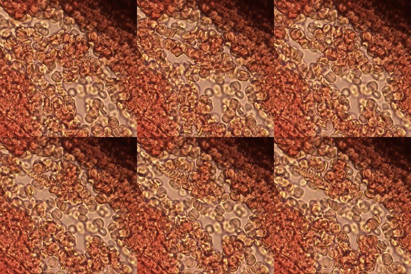
Stacked red blood cells flow across drying slide Human Red Blood Cells - Rouleau stacking.gif
Stacked red blood cells flow across drying slide

Erythrocytes aggregate in a special way, forming rouleaux. Rouleaux are stacks of erythrocytes which form because of the unique discoid shape of the cells in vertebrate body. The flat surface of the discoid RBCs give them a large surface area to make contact and stick to each other; thus, forming a rouleau. Rouleaux formation takes place only in suspensions of RBC containing high-molecular, fibrilar proteins or polymers in the suspending medium (often Dextran-2000 in-vitro). The most important protein causing rouleaux formation in plasma is fibrinogen. RBC suspended in simple salt solutions do not form rouleaux. [1] [2] [3]

Rouleaux

Rouleaux are stacks or aggregations of red blood cells (RBCs) which form because of the unique discoid shape of the cells in vertebrates. The flat surface of the discoid RBCs gives them a large surface area to make contact with and stick to each other; thus forming a rouleau. They occur when the plasma protein concentration is high, and because of them the ESR is also increased. This is a non-specific indicator of the presence of disease.

Vertebrate subphylum of chordates

Vertebrates comprise all species of animals within the subphylum Vertebrata. Vertebrates represent the overwhelming majority of the phylum Chordata, with currently about 69,276 species described. Vertebrates include such groups as the following:

Mechanism

Erythrocyte aggregation is a physiological phenomenon that takes places in normal blood under low-flow conditions or at stasis. The presence or increased concentrations of acute phase proteins, particularly fibrinogen, results in enhanced erythrocyte aggregation.

Fibrinogen Soluble protein complex in blood plasma and involved in clot formation

Fibrinogen is a glycoprotein that circulates in the blood of vertebrates. During tissue and vascular injury it is converted enzymatically by thrombin to fibrin and subsequently to a fibrin-based blood clot. Fibrinogen functions primarily to occlude blood vessels and thereby stop excessive bleeding. However, fibrinogen's product, fibrin, binds and reduces the activity of thrombin. This activity, sometimes referred to as antithrombin I, serves to limit blood clotting. Loss or reduction in this antithrombin 1 activity due to mutations in fibrinogen genes or hypo-fibrinogen conditions can lead to excessive blood clotting and thrombosis. Fibrin also mediates blood platelet and endothelial cell spreading, tissue fibroblast proliferation, capillary tube formation, and angiogenesis and thereby functions to promote tissue revascularization, wound healing, and tissue repair.

Current experimental and theoretical evidence supports the mechanism related to the depletion of high-molecular weight molecules (e.g., fibrinogen) for rouleaux formation. [4] This mechanism is also known as “chemiosmotic hypothesis” for aggregation. [5] Erythrocyte aggregation is determined by both suspending phase (blood plasma) and cellular properties. Surface properties of erythrocytes, such as surface charge density strongly influence the extent and time course of aggregation.

Blood plasma liquid component of blood

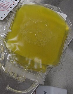
Blood plasma is a yellowish liquid component of blood that holds the blood cells in whole blood in suspension. It is the liquid part of the blood that carries cells and proteins throughout the body. It makes up about 55% of the body's total blood volume. It is the intravascular fluid part of extracellular fluid (all body fluid outside cells). It is mostly water (up to 95% by volume), and contains dissolved proteins (6–8%) (e.g. serum albumins, globulins, and fibrinogen), glucose, clotting factors, electrolytes (Na+, Ca2+, Mg2+, HCO3, Cl, etc.), hormones, carbon dioxide (plasma being the main medium for excretory product transportation) and oxygen. It plays a vital role in an intravascular osmotic effect that keeps electrolyte concentration balanced and protects the body from infection and other blood disorders.

Effects

Erythrocyte aggregation is the main determinant of blood viscosity at low shear rate. Rouleaux formation also determines Erythrocyte sedimentation rate which is a non-specific indicator of the presence of disease. [6]

Erythrocyte sedimentation rate physiological quantity

The erythrocyte sedimentation rate is the rate at which red blood cells in anticoagulated whole blood descend in a standardized tube over a period of one hour. It is a common hematology test, and is a non-specific measure of inflammation. To perform the test, anticoagulated blood is traditionally placed in an upright tube, known as a Westergren tube, and the distance which the red blood cells fall is measured and reported in mm at the end of one hour.

Influence of erythrocyte aggregation on in vivo blood flow is still a controversial issue. [7] Enhanced aggregation affects venous hemodynamics. [8] Erythrocyte aggregation also affects hemodynamic mechanisms in microcirculation and vascular control mechanisms. [9]

Causes

Conditions which cause increased rouleaux formation include infections, inflammatory and connective tissue disorders, and cancers (most common in multiple myeloma). It also occurs in diabetes mellitus and is one of the causative factors for microvascular occlusion in diabetic retinopathy.

Inflammation signs of activation of the immune system

Inflammation is part of the complex biological response of body tissues to harmful stimuli, such as pathogens, damaged cells, or irritants, and is a protective response involving immune cells, blood vessels, and molecular mediators. The function of inflammation is to eliminate the initial cause of cell injury, clear out necrotic cells and tissues damaged from the original insult and the inflammatory process, and initiate tissue repair.

Connective tissue type of biological tissue

Connective tissue (CT) is one of the four basic types of animal tissue, along with epithelial tissue, muscle tissue, and nervous tissue. It develops from the mesoderm. Connective tissue is found in between other tissues everywhere in the body, including the nervous system. In the central nervous system, the three outer membranes that envelop the brain and spinal cord are composed of connective tissue. They support and protect the body. All connective tissue consists of three main components: fibers, ground substance and cells. Not all authorities include blood or lymph as connective tissue because they lack the fiber component. All are immersed in the body water.

Multiple myeloma A myeloid neoplasm that is located in the plasma cells in bone marrow.

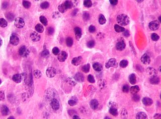
Multiple myeloma (MM), also known as plasma cell myeloma, is a cancer of plasma cells, a type of white blood cell which normally produces antibodies. Often, no symptoms are noticed initially. As it progresses, bone pain, bleeding, frequent infections, and anemia may occur. Complications may include amyloidosis.

Erythrocyte sedimentation rate closely reflects the extent of aggregation, therefore can be used as a measure of aggregation. Erythrocyte aggregation can also be quantitated by monitoring optical properties of blood during the time course of aggregation process. [10]

Measurement

blood film

syllectometry

intravital microscopy

high-frequency ultrasound

Optical coherence tomography

Related Research Articles

Red blood cell most common type of blood cell

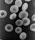
Red blood cells, also known as RBCs, red cells, red blood corpuscles, haematids, erythroid cells or erythrocytes (from Greek erythros for "red" and kytos for "hollow vessel", with -cyte translated as "cell" in modern usage), are the most common type of blood cell and the vertebrate's principal means of delivering oxygen (O2) to the body tissues—via blood flow through the circulatory system. RBCs take up oxygen in the lungs, or gills of fish, and release it into tissues while squeezing through the body's capillaries.

Disseminated intravascular coagulation pathological process characterized by the widespread activation of the clotting cascade that results in the formation of blood clots in the small blood vessels throughout the body

Disseminated intravascular coagulation (DIC) is a condition in which blood clots form throughout the body, blocking small blood vessels. Symptoms may include chest pain, shortness of breath, leg pain, problems speaking, or problems moving parts of the body. As clotting factors and platelets are used up, bleeding may occur. This may include blood in the urine, blood in the stool, or bleeding into the skin. Complications may include organ failure.

Hemorheology, also spelled haemorheology, or blood rheology, is the study of flow properties of blood and its elements of plasma and cells. Proper tissue perfusion can occur only when blood's rheological properties are within certain levels. Alterations of these properties play significant roles in disease processes. Blood viscosity is determined by plasma viscosity, hematocrit and mechanical properties of red blood cells. Red blood cells have unique mechanical behavior, which can be discussed under the terms erythrocyte deformability and erythrocyte aggregation. Because of that, blood behaves as a non-Newtonian fluid. As such, the viscosity of blood varies with shear rate. Blood becomes less viscous at high shear rates like those experienced with increased flow such as during exercise or in peak-systole. Therefore, blood is a shear-thinning fluid. Contrarily, blood viscosity increases when shear rate goes down with increased vessel diameters or with low flow, such as downstream from an obstruction or in diastole. Blood viscosity also increases with increases in red cell aggregability.

Hemolytic anemia is a form of anemia due to hemolysis, the abnormal breakdown of red blood cells (RBCs), either in the blood vessels or elsewhere in the human body. It has numerous possible consequences, ranging from relatively harmless to life-threatening. The general classification of hemolytic anemia is either inherited or acquired. Treatment depends on the cause and nature of the breakdown.

Conjunctiva Outer covering of sclera

The conjunctiva is a tissue that lines the inside of the eyelids and covers the sclera. It is composed of unkeratinized, stratified squamous epithelium with goblet cells, and stratified columnar epithelium. The conjunctiva is highly vascularised, with many microvessels easily accessible for imaging studies.

Dipyridamole anticoagulant drug

Dipyridamole is a medication that inhibits blood clot formation when given chronically and causes blood vessel dilation when given at high doses over a short time.

Autoagglutination represents clumping of an individual's red blood cells by his or her own serum due to the RBCs being coated on their surface by antibodies.

The dysfibrinogenemias consist of three types of fibrinogen disorders in which a critical blood clotting factor, fibrinogen, circulates at normal levels but is dysfunctional. Congenital dysfibrinogenemia is an inherited disorder in which one of the parental genes produces an abnormal fibrinogen. This fibrinogen interferes with normal blood clotting and/or lyses of blood clots. The condition therefore may cause pathological bleeding and/or thrombosis. Acquired dysfibrinogenemia is a non-hereditary disorder in which fibrinogen is dysfunctional due to the presence of liver disease, autoimmune disease, a plasma cell dyscrasias, or certain cancers. It is associated primarily with pathological bleeding. Hereditary fibrinogen Aα-Chain amyloidosis is a sub-category of congenital dysfibrinogenemia in which the dysfunctional fibrinogen does not cause bleeding or thrombosis but rather gradually accumulates in, and disrupts the function of, the kidney.

Cryofibrinogenemia refers to a condition classified as a fibrinogen disorder in which the chilling of an individual's blood plasma from the normal body temperature of 37 °C to the near-freezing temperature of 4 °C causes the reversible precipitation of a complex containing fibrinogen, fibrin, fibronectin, and, occasionally, small amounts of fibrin split products, albumin, immunoglobulins and other plasma proteins. Returning this plasma to 37 °C resolubilizes the precipitate.

Shu Chien, is a Chinese–American physiologist and bioengineer. His work on the fluid dynamics of blood flow has had a major impact on the diagnosis and treatment of cardiovascular diseases such as atherosclerosis. More recently, Chien's research has focused on the mechanical forces, such as pressure and flow, that regulate the behaviors of the cells in blood vessels. Chien is currently President of the Biomedical Engineering Society and is one of only 11 scholars who are members of all three U.S. national institutes: the National Academy of Sciences, National Academy of Engineering, and the Institute of Medicine.

Erythrocyte deformability refers to the ability of erythrocytes to change shape under a given level of applied stress, without hemolysing (rupturing). This is an important property because erythrocytes must change their shape extensively under the influence of mechanical forces in fluid flow or while passing through microcirculation. The extent and geometry of this shape change can be affected by the mechanical properties of the erythrocytes, the magnitude of the applied forces, and the orientation of erythrocytes with the applied forces. Deformability is an intrinsic cellular property of erythrocytes determined by geometric and material properties of the cell membrane, although as with many measurable properties the ambient conditions may also be relevant factors in any given measurement. No other cells of mammalian organisms have deformability comparable with erythrocytes; furthermore, non-mammalian erythrocytes are not deformable to an extent comparable with mammalian erythrocytes. In human RBC there are structural support that aids resilience in RBC which include the cytoskeleton- actin and spectrin that are held together by ankyrin.

Echinocyte, in human biology and medicine, refers to a form of red blood cell that has an abnormal cell membrane characterized by many small, evenly spaced thorny projections. A more common term for these cells is burr cells.

Laser diffraction analysis

Laser diffraction analysis, also known as laser diffraction spectroscopy, is a technology that utilizes diffraction patterns of a laser beam passed through any object ranging from nanometers to millimeters in size to quickly measure geometrical dimensions of a particle. This process does not depend on volumetric flow rate, the amount of particles that passes through a surface over time.

Erythrocyte fragility refers to the propensity of erythrocytes to hemolyse (rupture) under stress. It can be thought of as the degree or proportion of hemolysis that occurs when a sample of red blood cells are subjected to stress. Depending on the application as well as the kind of fragility involved, the amount of stress applied and/or the significance of the resultant hemolysis may vary.

Fåhræus effect

The Fåhræus effect is the decrease in average concentration of red blood cells in human blood as the diameter of the glass tube in which it is flowing decreases. In other words, in blood vessels with diameters less than 500 micrometers, the hematocrit decreases with decreasing capillary diameter. The Fåhræus effect definitely influences the Fåhræus–Lindqvist effect, which describes the dependence of apparent viscosity of blood on the capillary size, but the former is not the only cause of the latter.

Axel Radlach Pries is a German professor of physiology and, since 2015, Dean of the board of Charité hospital in Berlin, Germany. He is married to the photographer Gina Elisabeth Pries.
References

  1. Chien S, Sung LA (1987). "Physicochemical basis and clinical implications of red cell aggregation". Clinical Hemorheology. 7: 71–91.
  2. Chien S, Jan KM (1973). "Ultrastructural basis of the mechanism of rouleaux formation". Microvascular Research. 5 (2): 155–66. doi:10.1016/0026-2862(73)90068-X. PMID   4694282.
  3. Mesielman HJ (1993). "Red blood cell role in RBC aggregation: 1963-1993 and beyond". Clinical Hemorheology. 13: 575–592.
  4. Neu B, Meiselman HJ (2002). "Depletion-mediated red blood cell aggregation in polymer solutions". Biophysical Journal. 83 (5): 2482–2490. doi:10.1016/S0006-3495(02)75259-4. PMC   1302334 . PMID   12414682.
  5. Meiselman HJ (2009). "Red blood cell aggregation: 45 years being curious". Biorheology. 46 (1): 1–19. doi:10.3233/BIR-2009-0522. PMID   19252224.
  6. Oxford Textbook of Medicine
  7. Baskurt OK, Meiselman HJ (2008). "RBC Aggregation: More Important than RBC Adhesion to Endothelial Cells as a Determinant of In Vivo Blood Flow in Health and Disease". Microcirculation. 15 (7): 585–590. doi:10.1080/10739680802107447. PMID   18608991.
  8. Cabel M, Meiselman HJ, Popel AS, Johnson PC (1997). "Contribution of red blood cell aggregation to venous vascular resistance in skeletal muscle". American Journal of Physiology. 272 (2 Pt 2): H1020–H1032. PMID   9124410.
  9. Baskurt OK (2008). "In vivo correlates of altered blood rheology". Biorheology. 45 (6): 629–638. PMID   19065010.
  10. Baskurt OK, Uyuklu M, Ulker P, et al. (2009). "Comparison of three instruments for measuring red blood cell aggregation". Clinical Hemorheology and Microcirculation. 43 (4): 283–298. doi:10.3233/CH-2009-1240. PMID   19996518.