Evolution of human colour vision

Last updated

The evolution of human colour vision produced a trichromatic view of the world in comparison to a majority of other mammals that only have a dichromatic view. Early pre-primate mammals are believed to have viewed the world partly using ultraviolet (UV) light around 90 million years ago, but in the lineage leading to humans their spectral sensitivity shifted toward blue via a succession of mutations. [1] It is thought that the shift to trichromatic color vision evolved in primates as an adaptive trait over time. [2]

Contents

Primitive ancestral vision

It is believed that due to a number of environmental factors, ancient mammals lived with limited colour vision. This is believed to have also been influenced by life styles, including being predominantly nocturnal. [2] There is little data indicating the advantages of UV vision in early mammals. [2]

Early mammal ultraviolet vision, or ultraviolet sensitivity, included sensitivity in the wavelength ranges between 350nm and 430nm. [2] These wavelengths are shorter than visible light but longer than X-rays. In some rare cases, some modern day humans can see within the UV spectrum at wavelengths close to 310nm. [1]

In other animals that possess UV vision such as birds, ultraviolet sensitivity can be advantageous for courtship and reproductive success. This is because some birds have feathers with certain favourable colourations that can not be distinguished by human vision outside of the UV spectrum. [2]

Opsins and colour vision

Opsins function by acting as enzymes that are activated and change shape when light absorption causes chromophores to isomerize. [3] Opsins are responsible for adjusting wavelength dependence of the chromophore light induced isomerization reaction. [3] Therefore, opsins act by determining chromophore sensitivity to light at any given wavelength. Opsins that have different amino acid sequences but are bound to identical chromophores result in different absorption values at each wavelength. [4]

Opsin genes are used to encode the photoreceptor proteins responsible for color vision and dim light vision. [5] The photoreceptor proteins created can be further categorized in to rhodopsins, which are found in rod photoreceptor cells and assist with night vision; and photopsins, or cone opsins, which are responsible for colour vision and expressed in cone photoreceptor cells of the retina. [6]

Cone opsins are categorized further by their absorption maxima λ max which is the wavelength when the greatest amount of light absorption takes place. Further categorization of cone opsins also depends on the specific amino acid sequences each of the opsins uses, which may have an evolutionary basis. [5]

Evolution of cone opsins and human colour vision

Recent studies have shown that primitive nocturnal mammalian ancestors had dichromatic vision consisting of UV–sensitive and red–sensitive traits. [2] A change occurred approximately 30 million years ago where human ancestors evolved four classes of opsin genes, which enabled vision that included the full spectrum of visible light. [2] UV–sensitivity is said to have been lost at this time. [1]

Mutagenesis experiments involving the Boreoeutherian ancestor to humans have shown that seven genetic mutations are linked to losing UV vision and gaining the blue light vision that most humans have today over the course of millions of years. [1] These mutations: F46T, F49L, T52F, F86L, T93P, A114G and S118T, include 5040 potential pathways for the amino acid changes required to create genetic changes in the short wavelength sensitive, or blue opsin. [1] Of the 5040 pathways, 335 have been deemed as possible trajectories for the evolution of blue opsin. [1] It has been discovered that each individual mutation has no effect on its own, and that only multiple changes combined following an epistatic pattern in a specific order resulted in changes in the evolutionary direction of blue vision. [1]

Incomplete trajectories, or evolutionary pathways, are shown to be caused by T52F mutations occurring first because T52F does not have a peak for the absorption of light within the entire visible region. [1] T52F mutations are deemed to be structurally unstable, and the evolutionary path is immediately terminated. Having any of the other stable mutations occur first, including F46T, F49L, F86L, T93P, A114G or S118T, opens up the possibility of having 1032 out of 5042 potential trajectories open up to evolution. [1] This is because having any of the other mutations occur first would allow for 134, 74, 252, 348, 102 and 122 potential pathways for mutations involving each of the remaining 6 mutants, equal to 1032 potential pathways for the evolution of short wavelength sensitive opsins to take place. [1]

Studies using in vitro assays have shown that epistatic evolution took place in ancestral Boreoeutherian species with the 7 mutations on genetically reconstructed Boreoetherian short wavelength sensitive opsins. [1] λmax values were shifted from a value of 357nm to 411nm, an increase which indicated that human short wavelength sensitive opsins did indeed evolve from ancestral Boreoeutherian species using these 7 mutations. [1]

Further analysis has shown that 4008 out of the 5040 possible trajectories were terminated prematurely due to nonfunctional pigments that were dehydrated. [1] Mutagenesis results also reveal that ancestral human short wavelength sensitive opsin remained UV-sensitive until about 80 million years ago, before gradually increasing its λmax by 20nm 75 million years ago and 20nm 45 million years ago. It eventually reached the current λmax of 430nm 30 million years ago. [1]

It is believed that middle and long wave sensitive pigments appeared after the final stages of short wavelength sensitive opsin pigments evolved, and that trichromatic vision was formed through interprotein epistasis. [1]

Scientists believe that the slow rate of evolution of human ancestral vision can also be attributed to slow environmental changes. [2]

Evolutionary pathway of short wavelength opsins

It has been theorized that λ max-shifts might have been required as human ancestors started to switch from leading nocturnal lifestyles to more diurnal lifestyles. This caused their vision to adjust to various twilight settings over time. To identify the path from which short wavelength opsins evolved, increases in absolute max values were used by researchers with a limitation of approximately |Δλmax|<25nm per step. [1] This allows for subdivision of the 1032 potential pathways that were generated by analysis of first mutations beginning with any of the stable mutants: F46T, F49L, F86L, T93P, A114G or S118T to be narrowed down to 335 potential pathways. [1]

Evolutionary pathway of the medium and long wavelength opsins

It was found that the last two mutations, F46T and T52F, occurred between 45 million and 30 million years ago as the absolute max for short wave length opsins was increasing from 400nm to 430nm. [1] During this time, ancestral Boreotherian had two long wave length opsins created by gene duplication, with one retaining the absolute max value of 560nm, equal to the ancestral value. This led to the creation of the modern human long wavelength sensitive, or red opsin. [1]

The other short wavelength sensitive opsin increased its absolute max value to 530nm and became the middle wavelength sensitive, or green-sensitive opsin. This occurred through mutations involving S180A, Y277F and T285A. [1] The order that these mutations took place in the ancestral Boreotheria is currently not fully known. It is hypothesized that T285A was one of the first two mutations because absolute max values would be between 532-538nm which is close to the absolute value found in human middle wavelength opsins. [1]

See also

Related Research Articles

<span class="mw-page-title-main">Color blindness</span> Decreased ability to see color or color differences

Color blindness is the decreased ability to see color or differences in color. It can impair tasks such as selecting ripe fruit, choosing clothing, and reading traffic lights. Color blindness may make some academic activities more difficult. However, issues are generally minor, and the colorblind automatically develop adaptations and coping mechanisms. People with total color blindness (achromatopsia) may also be uncomfortable in bright environments and have decreased visual acuity.

<span class="mw-page-title-main">Color vision</span> Ability to perceive differences in light frequency

Color vision, a feature of visual perception, is an ability to perceive differences between light composed of different wavelengths independently of light intensity. Color perception is a part of the larger visual system and is mediated by a complex process between neurons that begins with differential stimulation of different types of photoreceptors by light entering the eye. Those photoreceptors then emit outputs that are propagated through many layers of neurons and then ultimately to the brain. Color vision is found in many animals and is mediated by similar underlying mechanisms with common types of biological molecules and a complex history of evolution in different animal taxa. In primates, color vision may have evolved under selective pressure for a variety of visual tasks including the foraging for nutritious young leaves, ripe fruit, and flowers, as well as detecting predator camouflage and emotional states in other primates.

<span class="mw-page-title-main">Photoreceptor cell</span> Type of neuroepithelial cell

A photoreceptor cell is a specialized type of neuroepithelial cell found in the retina that is capable of visual phototransduction. The great biological importance of photoreceptors is that they convert light into signals that can stimulate biological processes. To be more specific, photoreceptor proteins in the cell absorb photons, triggering a change in the cell's membrane potential.

<span class="mw-page-title-main">Tetrachromacy</span> Type of color vision with four types of cone cells

Tetrachromacy is the condition of possessing four independent channels for conveying color information, or possessing four types of cone cell in the eye. Organisms with tetrachromacy are called tetrachromats.

<span class="mw-page-title-main">Trichromacy</span> Possessing of three independent channels for conveying color information

Trichromacy or trichromatism is the possessing of three independent channels for conveying color information, derived from the three different types of cone cells in the eye. Organisms with trichromacy are called trichromats.

<span class="mw-page-title-main">Cone cell</span> Photoreceptor cells responsible for color vision made to function in bright light

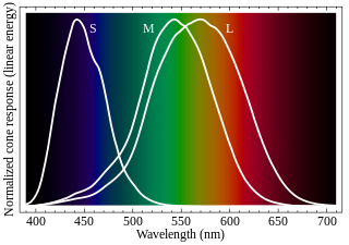
Cone cells, or cones, are photoreceptor cells in the retinas of vertebrate eyes including the human eye. They respond differently to light of different wavelengths, and the combination of their responses is responsible for color vision. Cones function best in relatively bright light, called the photopic region, as opposed to rod cells, which work better in dim light, or the scotopic region. Cone cells are densely packed in the fovea centralis, a 0.3 mm diameter rod-free area with very thin, densely packed cones which quickly reduce in number towards the periphery of the retina. Conversely, they are absent from the optic disc, contributing to the blind spot. There are about six to seven million cones in a human eye, with the highest concentration being towards the macula.

In visual physiology, adaptation is the ability of the retina of the eye to adjust to various levels of light. Natural night vision, or scotopic vision, is the ability to see under low-light conditions. In humans, rod cells are exclusively responsible for night vision as cone cells are only able to function at higher illumination levels. Night vision is of lower quality than day vision because it is limited in resolution and colors cannot be discerned; only shades of gray are seen. In order for humans to transition from day to night vision they must undergo a dark adaptation period of up to two hours in which each eye adjusts from a high to a low luminescence "setting", increasing sensitivity hugely, by many orders of magnitude. This adaptation period is different between rod and cone cells and results from the regeneration of photopigments to increase retinal sensitivity. Light adaptation, in contrast, works very quickly, within seconds.

<span class="mw-page-title-main">Monochromacy</span> Type of color vision

Monochromacy is the ability of organisms or machines to perceive only light intensity, without respect to spectral composition (color). Organisms with monochromacy are called monochromats.

<span class="mw-page-title-main">Photopsin</span> Photoreceptor proteins in the cone cells of the retina

Photopsins are the photoreceptor proteins found in the cone cells of the retina that are the basis of color vision. Photopsins bind the chromophore retinal to form iodopsins. Iodopsins are used in daylight vision and are analogous to rhodopsin that is used in night vision.

<span class="mw-page-title-main">Melanopsin</span> Mammalian protein found in Homo sapiens

Melanopsin is a type of photopigment belonging to a larger family of light-sensitive retinal proteins called opsins and encoded by the gene Opn4. In the mammalian retina, there are two additional categories of opsins, both involved in the formation of visual images: rhodopsin and photopsin in the rod and cone photoreceptor cells, respectively.

<span class="mw-page-title-main">Opsin</span> Class of light-sensitive proteins

Animal opsins are G-protein-coupled receptors and a group of proteins made light-sensitive via the chromophore retinal. Most prominently, they are found in photoreceptor cells of the retina. Five classical groups of opsins are involved in vision, mediating the conversion of a photon of light into an electrochemical signal, the first step in the visual transduction cascade. Another opsin found in the mammalian retina, melanopsin, is involved in circadian rhythms and pupillary reflex but not in vision. Humans have in total nine opsins. Beside vision and light perception, opsins may also sense temperature, sound, and chemicals.

Photoreceptor proteins are light-sensitive proteins involved in the sensing and response to light in a variety of organisms. Some examples are rhodopsin in the photoreceptor cells of the vertebrate retina, phytochrome in plants, and bacteriorhodopsin and bacteriophytochromes in some bacteria. They mediate light responses as varied as visual perception, phototropism and phototaxis, as well as responses to light-dark cycles such as circadian rhythm and other photoperiodisms including control of flowering times in plants and mating seasons in animals.

<span class="mw-page-title-main">OPN1SW</span> Protein-coding gene in the species Homo sapiens

Blue-sensitive opsin is a protein that in humans is encoded by the OPN1SW gene.

<span class="mw-page-title-main">OPN5</span> Protein-coding gene in the species Homo sapiens

Opsin-5, also known as G-protein coupled receptor 136 or neuropsin is a protein that in humans is encoded by the OPN5 gene. Opsin-5 is a member of the opsin subfamily of the G protein-coupled receptors. It is a photoreceptor protein sensitive to ultraviolet (UV) light. The OPN5 gene was discovered in mouse and human genomes and its mRNA expression was also found in neural tissues. Neuropsin is bistable at 0 °C and activates a UV-sensitive, heterotrimeric G protein Gi-mediated pathway in mammalian and avian tissues.

<span class="mw-page-title-main">OPN1MW</span> Protein-coding gene in the species Homo sapiens

Green-sensitive opsin is a protein that in humans is encoded by the OPN1MW gene. OPN1MW2 is a similar opsin.

<span class="mw-page-title-main">OPN1LW</span> Protein-coding gene in the species Homo sapiens

OPN1LW is a gene on the X chromosome that encodes for long wave sensitive (LWS) opsin, or red cone photopigment. It is responsible for perception of visible light in the yellow-green range on the visible spectrum. The gene contains 6 exons with variability that induces shifts in the spectral range. OPN1LW is subject to homologous recombination with OPN1MW, as the two have very similar sequences. These recombinations can lead to various vision problems, such as red-green colourblindness and blue monochromacy. The protein encoded is a G-protein coupled receptor with embedded 11-cis-retinal, whose light excitation causes a cis-trans conformational change that begins the process of chemical signalling to the brain.

<span class="mw-page-title-main">Evolution of color vision in primates</span> Loss and regain of colour vision during the evolution of primates

The evolution of color vision in primates is highly unusual compared to most eutherian mammals. A remote vertebrate ancestor of primates possessed tetrachromacy, but nocturnal, warm-blooded, mammalian ancestors lost two of four cones in the retina at the time of dinosaurs. Most teleost fish, reptiles and birds are therefore tetrachromatic while most mammals are strictly dichromats, the exceptions being some primates and marsupials, who are trichromats, and many marine mammals, who are monochromats.

Color vision, a proximate adaptation of the vision sensory modality, allows for the discrimination of light based on its wavelength components.

<span class="mw-page-title-main">Bird vision</span> Senses for birds

Vision is the most important sense for birds, since good eyesight is essential for safe flight. Birds have a number of adaptations which give visual acuity superior to that of other vertebrate groups; a pigeon has been described as "two eyes with wings". Birds are theropod dinosaurs, and the avian eye resembles that of other reptiles, with ciliary muscles that can change the shape of the lens rapidly and to a greater extent than in the mammals. Birds have the largest eyes relative to their size in the animal kingdom, and movement is consequently limited within the eye's bony socket. In addition to the two eyelids usually found in vertebrates, bird's eyes are protected by a third transparent movable membrane. The eye's internal anatomy is similar to that of other vertebrates, but has a structure, the pecten oculi, unique to birds.

Blue cone monochromacy (BCM) is an inherited eye disease that causes severely impaired color discrimination, low vision, nystagmus and photophobia due to the absence of functionality of red (L) and green (M) cone photoreceptor cells in the retina. This form of retinal disorder is a recessive X-linked disease and manifests its symptoms in early infancy.

References

  1. 1 2 3 4 5 6 7 8 9 10 11 12 13 14 15 16 17 18 19 20 21 Yokoyama et al. 2014.
  2. 1 2 3 4 5 6 7 8 Bowmaker 1998.
  3. 1 2 Terakita 2005.
  4. Cheng & Novales Flamarique 2004.
  5. 1 2 Shichida & Matsuyama 2009.
  6. Fernald 2006.

Sources