Fragmentation (mass spectrometry)

Last updated

In mass spectrometry, fragmentation is the dissociation of energetically unstable molecular ions formed from passing the molecules mass spectrum. These reactions are well documented over the decades and fragmentation patterns are useful to determine the molar weight and structural information of unknown molecules. [1] [2] Fragmentation that occurs in tandem mass spectrometry experiments has been a recent focus of research, because this data helps facilitate the identification of molecules. [3]

Contents

Toluene Fragmentation TolueneFragmentation.svg
Toluene Fragmentation

Mass spectrometry techniques

Fragmentation can occur in the ion source (in-source fragmentation) [4] [5] where it has been used with electron ionization [4] to help identify molecules and, recently (2020), with electrospray ionization it has been shown to provide the same benefit in facilitating molecular identification. [5] Prior to these experiments, [5] [6] electrospray ionization in-source fragmentation was generally considered an undesired effect [7] however, electrospray ionization using Enhanced In-Source Fragmentation/Annotation (EISA) has been shown to promote in-source fragmentation that creates fragment ions that are consistent with tandem mass spectrometers. [5] [6] Tandem mass spectrometry-generated fragmentation is typically made in the collision zone (post-source fragmentation) of a tandem mass spectrometer. EISA and collision-induced dissociation (CID) among other physical events that impact ions are a part of gas-phase ion chemistry. A few different types of mass fragmentation are collision-induced dissociation (CID) through collision with neutral molecule, surface-induced dissociation (SID) using fast moving ions collision with a solid surface, laser induced dissociation which uses laser to induce the ion formation, electron-capture dissociation (ECD) due to capturing of low energy electrons, electron-transfer dissociation (ETD) through electron transfer between ions, negative electron-transfer dissociation (NETD), electron-detachment dissociation (EDD), photodissociation, particularly infrared multiphoton dissociation (IRMPD) using IR radiation for the bombardment and blackbody infrared radiative dissociation (BIRD) which use IR radiation instead of laser, higher-energy C-trap dissociation (HCD), EISA, and charge remote fragmentation. [8] [9] [10]

Fragmentation reactions

Fragmentation is a type of chemical dissociation, in which the removal of the electron from the molecule results in ionization. Removal of electrons from either sigma bond, pi bond or nonbonding orbitals causes the ionization. [2] This can take place by a process of homolytic cleavage or homolysis or heterolytic cleavage or heterolysis of the bond. Relative bond energy and the ability to undergo favorable cyclic transition states affect the fragmentation process. Rules for the basic fragmentation processes are given by Stevenson's Rule.

Homolysis Homolysis (Chemistry) V.1.svg
Homolysis
Heterolysis Heterolysis (chemistry).svg
Heterolysis

Two major categories of bond cleavage patterns are simple bond cleavage reactions and rearrangement reactions. [2]

Simple bond cleavage reactions

Majority of organic compounds undergo simple bond cleavage reactions, in which direct cleavage of bond take place. Sigma bond cleavage, radical site-initiated fragmentation, and charge site-initiated fragmentation are few types of simple bond cleavage reactions. [2]

An example of sigma bond cleavage Sigma bond cleavage example.jpg
An example of sigma bond cleavage

Sigma bond cleavage / σ-cleavage

Sigma bond cleavage is most commonly observed in molecules, which can produce stable cations such as saturated alkanes, secondary and tertiary carbocations. This occurs when an alpha electron is removed. The C-C bond elongates and weakens causing fragmentation. Fragmentation at this site produces a charged and a radical fragment. [2]

An example of radical site-initiated fragmentation Radical site initiated.jpg
An example of radical site-initiated fragmentation

Radical site-initiated fragmentation

Sigma bond cleavage also occurs on radical cations remote from the site of ionization. This is commonly observed in alcohols, ethers, ketones, esters, amines, alkenes, and aromatic compounds with a carbon attached to ring. The cation has a radical on a heteroatom or an unsaturated functional group. The driving force of fragmentation is the strong tendency of the radical ion for electron pairing. Cleavage occurs when the radical and an odd electron from the bonds adjacent to the radical migrate to form a bond between the alpha carbon and either the heteroatom or the unsaturated functional group. The sigma bond breaks; hence this cleavage is also known as homolytic bond cleavage or α-cleavage. [2]

An example of charge site-initiated fragmentation Charged.jpg
An example of charge site-initiated fragmentation

Charge site-initiated cleavage

The driving force of charge site-initiated fragmentation is the inductive effect of the charge site in radical cations. The electrons from the bond adjacent to the charge-bearing atom migrate to that atom, neutralizing the original charge and causing it to move to a different site. This term is also called inductive cleavage and is an example of heterolytic bond cleavage. [2]

An example of McLafferty Rearrangement McLafferty rearrangement.gif
An example of McLafferty Rearrangement

Rearrangement reactions

Rearrangement reactions are fragmentation reactions that form new bonds producing an intermediate structure before cleavage. One of the most studied rearrangement reaction is the McLafferty rearrangement / γ-hydrogen rearrangement. This occurs in the radical cations with unsaturated functional groups, like ketones, aldehydes, carboxylic acids, esters, amides, olefins, phenylalkanes. During this reaction, γ-hydrogen will transfer to the functional group at first and then subsequent α, β-bond cleavage of the intermediate will take place. [2] Other rearrangement reactions include heterocyclic ring fission (HRF), benzofuran forming fission (BFF), quinone methide (QM) fission or Retro Diels-Alder (RDA). [11]

See also

Related Research Articles

Mass spectrometry (MS) is an analytical technique that is used to measure the mass-to-charge ratio of ions. The results are presented as a mass spectrum, a plot of intensity as a function of the mass-to-charge ratio. Mass spectrometry is used in many different fields and is applied to pure samples as well as complex mixtures.

<span class="mw-page-title-main">Ion source</span> Device that creates charged atoms and molecules (ions)

An ion source is a device that creates atomic and molecular ions. Ion sources are used to form ions for mass spectrometers, optical emission spectrometers, particle accelerators, ion implanters and ion engines.

<span class="mw-page-title-main">Electron ionization</span> Ionization technique

Electron ionization is an ionization method in which energetic electrons interact with solid or gas phase atoms or molecules to produce ions. EI was one of the first ionization techniques developed for mass spectrometry. However, this method is still a popular ionization technique. This technique is considered a hard ionization method, since it uses highly energetic electrons to produce ions. This leads to extensive fragmentation, which can be helpful for structure determination of unknown compounds. EI is the most useful for organic compounds which have a molecular weight below 600. Also, several other thermally stable and volatile compounds in solid, liquid and gas states can be detected with the use of this technique when coupled with various separation methods.

<span class="mw-page-title-main">Tandem mass spectrometry</span> Type of mass spectrometry

Tandem mass spectrometry, also known as MS/MS or MS2, is a technique in instrumental analysis where two or more mass analyzers are coupled together using an additional reaction step to increase their abilities to analyse chemical samples. A common use of tandem MS is the analysis of biomolecules, such as proteins and peptides.

<span class="mw-page-title-main">Chemical ionization</span> Ionization technique used in mass [[spectroscopy]]

Chemical ionization (CI) is a soft ionization technique used in mass spectrometry. This was first introduced by Burnaby Munson and Frank H. Field in 1966. This technique is a branch of gaseous ion-molecule chemistry. Reagent gas molecules are ionized by electron ionization to form reagent ions, which subsequently react with analyte molecules in the gas phase to create analyte ions for analysis by mass spectrometry. Negative chemical ionization (NCI), charge-exchange chemical ionization, atmospheric-pressure chemical ionization (APCI) and atmospheric pressure photoionization (APPI) are some of the common variants of the technique. CI mass spectrometry finds general application in the identification, structure elucidation and quantitation of organic compounds as well as some utility in biochemical analysis. Samples to be analyzed must be in vapour form, or else, must be vapourized before introduction into the source.

Infrared multiple photon dissociation (IRMPD) is a technique used in mass spectrometry to fragment molecules in the gas phase usually for structural analysis of the original (parent) molecule.

<span class="mw-page-title-main">Electron-capture dissociation</span>

Electron-capture dissociation (ECD) is a method of fragmenting gas-phase ions for structure elucidation of peptides and proteins in tandem mass spectrometry. It is one of the most widely used techniques for activation and dissociation of mass selected precursor ion in MS/MS. It involves the direct introduction of low-energy electrons to trapped gas-phase ions.

Gas phase ion chemistry is a field of science encompassed within both chemistry and physics. It is the science that studies ions and molecules in the gas phase, most often enabled by some form of mass spectrometry. By far the most important applications for this science is in studying the thermodynamics and kinetics of reactions. For example, one application is in studying the thermodynamics of the solvation of ions. Ions with small solvation spheres of 1, 2, 3... solvent molecules can be studied in the gas phase and then extrapolated to bulk solution.

Hydrogen–deuterium exchange is a chemical reaction in which a covalently bonded hydrogen atom is replaced by a deuterium atom, or vice versa. It can be applied most easily to exchangeable protons and deuterons, where such a transformation occurs in the presence of a suitable deuterium source, without any catalyst. The use of acid, base or metal catalysts, coupled with conditions of increased temperature and pressure, can facilitate the exchange of non-exchangeable hydrogen atoms, so long as the substrate is robust to the conditions and reagents employed. This often results in perdeuteration: hydrogen-deuterium exchange of all non-exchangeable hydrogen atoms in a molecule.

Alpha-cleavage (α-cleavage) in organic chemistry refers to the act of breaking the carbon-carbon bond adjacent to the carbon bearing a specified functional group.

<span class="mw-page-title-main">Electron-transfer dissociation</span>

Electron-transfer dissociation (ETD) is a method of fragmenting multiply-charged gaseous macromolecules in a mass spectrometer between the stages of tandem mass spectrometry (MS/MS). Similar to electron-capture dissociation, ETD induces fragmentation of large, multiply-charged cations by transferring electrons to them. ETD is used extensively with polymers and biological molecules such as proteins and peptides for sequence analysis. Transferring an electron causes peptide backbone cleavage into c- and z-ions while leaving labile post translational modifications (PTM) intact. The technique only works well for higher charge state peptide or polymer ions (z>2). However, relative to collision-induced dissociation (CID), ETD is advantageous for the fragmentation of longer peptides or even entire proteins. This makes the technique important for top-down proteomics. The method was developed by Hunt and coworkers at the University of Virginia.

<span class="mw-page-title-main">Protein mass spectrometry</span> Application of mass spectrometry

Protein mass spectrometry refers to the application of mass spectrometry to the study of proteins. Mass spectrometry is an important method for the accurate mass determination and characterization of proteins, and a variety of methods and instrumentations have been developed for its many uses. Its applications include the identification of proteins and their post-translational modifications, the elucidation of protein complexes, their subunits and functional interactions, as well as the global measurement of proteins in proteomics. It can also be used to localize proteins to the various organelles, and determine the interactions between different proteins as well as with membrane lipids.

<span class="mw-page-title-main">Mass spectral interpretation</span>

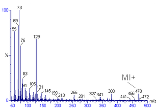
Mass spectral interpretation is the method employed to identify the chemical formula, characteristic fragment patterns and possible fragment ions from the mass spectra. Mass spectra is a plot of relative abundance against mass-to-charge ratio. It is commonly used for the identification of organic compounds from electron ionization mass spectrometry. Organic chemists obtain mass spectra of chemical compounds as part of structure elucidation and the analysis is part of many organic chemistry curricula.

<span class="mw-page-title-main">Triple quadrupole mass spectrometer</span>

A triple quadrupole mass spectrometer (TQMS), is a tandem mass spectrometer consisting of two quadrupole mass analyzers in series, with a (non-mass-resolving) radio frequency (RF)–only quadrupole between them to act as a cell for collision-induced dissociation. This configuration is often abbreviated QqQ, here Q1q2Q3.

Electron capture ionization is the ionization of a gas phase atom or molecule by attachment of an electron to create an ion of the form . The reaction is

Unimolecular ion decomposition is the fragmentation of a gas phase ion in a reaction with a molecularity of one. Ions with sufficient internal energy may fragment in a mass spectrometer, which in some cases may degrade the mass spectrometer performance, but in other cases, such as tandem mass spectrometry, the fragmentation can reveal information about the structure of the ion.

<span class="mw-page-title-main">Collision-induced dissociation</span> Mass spectrometry technique to induce fragmentation of selected ions in the gas phase

Collision-induced dissociation (CID), also known as collisionally activated dissociation (CAD), is a mass spectrometry technique to induce fragmentation of selected ions in the gas phase. The selected ions are usually accelerated by applying an electrical potential to increase the ion kinetic energy and then allowed to collide with neutral molecules. In the collision some of the kinetic energy is converted into internal energy which results in bond breakage and the fragmentation of the molecular ion into smaller fragments. These fragment ions can then be analyzed by tandem mass spectrometry.

<span class="mw-page-title-main">Atmospheric pressure photoionization</span> Soft ionization method

Atmospheric pressure photoionization (APPI) is a soft ionization method used in mass spectrometry (MS) usually coupled to liquid chromatography (LC). Molecules are ionized using a vacuum ultraviolet (VUV) light source operating at atmospheric pressure, either by direct absorption followed by electron ejection or through ionization of a dopant molecule that leads to chemical ionization of target molecules. The sample is usually a solvent spray that is vaporized by nebulization and heat. The benefit of APPI is that it ionizes molecules across a broad range of polarity and is particularly useful for ionization of low polarity molecules for which other popular ionization methods such as electrospray ionization (ESI) and atmospheric pressure chemical ionization (APCI) are less suitable. It is also less prone to ion suppression and matrix effects compared to ESI and APCI and typically has a wide linear dynamic range. The application of APPI with LC/MS is commonly used for analysis of petroleum compounds, pesticides, steroids, and drug metabolites lacking polar functional groups and is being extensively deployed for ambient ionization particularly for explosives detection in security applications.

<span class="mw-page-title-main">Gary Siuzdak</span> American chemist

Gary Siuzdak is an American chemist best known for his work in the field of metabolomics, activity metabolomics, and mass spectrometry. His lab discovered indole-3-propionic acid as a gut bacteria derived metabolite in 2009. He is currently the Professor and Director of The Center for Metabolomics and Mass Spectrometry at Scripps Research in La Jolla, California. Siuzdak has also made contributions to virus analysis, viral structural dynamics, as well as developing mass spectrometry imaging technology using nanostructured surfaces. The Siuzdak lab is also responsible for creating the research tools eXtensible Computational Mass Spectrometry (XCMS), METLIN, METLIN Neutral Loss and Q-MRM. As of January 2021, the XCMS/METLIN platform has over 50,000 registered users.

References

  1. McLafferty FW (1 January 1993). Interpretation of Mass Spectra. University Science Books. ISBN   978-0-935702-25-5.
  2. 1 2 3 4 5 6 7 8 Dass C (2007). Fundamentals of contemporary mass spectrometry ([Online-Ausg.]. ed.). Hoboken, NJ [u.a.]: Wiley. ISBN   978-0-471-68229-5.
  3. Xue J, Guijas C, Benton HP, Warth B, Siuzdak G (October 2020). "2 molecular standards database: a broad chemical and biological resource". Nature Methods. 17 (10): 953–954. doi:10.1038/s41592-020-0942-5. PMC   8802982 . PMID   32839599. S2CID   221285246.
  4. 1 2 Gohlke RS, McLafferty FW (1993-05-01). "Early gas chromatography/mass spectrometry". Journal of the American Society for Mass Spectrometry. 4 (5): 367–371. doi:10.1016/1044-0305(93)85001-E. PMID   24234933. S2CID   33972992.
  5. 1 2 3 4 Xue J, Domingo-Almenara X, Guijas C, Palermo A, Rinschen MM, Isbell J, et al. (April 2020). "Enhanced in-Source Fragmentation Annotation Enables Novel Data Independent Acquisition and Autonomous METLIN Molecular Identification". Analytical Chemistry. 92 (8): 6051–6059. doi:10.1021/acs.analchem.0c00409. PMC   8966047 . PMID   32242660. S2CID   214768212.
  6. 1 2 Domingo-Almenara X, Montenegro-Burke JR, Guijas C, Majumder EL, Benton HP, Siuzdak G (March 2019). "Autonomous METLIN-Guided In-source Fragment Annotation for Untargeted Metabolomics". Analytical Chemistry. 91 (5): 3246–3253. doi:10.1021/acs.analchem.8b03126. PMC   6637741 . PMID   30681830.
  7. Lu W, Su X, Klein MS, Lewis IA, Fiehn O, Rabinowitz JD (June 2017). "Metabolite Measurement: Pitfalls to Avoid and Practices to Follow". Annual Review of Biochemistry. 86 (1): 277–304. doi:10.1146/annurev-biochem-061516-044952. PMC   5734093 . PMID   28654323.
  8. Yost RA, Enke CG (1978). "Selected ion fragmentation with a tandem quadrupole mass spectrometer". Journal of the American Chemical Society. 100 (7): 2274–2275. doi:10.1021/ja00475a072.
  9. Lermyte F, Valkenborg D, Loo JA, Sobott F (November 2018). "Radical solutions: Principles and application of electron-based dissociation in mass spectrometry-based analysis of protein structure" (PDF). Mass Spectrometry Reviews. 37 (6): 750–771. doi:10.1002/mas.21560. PMC   6131092 . PMID   29425406.
  10. Chen X, Wang Z, Wong YE, Wu R, Zhang F, Chan TD (November 2018). "Electron-ion reaction-based dissociation: A powerful ion activation method for the elucidation of natural product structures". Mass Spectrometry Reviews. 37 (6): 793–810. doi:10.1002/mas.21563. PMID   29603345.
  11. Li HJ, Deinzer ML (February 2007). "Tandem mass spectrometry for sequencing proanthocyanidins". Analytical Chemistry. 79 (4): 1739–48. doi:10.1021/ac061823v. PMID   17297981.