Giardia lamblia, also known as Giardia intestinalis, is a flagellatedparasiticmicroorganism, that colonizes and reproduces in the small intestine, causing giardiasis. The parasite attaches to the epithelium by a ventraladhesive disc or sucker, and reproduces via binary fission.[1] Giardiasis does not spread via the bloodstream, nor does it spread to other parts of the gastrointestinal tract, but remains confined to the lumen of the small intestine.[2]Giardiatrophozoites absorb their nutrients from the lumen of the small intestine, and are anaerobes. If the organism is split and stained, its characteristic pattern resembles the familiar "smiley face" symbol.[3] Chief pathways of human infection include ingestion of untreated sewage, a phenomenon particularly common in many developing countries;[4] contamination of natural waters also occurs in watersheds where intensive grazing occurs. Giardia infections occur worldwide, however Giardia lamblia is the most commonly identified intestinal parasite in the United States and Canada among children in day care centers, hikers, family members and immunocompromised adults. Approximately 20,000 cases per year in the United States are reported.[5]
A flagellate is a cell or organism with one or more whip-like appendages called flagella. The word flagellate also describes a particular construction characteristic of many prokaryotes and eukaryotes and their means of motion. The term presently does not imply any specific relationship or classification of the organisms that possess flagellae. However, the term "flagellate" is included in other terms which are more formally characterized.
In evolutionary biology, parasitism is a relationship between species, where one organism, the parasite, lives on or in another organism, the host, causing it some harm, and is adapted structurally to this way of life. The entomologist E. O. Wilson has characterised parasites as "predators that eat prey in units of less than one". Parasites include protozoans such as the agents of malaria, sleeping sickness, and amoebic dysentery; animals such as hookworms, lice, mosquitoes, and vampire bats; fungi such as honey fungus and the agents of ringworm; and plants such as mistletoe, dodder, and the broomrapes. There are six major parasitic strategies of exploitation of animal hosts, namely parasitic castration, directly transmitted parasitism, trophically transmitted parasitism, vector-transmitted parasitism, parasitoidism, and micropredation.
A microorganism, or microbe, is a microscopic organism, which may exist in its single-celled form or in a colony of cells.
G. lamblia takes on two morphologically distinct forms during its life cycle. The replicative form is a motile pear-shaped cell that survives only in host small intestines called a trophozoite.[6] Trophozoites swim through the intestinal mucus until they eventually adhere to the host intestinal epithelium.[7][6] Adhered trophozoites then divide by binary fission, forming either more trophozoites or the non-replicative cyst stage.[6] Cysts pass through the host large intestine and are shed in the feces.[6]G. lamblia cysts are resistant to environment stressors, and can survive in the environment for weeks to months if kept moist.[7][8][6] Cysts remain dormant until ingested by a host animal. In the new host, environmental conditions trigger the cyst to produce two trophozoites, which then attach to epithelial cells, starting the cycle anew.[6]
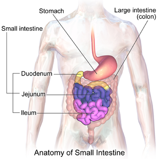
The small intestine or small bowel is an organ in the gastrointestinal tract where most of the end absorption of nutrients and minerals from food takes place. It lies between the stomach and large intestine, and receives bile and pancreatic juice through the pancreatic duct to aid in digestion.
A trophozoite is the activated, feeding stage in the life cycle of certain protozoa such malaria-causing Plasmodium falciparum and those of the Giardia group..
The large intestine, also known as the large bowel, is the last part of the gastrointestinal tract and of the digestive system in vertebrates. Water is absorbed here and the remaining waste material is stored as feces before being removed by defecation.
Ecology and distribution
The cyst can survive for weeks to months in cold water,[9] so can be present in contaminated wells and water systems, especially stagnant water sources, such as naturally occurring ponds, storm water storage systems, and even clean-looking mountain streams. Can also be found on surfaces, soil, food, or water that has been contaminated with feces from infected humans or animals.[10] They may also occur in city reservoirs and persist after water treatment, as the cysts are resistant to conventional water treatment methods, such as chlorination and ozonolysis.[9]Zoonotic transmission is also possible, so Giardia infection is a concern for people camping in the wilderness or swimming in contaminated streams or lakes, especially the artificial lakes formed by beaver dams (hence the popular name for giardiasis, "beaver fever").
Water chlorination is the process of adding chlorine or chlorine compounds such as sodium hypochlorite to water. This method is used to kill certain bacteria and other microbes in tap water as chlorine is highly toxic. In particular, chlorination is used to prevent the spread of waterborne diseases such as cholera, dysentery, and typhoid.
Ozonolysis is an organic reaction where the unsaturated bonds of alkenes, alkynes, or azo compounds are cleaved with ozone. Alkenes and alkynes form organic compounds in which the multiple carbon–carbon bond has been replaced by a carbonyl group while azo compounds form nitrosamines. The outcome of the reaction depends on the type of multiple bond being oxidized and the work-up conditions.
The beaver is a large, primarily nocturnal, semiaquatic rodent. Castor includes two extant species, the North American beaver and Eurasian beaver (Eurasia). Beavers are known for building dams, canals, and lodges (homes). They are the second-largest rodent in the world. Their colonies create one or more dams to provide still, deep water to protect against predators, and to float food and building material. The North American beaver population was once more than 60 million, but as of 1988 was 6–12 million. This population decline is the result of extensive hunting for fur, for glands used as medicine and perfume, and because the beavers' harvesting of trees and flooding of waterways may interfere with other land uses.
In addition to waterborne sources, fecal–oral transmission can also occur, for example in day-care centers, where children may have poor hygiene practices. Those who work with children are also at risk of being infected, as are family members of infected individuals. Not all Giardia infections are symptomatic, and many people can unknowingly serve as carriers of the parasite.
Giardia infects humans, but is also one of the most common parasites infecting cats, dogs and birds. Mammalian hosts also include dozens of species,[11] including cattle, sheep,[12] and goats.[12]
Cattle—colloquially cows—are the most common type of large domesticated ungulates. They are a prominent modern member of the subfamily Bovinae, are the most widespread species of the genus Bos, and are most commonly classified collectively as Bos taurus.
Domestic sheep are quadrupedal, ruminant mammals typically kept as livestock. Like most ruminants, sheep are members of the order Artiodactyla, the even-toed ungulates. Although the name sheep applies to many species in the genus Ovis, in everyday usage it almost always refers to Ovis aries. Numbering a little over one billion, domestic sheep are also the most numerous species of sheep. An adult female sheep is referred to as a ewe, an intact male as a ram or occasionally a tup, a castrated male as a wether, and a younger sheep as a lamb.
Cats can be cured easily and lambs usually simply lose weight, but in calves, the parasites can be fatal and often are not responsive to antibiotics or electrolytes. Carriers among calves can also be asymptomatic. This parasite is deadly for chinchillas, so extra care must be taken by providing them with safe water. Dogs have a high infection rate, as 30% of the population under one year old are known to be infected in kennels. The infection is more prevalent in puppies than in adult dogs. Infected dogs can be isolated and treated, or the entire pack at a kennel can be treated together regardless. Kennels should also be then cleaned with bleach or other cleaning disinfectants. The grass areas used for exercise should be considered contaminated for at least one month after dogs show signs of infection, as cysts can survive in the environment for long periods of time. Prevention can be achieved by quarantine of infected dogs for at least 20 days and careful management and maintenance of a clean water supply.
A quarantine is a restriction on the movement of people and goods which is intended to prevent the spread of disease or pests. It is often used in connection to disease and illness, preventing the movement of those who may have been exposed to a communicable disease, but do not have a confirmed medical diagnosis. The term is often used synonymously with medical isolation, in which those confirmed to be infected with a communicable disease are isolated from the healthy population.
Cell biology
Giardia trophozoites stained with Giemsa; 100x magnification.
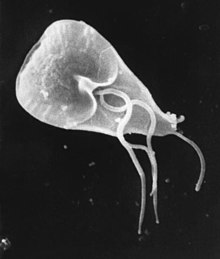
G. lamblia trophozoites are pear-shaped cells, 10 to 20 micrometers long, 7 to 10 micrometers across, and 2 to 4 micrometers thick.[6][7] They are motile by way of four pairs of flagella, which propel the trophozoites through the intestine.[7] Notably, each G. lamblia cell has two nuclei, both of which actively transcribe genes.[6] Adjacent to the nucleus, G. lamblia cells have an endoplasmic reticulum that extends through much of the cell.[13] Trophozoites about to differentiate into cysts also contain prominent vesicles termed encystation-specific vesicles that disappear once cyst wall construction begins.[13] Unlike most other eukaryotes, G. lamblia cells contain no visible mitochondria, but instead contains a substantially reduced metabolic organelle termed a mitosome.[7] Additionally, cells appear to contain no Golgi bodies, and instead the secretory system consists entirely of the endoplasmic reticulum and numerous vesicles spread throughout the cell, termed peripheral vesicles.[13] Peripheral vesicles are responsible both for taking up extracellular nutrients, and expelling waste outside the cell.[8] Each cell also contains a pair of rigid structures called median bodies which make up part of the G. lambliacytoskeleton.[6] Trophozoites adhere to host epithelial cells via a specialized disk-shaped organelle called the ventral disk.[6]
A micrometer, sometimes known as a micrometer screw gauge, is a device incorporating a calibrated screw widely used for accurate measurement of components in mechanical engineering and machining as well as most mechanical trades, along with other metrological instruments such as dial, vernier, and digital calipers. Micrometers are usually, but not always, in the form of calipers. The spindle is a very accurately machined screw and the object to be measured is placed between the spindle and the anvil. The spindle is moved by turning the ratchet knob or thimble until the object to be measured is lightly touched by both the spindle and the anvil.
In cell biology, the nucleus is a membrane-bound organelle found in eukaryotic cells. Eukaryotes usually have a single nucleus, but a few cell types, such as mammalian red blood cells, have no nuclei, and a few others including osteoclasts have many.
Transcription is the first step of DNA based gene expression, in which a particular segment of DNA is copied into RNA by the enzyme RNA polymerase. Both DNA and RNA are nucleic acids, which use base pairs of nucleotides as a complementary language. During transcription, a DNA sequence is read by an RNA polymerase, which produces a complementary, antiparallel RNA strand called a primary transcript.
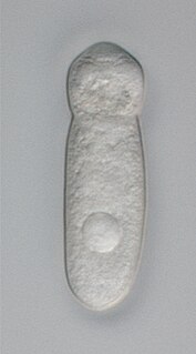
Cysts are oval-shaped cells slightly smaller than trophozoites.[7] They lack flagella, and are covered by a smooth, clear cyst wall.[7] Each cyst contains the organelles for two trophzoites: four nuclei, two ventral disks, etc.[7]
Multiple views of a Giardia lamblia cyst imaged by confocal microscopy. Bar = 10 micrometers (A) Cyst imaged by transmission (differential interference contrast). (B) Cyst wall selectively imaged through use of fluorescent-labelled antibody. (C) Cyst imaged through use of carboxy fluorescein diacetate, a viability stain. (D) Composite image of (B) and (C). (E) Composite image of (A), (B), and (C).
Metabolism
G. lamblia primarily generates its energy by breaking down glucose via glycolysis as well as the arginine dihydrolase pathway.[14] It is unable to synthesize nucleotides on its own, instead salvaging them from its host.[14] Synthesis of iron-sulfur clusters is done in a double-membrane-bound compartment called the mitosome, which is likely a remnant of mitochondria.[14] Each cell contains 25 to 100 mitosomes divided into two categories: peripheral mitosomes which are scattered throughout the cell, and central mitosomes which gather at the center of the cell for unknown reasons.[15] Like in mitochondria, proteins with a certain peptide signal sequence are trafficked to and imported into the mitosome.[14] Unlike mitochondria, mitosomes have no genome of their own. All mitosomal genes are encoded by the Giardia nuclear genome.[14]
Genetics
Giardia and the other diplomonads are unique in their possession of two nuclei that are similar in appearance, DNA content, transcription and time of replication. There are five chromosomes per the haploid genome. The genome has been sequenced and was published in 2007, although the sequence contains several gaps. The sequence is about 12 million base pairs and contains about 5000 protein-coding genes.[16] The GC content is 46%. Trophozoites have a ploidy of four and the ploidy of cysts is eight, which in turn raises the question of how Giardia maintains homogeneity between the chromosomes of the same and opposite nuclei. Modern sequencing technologies have been used to resequence different strains.[17]
Evolution
Giardia had been assumed to be primitively asexual and with no means of transferring DNA between nuclei. These assumptions made it very difficult to explain the remarkably low level of allelic heterozygosity (< 0.01%) in the genome isolate, WB. However, all those assumptions of asexuality are now in doubt, with population genetics providing evidence for recombination[18] and the identification of meiotic genes, evidence for recombination among isolates and the evidence for exchange of genetic material between nuclei during the process of encystation.[19]
These findings on sexuality in Giardia, above, have important implications for understanding the origin of sexual reproduction in eukaryotes. Even though sexual reproduction is widespread among extant eukaryotes, it seemed unlikely, until recently, that sex is a primordial and fundamental feature of eukaryotes. A probable reason for the view that sex may not be fundamental to eukaryotes was that sexual reproduction previously appeared to be lacking in certain human pathogenic single-celled eukaryotes (e.g. Giardia) that diverged from early ancestors in the eukaryotic lineage.
In addition to the evidence cited above for recombination in Giardia, Malik et al.[20] reported that many meiosis specific genes occur in the Giardia genome, and further that homologs of these genes also occur in another unicellular eukaryote, Trichomonas vaginalis. Because these two species are descendants of lineages that are highly divergent among eukaryotes, Malik et al.[20] suggested that these meiotic genes were present in a common ancestor of all eukaryotes. Thus, on this view, the earliest ancestor of eukaryotes was likely capable of sexual reproduction. Furthermore, Dacks and Roger[21] proposed, based on phylogenetic analysis, that facultative sex was present in the common ancestor of all eukaryotes. Bernstein et al. also reviewed evidence in support of this view.[22]
Eight genotypes assemblages of Giardia duodenalis have been recognized to date (A-H).[11] Genotyping of G. duodenalis isolated from various hosts has shown that assemblages A and B infect the largest range of host species, and appear to be the main (or possibly only) G. duodenalis assemblages that undeniably infect human subjects.[11]
Research
Dr. Frances Gillin of the University of California, San Diego and her colleagues cultivated the entire life cycle of this parasite in the laboratory, and identified biochemical cues in the host's digestive system which trigger Giardia's life cycle transformations.[23][24] They also uncovered several ways in which the parasite evades the defences of the infected organism. One of these is by altering the proteins on its surface, which confounds the ability of the infected animal's immune system to detect and combat the parasite (called antigenic variation). Gillin's work reveals why Giardia infections are extremely persistent and prone to recur. In addition, these insights into its biology and survival techniques may enable scientists to develop better strategies to understand, prevent, and treat Giardia infections.
In December 2008, Nature published an article showing the discovery of an RNA interference mechanism that allows Giardia to switch variant-specific surface proteins to avoid host immune response.[25] The discovery was made by the team working at the Biochemistry and Molecular Biology Laboratory, School of Medicine, Catholic University of Cordoba, Argentina, led by Dr. Hugo Lujan.
History
A Giardia trophozoite, drawn by Vilém Lambl and published in 1859.Drawings of a Giardia trophozoite and Cyst by Charles E. Simon in 1921
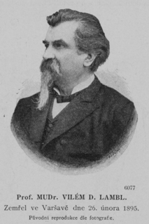
The first likely description of Giardia was in 1681 by Antonie van Leeuwenhoek who, in a letter to Robert Hooke, described "animalcules" resembling Giardia trophozoites in his stool.[6][26] The next known description of Giardia wasn't until 1859, when Czech physician Vilém Lambl published a description of the trophozoite stages he saw in the stool of a pediatric patient. Lambl termed the organism Cercomonas intestinalis.[27] In 1888, Raphaël Blanchard renamed the parasite Lamblia intestinalis in Lambl's honor.[27] In 1915, Charles Stiles renamed the organism Giardia lamblia in honor of both Lambl and Professor Alfred Mathieu Giard of Paris.[27][28] In 1921, Charles E. Simon published a detailed description of the parasite's morphology.[6]
Entamoeba is a genus of Amoebozoa found as internal parasites or commensals of animals.
Giardia is a genus of anaerobic flagellated protozoan parasites of the phylum metamonada that colonise and reproduce in the small intestines of several vertebrates, causing giardiasis. Their life cycle alternates between a swimming trophozoite and an infective, resistant cyst. Giardia were first described by the Dutch microscopist Antonie van Leeuwenhoek in 1681. The genus is named after French zoologist Alfred Mathieu Giard.
Entamoeba histolytica is an anaerobic parasitic amoebozoan, part of the genus Entamoeba. Predominantly infecting humans and other primates causing amoebiasis, E. histolytica is estimated to infect about 50 million people worldwide. E. histolytica infection is estimated to kill more than 55,000 people each year. Previously, it was thought that 10% of the world population was infected, but these figures predate the recognition that at least 90% of these infections were due to a second species, E. dispar. Mammals such as dogs and cats can become infected transiently, but are not thought to contribute significantly to transmission.
Giardiasis, popularly known as beaver fever, is a parasitic disease caused by Giardia lamblia. About 10% of those infected have no symptoms. When symptoms occur they may include diarrhea, abdominal pain, and weight loss. Vomiting, blood in the stool, and fever are less common. Symptoms usually begin 1 to 3 weeks after exposure and without treatment may last up to six weeks.
Trichomonas vaginalis is an anaerobic, flagellated protozoan parasite and the causative agent of trichomoniasis. It is the most common pathogenic protozoan infection of humans in industrialized countries. Infection rates between men and women are similar with women usually being symptomatic, while infections in men are usually asymptomatic. Transmission usually occurs via direct, skin-to-skin contact with an infected individual, most often through vaginal intercourse. The WHO has estimated that 160 million cases of infection are acquired annually worldwide. The estimates for North America alone are between 5 and 8 million new infections each year, with an estimated rate of asymptomatic cases as high as 50%. Usually treatment consists of metronidazole and tinidazole.
Entamoeba coli is a non-pathogenic species of Entamoeba that frequently exists as a commensal parasite in the human gastrointestinal tract. E. coli is important in medicine because it can be confused during microscopic examination of stained stool specimens with the pathogenic Entamoeba histolytica. This amoeba does not move much by the use of its pseudopod, and creates a "sur place (non-progressive) movement" inside the large intestine. Usually, the amoeba is immobile, and keeps its round shape. This amoeba, in its trophozoite stage, is only visible in fresh, unfixed stool specimens. Sometimes the Entamoeba coli have parasites as well. One is the fungus Sphaerita spp. This fungus lives in the cytoplasm of the E. coli. While this differentiation is typically done by visual examination of the parasitic cysts via light microscopy, new methods using molecular biology techniques have been developed.
The diplomonads are a group of flagellates, most of which are parasitic. They include Giardia lamblia, which causes giardiasis in humans. They are placed among the metamonads, and appear to be particularly close relatives of the retortamonads.
Balantidium coli is a parasitic species of ciliate alveolates that causes the disease balantidiasis. It is the only member of the ciliate phylum known to be pathogenic to humans.
The gregarines are a group of Apicomplexan alveolates, classified as the Gregarinasina or Gregarinia. The large parasites inhabit the intestines of many invertebrates. They are not found in any vertebrates. However, gregarines are closely related to both Toxoplasma and Plasmodium, which cause toxoplasmosis and malaria, respectively. Both protists use protein complexes similar to those that are formed by the gregarines for gliding motility and invading target cells. This makes them an excellent model for studying gliding motility with the goal of developing treatment options for toxoplasmosis and malaria.
Vilém Dušan Lambl was a Czech physician from Letina, Kreis Pilsen, Bohemia.
The discovery of disease-causing pathogens is an important activity in the field of medical science. Many viruses, bacteria, protozoa, fungi, helminthes and prions are identified as a confirmed or potential pathogen. In the United States, a Centers for Disease Control program, begun in 1995, identified over a hundred patients with life-threatening illnesses that were considered to be of an infectious cause, but that could not be linked to a known pathogen. The association of pathogens with disease can be a complex and controversial process, in some cases requiring decades or even centuries to achieve.
Protozoan infections are parasitic diseases caused by organisms formerly classified in the Kingdom Protozoa. They include organisms classified in Amoebozoa, Excavata, and Chromalveolata.
Spironucleus salmonicida is a species of fish parasite. It is a flagellate adapted to micro-aerobic environments that causes systemic infections in salmonid fish. The species creates foul-smelling, pus-filled abscesses in muscles and internal organs of aquarium fish. In the late 1980s when the disease was first reported, it was believed to be caused by Spironucleus barkhanus. Anders Jørgensen was the person that found out what species really caused the disease.
Spironucleus is a diplomonad genus that is bilaterally symmetrical and can be found in various animal hosts. This genus is a binucleate flagellate, which is able to live in the anaerobic conditions of animal intestinal tracts. A characteristic of Spironucleus that is common to all metamonads is that it does not have aerobic mitochondria, but instead rely on hydrogenosomes to produce energy. Spironucleus has six anterior and two posterior flagella. The life cycle of Spironucleus involves one active trophozoite stage and one inactive cyst stage. Spironucleus undergoes asexual reproduction via longitudinal binary fission. Spironucleusvortens can cause lateral line erosion in freshwater anglefish. Spironucleuscolumbae is found to cause hexamitiasis in pigeons. Finally, Spironucleusmuris is found to cause illnesses of the digestive system in mice, rats, and hamsters. The genome of Spironucleus has been studied to exhibit the role of lateral gene transfer from prokaryotes in allowing for anaerobic metabolic processes in diplomonads.
Amoebiasis, also known amoebic dysentery, is an infection caused by any of the amobae of the Entamoeba group. Symptoms are most common during infection by Entamoeba histolytica. Amoebiasis can be present with no, mild, or severe symptoms. Symptoms may include abdominal pain, diarrhea, or bloody diarrhea. Complications can include inflammation and ulceration of the colon with tissue death or perforation, which may result in peritonitis. People affected may develop anemia due to loss of blood.
Dientamoeba fragilis is a species of single-celled excavates found in the gastrointestinal tract of some humans, pigs and gorillas. It causes gastrointestinal upset in some people, but not in others. It is an important cause of travellers diarrhoea, chronic diarrhoea, fatigue and, in children, failure to thrive. Despite this, its role as a "commensal, pathobiont, or pathogen" is still debated. D. fragilis is one of the smaller parasites that are able to live in the human intestine. Dientamoeba fragilis cells are able to survive and move in fresh feces but are sensitive to aerobic environments. They dissociate when in contact or placed in saline, tap water or distilled water.
Entamoeba invadens is an amoebozoa parasite of reptiles, within the genus Entamoeba. It is closely related to the human parasite Entamoeba histolytica, causing similar invasive disease in reptiles, in addition to a similar morphology and lifecycle.
References
↑ Oxford textbook of Medicine. 1 (4th ed.). Oxford University Press. 2003. pp.759–760. ISBN978-0-19-262922-7.
↑ Hogan, C. Michael (2010). "Water pollution". In McGinley, Mark; Cleveland, C. (eds.). Encyclopedia of Earth. Washington DC: National Council for Science and the Environment.
1 2 Huang DB, White AC (2006). "An updated review on Cryptosporidium and Giardia". Gastroenterol. Clin. North Am. 35 (2): 291–314, viii. doi:10.1016/j.gtc.2006.03.006. PMID16880067.
1 2 3 Faso C, Hehl AB (April 2011). "Membrane trafficking and organelle biogenesis in Giardia lamblia:Use it or lose it". International Journal for Parasitology. 41 (5): 471–480. doi:10.1016/j.ijpara.2010.12.014. PMID21296082.
↑ Hetsko ML, McCaffery JM, Svärd SG, Meng TC, Que X, Gillin FD (1998). "Cellular and transcriptional changes during excystation of Giardia lamblia in vitro". Experimental Parasitology. 88 (3): 172–83. doi:10.1006/expr.1998.4246. PMID9562420.
↑ Svärd SG, Meng TC, Hetsko ML, McCaffery JM, Gillin FD (1998). "Differentiation-associated surface antigen variation in the ancient eukaryote Giardia lamblia". Molecular Microbiology. 30 (5): 979–89. doi:10.1046/j.1365-2958.1998.01125.x. PMID9988475.
↑ Prucca CG, Slavin I, Quiroga R, Elias EV, Rivero FD, Saura A, Carranza PG, Lujan HD (2008). "Antigenic variation in Giardia lamblia is regulated by RNA interference". Nature. 456 (7223): 750–754. doi:10.1038/nature07585. PMID19079052.
↑ Feely, Dennis E.; Erlandsen, Stanley L.; Chase, David G. (2013). "Structure of the trophozoite and cyst". In Erlandsen, Stanley L.; Meyer, Ernest A. (eds.). Giardia and Giardiasis: Biology, Pathogenesis, and Epidemiology. Springer Science. p.3. ISBN9781489905949.
This page is based on this Wikipedia article Text is available under the CC BY-SA 4.0 license; additional terms may apply. Images, videos and audio are available under their respective licenses.