Gingival graft

Last updated
Gingival graft
Gingival tissue.png
ICD-9-CM 24.2-24.3

A gingival graft, also called gum graft or periodontal plastic surgery, [1] [2] [3] is a generic name for any of a number of periodontal surgical procedures in which the gum tissue is grafted. The aim may be to cover exposed root surfaces or merely to augment the band of keratinized tissue.

Contents

Anatomy

Gums showing recession Gum graft fig 1.png
Gums showing recession

The soft tissue in the oral cavity is classified as either keratinized or nonkeratinized based on the presence of keratin in the epithelium. In health, the soft tissue immediately around the teeth is keratinized and is referred to as keratinized tissue or gingiva. Alveolar mucosa is non keratinized oral epithelium and is located apical to the keratinized tissue, delineated by the mucogingival junction (MGJ). It should also be pointed out that mucosa can surround a tooth in health. (Dorfman and Kennedy et al.) Nonkeratinized tissue also lines the cheeks (buccal mucosa), underside of the tongue and floor of the mouth. The lips contain both non-keratinized tissue (on the inside) and keratinized tissue on the outside, demarcated by the vermillion border. The dorsum of the tongue is keratinized and features many papillae, some of which contain taste buds.

Exposure of the tooth root due to loss of keratinized tissue around the neck of a tooth is referred to as gingival recession. This can result in sensitivity or pain from the exposed tooth root surface (dentin is more permeable and soft compared to enamel and dentin is what makes up the tooth root). Recession may also cause there to be an unasthetic appearance especially if located in the anterior dentition (front teeth). While not all cases of gingival recession require surgical correction, there are various options if that is what the patient desires. It should be reinforced that recession left untreated will not result in tooth loss, contrary to popular belief. Also, recession that is left untreated can be maintained and the inflammation kept at bay with proper brushing and oral hygiene technique (Kennedy and Dorfman et al.) On the other hand, if one desires to pursue corrective therapy, there are a wide variety of techniques ranging from autograft (your own tissue, usually taken from the palate), allograft (someone else's tissue, cadaver), xenograft (animal, usually porcine or bovine) or simply repositioning of the tissue native to the site. The benefits of corrective therapy often result in decreased sensitivity through coverage of the root surface in addition to a gain in the keratinized tissue mentioned beforehand.

Rationale

Gum grafting, also known as a gingival graft or periodontal plastic surgery, is a surgical procedure to reverse gum recession. Gum recession exposes the roots of teeth, [4] which can lead to sensitivity and put teeth at a higher risk of damage or disease [5] due to the loosening of their attachment within the gums and bones of the jaw. Should gum recession continue, bone and keratinized tissue will be at a greater risk of being damaged and permanently lost around the teeth. The aim of a gum graft is to extend keratinized tissue of the gums to cover tooth roots, [6] which restores their firm placement within the jaw and prevents further damage.

Options in gum grafting

Traditional gum grafting will have a piece of the gums harvested from the roof of the mouth and sutured facing the exposed root to increase the lost keratinized tissue. [6] The limitation in quantity and the morbidity are the limiting factor of this technique. Allografting techniques (skin from cadavers bought from tissue banks) are used as well to supply the surgeon with larger amount of tissues when needed in larger cases, [7] but the type of healing and the risk of possible disease transmission should be considered and disclosed to the patient when opting for such technique.

Advantages of APRF in gum grafting

Blood-derived growth factors have been used in medicine and oral surgery for more than twenty years with an abundance of scientific data supporting its role in soft and hard tissue regenerations. APRF introduced by Dr. Choukroun represents the fourth improved generation of such technology and has been widely use in the field of dentistry and oral surgery. The advantages of APRF are of multiple folds: Unlimited amount (only 10 millilitres (0.35 imp fl oz; 0.34 US fl oz) per tube harvested), no risk of rejection or disease transmission (using your own blood), high noble type of healing (autogenous growth factors and hematopoietic stem cells).[ citation needed ]

Technique

Platelet-rich fibrin clots being prepared for use Aprf-clots.png
Platelet-rich fibrin clots being prepared for use
Gums sutured during a graft Sutures-fig-5.png
Gums sutured during a graft

A small amount of blood (10 millilitres (0.35 imp fl oz; 0.34 US fl oz) per tube) is harvested and spun in a centrifuge for eight minutes at 1300 rpm. A fibrin clot packed with blood-derived growth factors, extra cellular matrix and hematopoietic stem cells is fabricated and implanted into the gums above the area of gum recession. Advanced platelet-rich fibrin will promote the patient's own gums to fabricate more gum thus eliminating the need to harvest gums from the roof of the mouth or the use of allografting tissue.

Post-gingival graft Final-result.png
Post-gingival graft

Following this procedure, patients require less recovery time due to enhanced healing factors. And improve quality. [8] [9] [10] [11]

Specific procedures

Coronally and apically positioned flaps, although technically not grafting procedures, are other forms of a pedicle grafts in that gingival tissue is freed up and moved either coronally or apically. This requires adequate thickness and width of gingival tissue at the base of the recession defect.

A free gingival graft is a dental procedure where a small layer of tissue is removed from the palate of the patient's mouth and then relocated to the site of gum recession. It is sutured (stitched) into place and will serve to protect the exposed root as living tissue. The donor site will heal over a period of time without damage. This procedure is often used to increase the thickness of very thin gum tissue.

A subepithelial connective tissue graft takes tissue from under healthy gum tissue in the palate, which may be placed at the area of gum recession. This procedure has the advantage of excellent predictability of root coverage, [12] as well as decreased pain at the palatal donor site compared to the free gingival graft. The subepithelial connective tissue graft is a very common procedure for covering exposed roots.

A lateral pedicle graft, or pedicle graft, takes tissue from the area immediately adjacent to the damaged gingiva. This is not always an option, as the constraint that there must be sufficient tissue immediately lateral to the area of interest is an onerous one. When this procedure is performed, the transplant tissue is cut away and rotated over the damaged area. This can place the donor area at risk of recession as well.

An acellular dermal matrix (such as Alloderm) graft uses donated medically processed human skin tissue as a source for the graft. The advantage of this procedure is no need for a palatal donor site, and though some periodontists consider it equally successful as a subepithelial connective tissue graft, [13] others consider it less successful. [14]

Guided bone reconstruction is a technique in which bone growth is enhanced by preventing soft tissue ingrowth into the desired area and utilizes either resorbable or nonresorbable membranes. Metallic membranes [15] or membranes supported by a titanium frame [16] have been tested and have been successful. The acellular dermal matrix has been used as a barrier membrane with demineralized freeze-dried bone allograft.

Through the advent of micro-surgical procedures these procedures have become more predictable and comfortable for the patients. Gum grafts are usually performed by periodontists who are trained in these procedures, though general-purpose dentists may offer the procedures themselves. Outcome comparisons between both are highly variable, though with periodontists being specially-trained, periodontists generally recommend patients seeking their services over general-practice dentists.

See also

Related Research Articles

Periodontal disease human disease of the tissues surrounding the teeth

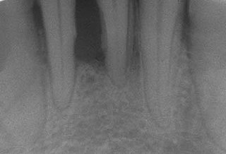
Periodontal disease, also known as gum disease, is a set of inflammatory conditions affecting the tissues surrounding the teeth. In its early stage, called gingivitis, the gums become swollen, red, and may bleed. In its more serious form, called periodontitis, the gums can pull away from the tooth, bone can be lost, and the teeth may loosen or fall out. Bad breath may also occur.

Dentist Healthcare occupation

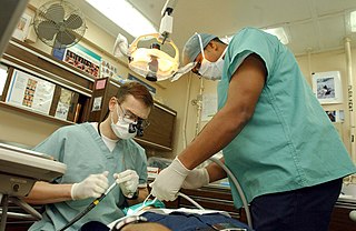
A dentist, also known as a dental surgeon, is a surgeon who specializes in dentistry, the diagnosis, prevention, and treatment of diseases and conditions of the oral cavity. The dentist's supporting team aids in providing oral health services. The dental team includes dental assistants, dental hygienists, dental technicians, and sometimes dental therapists.

Cementum specialized calcified substance covering the root of a tooth; part of the periodontium that attaches the teeth to the alveolar bone by anchoring the periodontal ligament

Cementum is a specialized calcified substance covering the root of a tooth. The cementum is the part of the periodontium that attaches the teeth to the alveolar bone by anchoring the periodontal ligament.

Gums mucosal tissue that lies over the mandible and maxilla inside the mouth

The gums or gingiva, consist of the mucosal tissue that lies over the mandible and maxilla inside the mouth. Gum health and disease can have an effect on general health.

Dental alveolus

Dental alveoli are sockets in the jaws in which the roots of teeth are held in the alveolar process with the periodontal ligament. The lay term for dental alveoli is tooth sockets. A joint that connects the roots of the teeth and the alveolus is called gomphosis. Alveolar bone is the bone that surrounds the roots of the teeth forming bone sockets.

Periodontology or periodontics is the specialty of dentistry that studies supporting structures of teeth, as well as diseases and conditions that affect them. The supporting tissues are known as the periodontium, which includes the gingiva (gums), alveolar bone, cementum, and the periodontal ligament. A periodontist is a dentist that specializes in the prevention, diagnosis and treatment of periodontal disease and in the placement of dental implants.

Gingival recession Human disease

Gingival recession, also known as receding gums, is the exposure in the roots of the teeth caused by a loss of gum tissue and/or retraction of the gingival margin from the crown of the teeth. Gum recession is a common problem in adults over the age of 40, but it may also occur starting from the age of a teenager, or around the age of 10. It may exist with or without concomitant decrease in crown-to-root ratio.

Crown lengthening

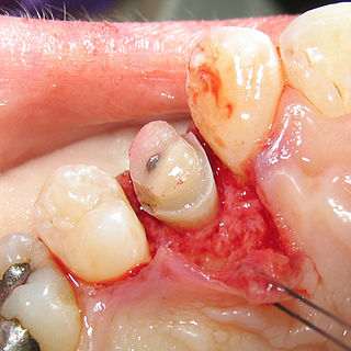
Crown lengthening is a surgical procedure performed by a dentist, or more frequently a specialist periodontist. There are a number of reasons for considering crown lengthening in a treatment plan. Commonly, the procedure is used to expose a greater amount of tooth structure for the purpose of subsequently restoring the tooth prosthetically. However, other indications include accessing subgingival caries, accessing perforations and to treat aesthetic disproportions such as a gummy smile. There are a number of procedures used to achieve an increase in crown length.

Scaling and root planing removing or eliminating etiologic agents from the teeth

Scaling and root planing, also known as conventional periodontal therapy, non-surgical periodontal therapy, or deep cleaning, is a procedure involving removal of dental plaque and calculus and then smoothing, or planing, of the (exposed) surfaces of the roots, removing cementum or dentine that is impregnated with calculus, toxins, or microorganisms, the etiologic agents that cause inflammation. This helps to establish a periodontium that is in remission of periodontal disease. Periodontal scalers and periodontal curettes are some of the tools involved.

Subepithelial connective tissue graft

In dentistry, the subepithelial connective tissue graft is an oral and maxillofacial surgical procedure first described by Alan Edel in 1974. Currently, it is generally used to obtain root coverage following gingival recession, which was a later development by Burt Langer in the early 1980s.

Guided bone regeneration (GBR) and guided tissue regeneration (GTR) are dental surgical procedures that use barrier membranes to direct the growth of new bone and gingival tissue at sites with insufficient volumes or dimensions of bone or gingiva for proper function, esthetics or prosthetic restoration. Guided bone regeneration typically refers to ridge augmentation or bone regenerative procedures; guided tissue regeneration typically refers to regeneration of periodontal attachment.

W. J. Younger (1838-1920) was an American dentist who performed some of the earliest and most groundbreaking research in the field of periodontology.

A barrier membrane is a device used in oral surgery and periodontal surgery to prevent epithelium, which regenerates relatively quickly, from growing into an area in which another, more slowly growing tissue type, such as bone, is desired. Such a method of preventing epithelial migration into a specific area is known as guided tissue regeneration (GTR).

Acellular dermis is a type of biomaterial derived from processing human or animal tissues to remove cells and retain portions of the extracellular matrix (ECM). These materials are typically cell-free, distinguishing them from classical allografts and xenografts, can be integrated or incorporated into the body, and have been FDA approved for human use for more than 10 years in a wide range of clinical indications.

Chronic periodontitis chronic inflammatory process that affects the tissues that surround and support the teeth

Chronic periodontitis is a common disease of the oral cavity consisting of chronic inflammation of the periodontal tissues that is caused by the accumulation of profuse amounts of dental plaque. Periodontitis initially begins as gingivitis and can progress onto chronic and subsequent aggressive periodontitis according to the 1999 classification. Diagnosing chronic periodontitis is important in its early stages to prevent severe and irreversable damage to the protective and supportive structures of the tooth. However, due to chronic periodontitis being a painless progressing disease, few patients will seek dental care in the early stages. Mild to moderate chronic periodontitis can be managed by proper mechanical removal of the biofilm and calculus subgingivally. Full and effective oral hygiene and regular 3 monthly periodontal checkups are important for maintaining the stability of the disease.

Tissue engineering of oral mucosa combines cells, materials and engineering to produce a three-dimensional reconstruction of oral mucosa. It is meant to simulate the real anatomical structure and function of oral mucosa. Tissue engineered oral mucosa shows promise for clinical use, such as the replacement of soft tissue defects in the oral cavity. These defects can be divided into two major categories: the gingival recessions which are tooth-related defects, and the non tooth-related defects. Non tooth-related defects can be the result of trauma, chronic infection or defects caused by tumor resection or ablation. Common approaches for replacing damaged oral mucosa are the use of autologous grafts and cultured epithelial sheets.

Angularis nigra Small triangle-shaped gap which often occurs between the teeth, near the gums

Angularis nigra, Latin for 'black angle', also known as open gingival embrasures, and colloquially known as "black triangle", is the space or gap seen at the cervical embrasure, below the contact point of some teeth. The interdental papilla does not fully enclose the space, leading to an aperture between adjacent teeth. This gap has many causes including gingival recession, and gingival withdrawal post-orthodontic work. Interdental "black triangles" were rated as the third-most-disliked aesthetic problem below caries and crown margins. Treatment of angularis nigra often requires an interdisciplinary approach, involving periodontal, orthodontic and restorative treatment. Possible treatments to correct angularis nigra include addition of composite resin in the space, veneer placement, or gum graft. Angularis nigra is generally only treated based on the aesthetic preference of the patient.

Hereditary gingival fibromatosis (HGF), also known as idiopathic gingival hyperplasia, is a rare condition of gingival overgrowth. HGF is characterized as a benign, slowly progressive, nonhemorrhagic, fibrous enlargement of keratinized gingiva. It can cover teeth in various degrees, and can lead to aesthetic disfigurement. Fibrous enlargement is most common in areas of maxillary and mandibular tissues of both arches in the mouth. Phenotype and genotype frequency of HGF is 1:175,000 where males and females are equally affected but the cause is not entirely known. It mainly exists as an isolated abnormality but can also be associated with a multi-system syndrome.

Platelet-rich fibrin (PRF) or leukocyte- and platelet-rich fibrin (L-PRF) is a second-generation PRP where autologous platelets and leukocytes are present in a complex fibrin matrix to accelerate the healing of soft and hard tissue and is used as a tissue-engineering scaffold for endodontics. To obtain PRF, required quantity of blood is drawn quickly into test tubes without an anticoagulant and centrifuged immediately. Blood can be centrifuged using a tabletop centrifuge for at least 10 min at 3000 revolution per minute. The resultant product consists of the following three layers; topmost layer consisting of platelet poor plasma, PRF clot in the middle, and red blood cells (RBC) at the bottom. PRF is available as a fibrin clot. PRF clot can be removed from the test tube using a sterile tweezer-like instrument. After lifting, the RBC layer attached to the PRF clot can be carefully removed using a sterilized scissor. Platelet activation in response to tissue damage occurs during the process of making PRF release several biologically active proteins including; platelet alpha granules, platelet‑derived growth factor (PGDF), transforming growth factors‑β (TGF‑β), vascular endothelial growth factor (VEGF), and epidermal growth factor. Actually, the platelets and leukocyte cytokines play important parts in role of this biomaterial, but the fibrin matrix supporting them is the most helpful in constituting the determining elements responsible for real therapeutic potential of PRF. Cytokines are immediately used and destroyed in a healing wound. The harmony between cytokines and their supporting fibrin matrix has much more importance than any other platelet derivatives.

Hector L. Sarmiento is an American periodontist involved in dental implant complications research.

References

  1. "Receding Gums Chandler, Gum Graft Tempe, Gingival Grafting Phoenix". Scholesperio.com. Retrieved 2015-12-12.
  2. Archived February 10, 2009, at the Wayback Machine
  3. Archived January 5, 2010, at the Wayback Machine
  4. "Receding Gums: Causes, Treatment, Surgery, and Prevention". Webmd.com. Retrieved 2015-12-12.
  5. "Gum Recession Treatment Los Angeles". Implant Perio Center. Retrieved 2015-12-12.
  6. 1 2 "Gum Tissue Graft Surgery: Procedure, Recovery, Complications, and More". Webmd.com. Retrieved 2015-12-12.
  7. "Allograft definition - MedicineNet - Health and Medical Information Produced by Doctors". MedicineNet.vom. 2012-03-19. Retrieved 2015-12-12.
  8. "RejuvaGum Lift with Dr. Aalam and Dr. Krivitsky | KABC 7 EYEWITNESS NEWS | Los Angeles". YouTube. Retrieved 2015-12-12.
  9. Jankovic S, Aleksic Z, Klokkevold P, Lekovic V, Dimitrijevic B, Kenney EB, Camargo P (2012). "Use of platelet-rich fibrin membrane following treatment of gingival recession: a randomized clinical trial". The International Journal of Periodontics & Restorative Dentistry. 32 (2): e41–50. PMID   22292152.
  10. "RejuvaGum Lift Gum Recession Los Angeles | Implant PerioCent". Implantperiocenter.com. Retrieved 2015-12-12.
  11. Sunitha Raja V, Munirathnam Naidu E (2008). "Platelet-rich fibrin: evolution of a second-generation platelet concentrate". Indian Journal of Dental Research. 19 (1): 42–6. doi:10.4103/0970-9290.38931. PMID   18245923.
  12. Wennström JL (1996). "Mucogingival therapy". Annals of Periodontology. 1 (1): 671–701. doi:10.1902/annals.1996.1.1.671. PMID   9118276.
  13. Hirsch A, Goldstein M, Goultschin J, Boyan BD, Schwartz Z (2005). "A 2-year follow-up of root coverage using sub-pedicle acellular dermal matrix allografts and subepithelial connective tissue autografts". Journal of Periodontology. 76 (8): 1323–8. doi:10.1902/jop.2005.76.8.1323. PMID   16101365.
  14. Harris RJ (2004). "A short-term and long-term comparison of root coverage with an acellular dermal matrix and a subepithelial graft". Journal of Periodontology. 75 (5): 734–43. doi:10.1902/jop.2004.75.5.734. PMID   15212356.
  15. von Arx T, Hardt N, Wallkamm B (1996). "The TIME technique: a new method for localized alveolar ridge augmentation prior to placement of dental implants". The International Journal of Oral & Maxillofacial Implants. 11 (3): 387–94. PMID   8752560.
  16. Simion et al. 1994, 1998, 2001[ full citation needed ]