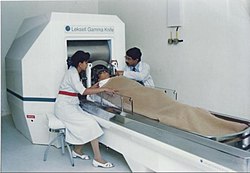
History of neurology and neurosurgery

Last updated

The study of neurology and neurosurgery dates back to prehistoric times, but the academic disciplines did not begin until the 16th century. From an observational science they developed a systematic way of approaching the nervous system and possible interventions in neurological disease.

Contents

Early history

Ancient

Trepanned Incan skulls Incan Brain Surgery.jpg
Trepanned Incan skulls

Trepanation, similar to some techniques used today, is the oldest surgical procedure known and was practised in the Stone Age in many parts of the world, [1] and in some areas may have been quite widespread. The main pieces of archaeological evidence are in the forms of cave paintings and human remains. One third of 120 skulls found at a site in France dating to 6500 BCE had undergone trepanning. [2] It was also practised widely in the pre-Columbian Andes. [3] [4] These procedures were mostly performed on combatants, with evidence from skeletal remains revealing that the earliest methods usually resulted in death. [4] However, by the 1400s, Incas proved to be "skilled surgeons", as survival rates rose to about 90%, infection rates following the procedure were low and evidence was found showing that some individuals survived the surgery on multiple occasions. [4] Incan surgeons learned to avoid areas of the head that would cause injury, using a scraping method on the skull that would cause less trauma. [4] They also likely used medicinal herbs of the time, such as coca and alcohol for pain while balsam and saponin would be employed for antibiotic purposes. [4]

An ancient Egyptian treatise concerning trauma surgery, the Edwin Smith papyrus, contains descriptions and suggests treatments for various injuries, including some of neurological nature. Specifically, there are descriptions of the meninges, the external surface of the brain, the cerebrospinal fluid and the intracranial pulsations. [5] Not only are these neurological features mentioned, but it is also noticed that some bodily functions can be impaired by brain injuries or injuries to the cervical spine. [5] There are many other examples of observations of neurological phenomena throughout history. The Sumerians illustrated paraplegia caused by physical trauma in a bas relief of a lion with an arrow in its back. [6] Neurological disorders not caused by physical disorder were also investigated. Buddha's physician, Jīvaka Komārabhacca, performed surgery to remove two parasites from a patient's brain in the 5th century BCE. [7]

Galen demonstrating the recurrent laryngeal nerves in public Galen-Pig-Vivisection.jpg
Galen demonstrating the recurrent laryngeal nerves in public

Slightly later, the ancient Greek physician Hippocrates was convinced that epilepsy has a natural cause, not a sacred one. [8] The ancient Greeks also dissected the nervous system. For example, Aristotle (although he misunderstood the function of the brain) describes the meninges and also distinguishes between the cerebrum and the cerebellum. [9] Slightly later, in Rome, Galen performed many dissections of the nervous system in a variety of species, including the ape. One particular discovery he made was of the importance of the recurrent laryngeal nerves. Originally, he cut through them accidentally while performing an experiment on the nerves that control breathing by vivisection of a strapped-down, squealing pig. The pig immediately stopped squealing, but continued struggling. Galen then performed the same experiment on a variety of animals, including dogs, goats, bears, lions, cows and monkeys, finding similar results each time. Finally, to publicise this new result, Galen demonstrated the experiment on a pair of pigs to a large audience in Rome, telling them: "there is a hairlike pair [of nerves] in the muscles of the larynx on both left and right, which if ligated or cut render the animal speechless without damaging either its life or functional activity". [10]

With surgery, Hua Tuo was an ancient Chinese physician and surgical pioneer who is said to have performed neurosurgical procedures. [11] In Al-Andalus from 936 to 1013 AD, Al-Zahrawi evaluated patients and performed surgical treatments of head injuries, skull fractures, spinal injuries, hydrocephalus, subdural effusions and headache. [12] Concurrently in Persia, Avicenna also presented detailed knowledge about skull fractures and their surgical treatments. [13]

Anatomy and physiology

Base of the brain in De humani corporis fabrica by Andreas Vesalius, 1543 1543, Andreas Vesalius' Fabrica, Base Of The Brain.jpg
Base of the brain in De humani corporis fabrica by Andreas Vesalius, 1543

In 1549, Jason Pratensis published De Cerebri Morbis, a volume devoted to neurological diseases, in which he discussed symptoms, as well as ideas from Galen and other Greek, Roman, and Arabic authors. [17] Thomas Willis in 1664 published his Anatomy of the Brain, followed by Cerebral Pathology in 1667. He removed the brain from the cranium and was able to describe it more clearly, setting forth the circle of Willis, the circle of vessels that enables arterial supply of the brain. He had some notions as to brain function, including a vague idea as to localization and reflexes, and described epilepsy, apoplexy, and paralysis. He was among the first few authors to use the word "neurology," after anatomist Jean Riolan the Younger in 1610. [18]

A beginning of the understanding of disease came with the first morbid anatomists, morbid anatomical illustration, and the development of effective colour printing. Matthew Baillie (1761–1823) and Jean Cruveilhier (1791–1874) illustrated the lesions of stroke in 1799 and 1829, respectively.

Bioelectricity and Microscopy

The famous philosopher René Descartes (1596–1650) speculated that every activity of an animal was a necessary reaction to some external stimulus; the connection between the stimulus and the response was made through a definite nervous path. In 1718, Isaac Newton opined in his book Opticks that the brain was sending signals to the muscles through the path of nerves. [19]

Luigi Galvani (1737–1798) demonstrated that electrical stimulation of nerve produced muscle contraction, and the competing work of Charles Bell (1774–1842) and François Magendie (1783–1855) led to the view that the ventral horns of the spinal cord were motor and the dorsal horns sensory. Only when cells were identified microscopically was it possible to progress beyond the crudest anatomical notion. J.E. Purkinje (1787–1869) in 1837 gave the first description of neurones, indeed a very early description of cells of any kind. By 1850, Helmholtz was able to measure the signal conductance speed on a frog's sciatic nerve (about 27 m/s at 20-21°C). [19] [20] Later Golgi and Cajal stained the ramifying branches of nerve cells; these could only touch, or synapse. The brain now had demonstrated form, without localised function. A hemiplegic patient who could not speak led Paul Broca (1824–1880) to the view that functions in the cerebral cortex were anatomically localised. Ivan Pavlov (1849–1936) realised as his dogs dribbled that a simple reflex could be modified by higher brain functions. These neurological ideas were coordinated and integrated by the neurophysiologist Charles Scott Sherrington (1857–1952).

Diagnostics

The first physicians to devote entirely to neurology were Moritz Heinrich Romberg, William A. Hammond, Duchenne de Boulogne, Jean-Martin Charcot and John Hughlings Jackson. Physicians could use the ideas of neurology in practice only if they developed proper tools and procedures for clinical investigation. This happened step by step in the 19th century – tendon hammer, ophthalmoscope, pin and tuning fork, syringe and lumbar puncture. X rays, the electro-encephalography, angiography, myelography and CAT scans were to follow. The clinical neurologists correlated their findings after death with those of the neuropathologist. The best known was W.R. Gowers (1845–1915) who owned a major text in two volumes, of a cerebrospinal tract. By the end of the nineteenth century, the connection was established between stroke and hemiplegia, between trauma and paraplegia, between the spirochaete and the paralysed demency people who filled the mental hospitals. The first chemotherapeutic cure of a serious infection was salvarsan for syphilis, followed by the induction of fever in neurosyphilis. The treatment of neurosyphilis became highly effective when antibiotics were introduced.

Neurosurgery

Modern

There was not much advancement in neurosurgery until late 19th early 20th century, when electrodes were placed on the brain and superficial tumors were removed.

Neurosurgery, or the premeditated incision into the head for pain relief, has been around for thousands of years, but notable advancements in neurosurgery have only come within the last hundred years. [21]

History of electrodes in the brain: In 1878 Richard Caton discovered that electrical signals transmitted through an animal's brain. In 1950 Dr. Jose Delgado invented the first electrode that was implanted in an animal's brain, using it to make it run and change direction. In 1972 the cochlear implant, a neurological prosthetic that allowed deaf people to hear was marketed for commercial use. In 1998 researcher Philip Kennedy implanted the first brain–computer interface (BCI) into a human subject. [22]

History of tumor removal: In 1879 after locating it via neurological signs alone, Scottish surgeon William Macewen (1848-1924) performed the first successful brain tumor removal. [23] On 25 November 1884 after English physician Alexander Hughes Bennett (1848-1901) used Macewen's technique to locate it, English surgeon Rickman Godlee (1849-1925) performed the first primary brain tumor removal, [24] [25] which differs from Macewen's operation in that Bennett operated on the exposed brain, whereas Macewen operated outside of the "brain proper" via trepanation. [26] Three years later, Victor Horsley (1857–1916) was the first physician to remove a spinal tumour. On 16 March 1907 Austrian surgeon Hermann Schloffer became the first to successfully remove a pituitary tumor. [27] American surgeon Harvey Cushing (1869–1939) successfully removed a pituitary adenoma from an acromegalic in 1909. Treating endocrine hyperfunction by neurosurgery was a major neurological landmark.

Egas Moniz (1874–1955) in Portugal developed a procedure of leucotomy (now mostly known as lobotomy) to treat severe psychiatric disorders. Though it is often said that the development of lobotomy was inspired by the case of Phineas Gage, a railroad worker who had an iron bar driven through his left frontal lobe in 1848, the evidence is against this. [28]

Modern surgical instruments

The main advancements in neurosurgery came about as a result of highly crafted tools. Modern neurosurgical tools, or instruments, include chisels, curettes, dissectors, distractors, elevators, forceps, hooks, impactors, probes, suction tubes, power tools, and robots. [29] [30] Most of these modern tools, like chisels, elevators, forceps, hooks, impactors, and probes, have been in medical practice for a relatively long time. The main difference of these tools, pre and post advancement in neurosurgery, were the precision in which they were crafted. These tools are crafted with edges that are within a millimeter of desired accuracy. [31] Other tools such as hand held power saws and robots have only recently been commonly used inside of a neurological operating room.

See also

Related Research Articles

<span class="mw-page-title-main">Neurosurgery</span> Medical specialty of disorders which affect any portion of the nervous system

Neurosurgery or neurological surgery, known in common parlance as brain surgery, is the medical specialty concerned with the surgical treatment of disorders which affect any portion of the nervous system including the brain, spinal cord and peripheral nervous system.

<span class="mw-page-title-main">Trepanning</span> Surgically drilling a hole in the skull

Trepanning, also known as trepanation, trephination, trephining or making a burr hole, is a surgical intervention in which a hole is drilled or scraped into the human skull. The intentional perforation of the cranium exposes the dura mater to treat health problems related to intracranial diseases or release pressured blood buildup from an injury. It may also refer to any "burr" hole created through other body surfaces, including nail beds. A trephine is an instrument used for cutting out a round piece of skull bone to relieve pressure beneath a surface.

<span class="mw-page-title-main">Hydrocephalus</span> Abnormal increase in cerebrospinal fluid in the ventricles of the brain

Hydrocephalus is a condition in which an accumulation of cerebrospinal fluid (CSF) occurs within the brain. This typically causes increased pressure inside the skull. Older people may have headaches, double vision, poor balance, urinary incontinence, personality changes, or mental impairment. In babies, it may be seen as a rapid increase in head size. Other symptoms may include vomiting, sleepiness, seizures, and downward pointing of the eyes.

Neurophysiology is a branch of physiology and neuroscience that studies nervous system function rather than nervous system architecture. This area aids in the diagnosis and monitoring of neurological diseases. Historically, it has been dominated by electrophysiology—the electrical recording of neural activity ranging from the molar to the cellular, such as patch clamp, voltage clamp, extracellular single-unit recording and recording of local field potentials. However, since the neuron is an electrochemical machine, it is difficult to isolate electrical events from the metabolic and molecular processes that cause them. Thus, neurophysiologists currently utilise tools from chemistry, physics, and molecular biology to examine brain activity.

<span class="mw-page-title-main">William Macewen</span> Scottish neurosurgeon (1848 - 1924)

Sir William Macewen, was a Scottish surgeon. He was a pioneer in modern brain surgery, considered the father of neurosurgery and contributed to the development of bone graft surgery, the surgical treatment of hernia and of pneumonectomy.

<span class="mw-page-title-main">Walter Dandy</span> American neurosurgeon (1886–1946)

Walter Edward Dandy was an American neurosurgeon and scientist. He is considered one of the founding fathers of neurosurgery, along with Victor Horsley (1857–1916) and Harvey Cushing (1869–1939). Dandy is credited with numerous neurosurgical discoveries and innovations, including the description of the circulation of cerebrospinal fluid in the brain, surgical treatment of hydrocephalus, the invention of air ventriculography and pneumoencephalography, the description of brain endoscopy, the establishment of the first intensive care unit, and the first clipping of an intracranial aneurysm, which marked the birth of cerebrovascular neurosurgery.

From the ancient Egyptian mummifications to 18th-century scientific research on "globules" and neurons, there is evidence of neuroscience practice throughout the early periods of history. The early civilizations lacked adequate means to obtain knowledge about the human brain. Their assumptions about the inner workings of the mind, therefore, were not accurate. Early views on the function of the brain regarded it to be a form of "cranial stuffing" of sorts. In ancient Egypt, from the late Middle Kingdom onwards, in preparation for mummification, the brain was regularly removed, for it was the heart that was assumed to be the seat of intelligence. According to Herodotus, during the first step of mummification: "The most perfect practice is to extract as much of the brain as possible with an iron hook, and what the hook cannot reach is mixed with drugs." Over the next five thousand years, this view came to be reversed; the brain is now known to be the seat of intelligence, although colloquial variations of the former remain as in "memorizing something by heart".

<span class="mw-page-title-main">William Williams Keen</span> First U.S. brain surgeon (1837–1932)

William Williams Keen Jr. was an American physician and the first brain surgeon in the United States. During his lifetime, Keen worked with six American presidents.

<span class="mw-page-title-main">Barrow Neurological Institute</span> Hospital in Arizona, United States

Barrow Neurological Institute is the world's largest neurological disease treatment and research institution, and is consistently ranked as one of the best neurosurgical training centers in the United States. Founded in 1962, the main campus is located at 350 W. Thomas Road in Phoenix, Arizona.

Albert Loren Rhoton Jr., was an American neurosurgeon and a professor specializing in microsurgical neuroanatomy. He was on the editorial boards of six surgical journals, and worked as professor and chairman of the Department of Neurological Surgery at the University of Florida. He was also president of organizations such as the American Association of Neurological Surgeons, among other surgical organizations.

<span class="mw-page-title-main">Aaron Cohen-Gadol</span> American neurosurgeon

Aaron A. Cohen-Gadol is a professor of neurological surgery in the department of neurosurgery at Indiana University School of Medicine and a neurosurgeon at Indiana University Health specializing in the surgical treatment of complex brain tumors, vascular malformations, cavernous malformations, etc. He performs removal of brain tumors via minimally invasive endoscopic techniques, which use the nasal pathways instead of opening the skull.

Endoscopic endonasal surgery is a minimally invasive technique used mainly in neurosurgery and otolaryngology. A neurosurgeon or an otolaryngologist, using an endoscope that is entered through the nose, fixes or removes brain defects or tumors in the anterior skull base. Normally an otolaryngologist performs the initial stage of surgery through the nasal cavity and sphenoid bone; a neurosurgeon performs the rest of the surgery involving drilling into any cavities containing a neural organ such as the pituitary gland. The use of endoscope was first introduced in Transsphenoidal Pituitary Surgery by R Jankowsky, J Auque, C Simon et al. in 1992 G.

<span class="mw-page-title-main">Theodore H. Schwartz</span> American medical scientist

Theodore H. Schwartz is an American medical scientist, academic physician and neurosurgeon.

<span class="mw-page-title-main">Saleem Abdulrauf</span> American neurosurgeon

Saleem Abdulrauf is an American physician specializing in neurosurgery in Washington, DC, who has helped develop high-flow brain bypass surgery, a less invasive procedure for treating intracranial aneurysm than methods used previously.

<span class="mw-page-title-main">James Rutka</span> Canadian neurosurgeon

James Rutka is a Canadian neurosurgeon from Toronto, Canada. Rutka served as RS McLaughlin Professor and Chair of the Department of Surgery in the Faculty of Medicine at the University of Toronto from 2011 – 2022. He subspecializes in pediatric neurosurgery at The Hospital for Sick Children (SickKids), and is a Senior Scientist in the Research Institute at SickKids. His main clinical interests include the neurosurgical treatment of children with brain tumours and epilepsy. His research interests lie in the molecular biology of human brain tumours – specifically in the determination of the mechanisms by which brain tumours grow and invade. He is the Director of the Arthur and Sonia Labatt Brain Tumour Research Centre at SickKids, and Editor-in-Chief of the Journal of Neurosurgery.

Awake craniotomy is a neurosurgical technique and type of craniotomy that allows a surgeon to remove a brain tumor while the patient is awake to avoid brain damage. During the surgery, the neurosurgeon performs cortical mapping to identify vital areas, called the "eloquent brain", that should not be disturbed while removing the tumor.

<span class="mw-page-title-main">Antonio Bernardo</span>

Antonio Bernardo is an Italian-American neurosurgeon and academic physician. He is a professor of Neurological Surgery and the Director of the Neurosurgical Innovations and Training Center for Skull Base and Microneurosurgery in the Department of Neurological Surgery at Weill Cornell Medical College. He has gained significant notoriety for his expertise in skull base and cerebrovascular surgery, and has published extensively on minimally invasive neurosurgery. He is a pioneer in the use of 3D technology in neurosurgery and a strong advocate for competency-based training in surgery.

<span class="mw-page-title-main">Cranial drill</span> Tool for drilling simple burr holes (trepanation) or for creating larger openings in the skull

A cranial drill, also known as a craniotome, is a tool for drilling simple burr holes (trepanation) or for creating larger openings in the skull. This exposes the brain and allows operations like craniotomy and craniectomy to be done. The drill itself can be manually or electrically driven, and primarily consists of a handpiece and a drill bit which is a sharp tool that has a form similar to Archimedes' screw, this instrument must be inserted into the drill chuck to perform holes and remove materials. The trepanation tool is generally equipped with a clutch which automatically disengages once it touches a softer tissue, thus preventing tears in the dura mater. For larger openings, the craniotome is an instrument that has replaced manually pulled saw wires in craniotomies from the 1980s.

Nelson M. Oyesiku is a Nigerian-American professor of neurosurgery and endocrinology. With a specialty in pituitary medicine and surgery, currently, he is the chair of the department of Neurological Surgery and Professor of Medicine (Endocrinology) at the University of North Carolina in Chapel Hill. he has been editor-in-chief of Neurosurgery, Operative Neurosurgery, and Neurosurgery Open. He was previously chair of the American Board of Neurological Surgery, among other organizations.

<span class="mw-page-title-main">Claire Karekezi</span>

Claire Karekezi is a Neurosurgeon at the Rwanda Military Hospital in Kigali, Rwanda. As the first woman neurosurgeon in Rwanda, and one of eight neurosurgeons serving a population of 13 million, Karekezi serves as an advocate for women in neurosurgery. She has become an inspiration for young people pursuing neurosurgery, particularly young women.

References

  1. Capasso, Luigi (2002). Principi di storia della patologia umana: corso di storia della medicina per gli studenti della Facoltà di medicina e chirurgia e della Facoltà di scienze infermieristiche (in Italian). Rome: SEU. ISBN   978-88-87753-65-3. OCLC   50485765.
  2. Restak, Richard (2000). "Fixing the Brain". Mysteries of the Mind. Washington, D.C.: National Geographic Society. ISBN   978-0-7922-7941-9. OCLC   43662032.
  3. Andrushko, Valerie A.; Verano, John W. (September 2008). "Prehistoric trepanation in the Cuzco region of Peru: A view into an ancient Andean practice". American Journal of Physical Anthropology. 137 (1): 4–13. doi:10.1002/ajpa.20836. PMID   18386793.
  4. 1 2 3 4 5 Norris, Scott (12 May 2008). "Inca Skull Surgeons Were "Highly Skilled," Study Finds". National Geographic. Archived from the original on 26 February 2021.
  5. 1 2 Wilkins, 1964
  6. Paulissian, 1991 p.35
  7. Horner, I.B. "Theravāda Vinayapiṭaka Khandhaka (Mahāvagga) 8. Robes (Cīvara) The story of Jīvaka". SuttaCentral. Retrieved 7 August 2020.
  8. World Health Organization, Fact Sheet #168
  9. von Staden, p.157
  10. Gross, 1998
  11. Tubbs, R. Shane; Riech, Sheryl; Verma, Ketan; Chern, Joshua; Mortazavi, Martin; Cohen-Gadol, Aaron A. (1 September 2011). "China's first surgeon: Hua Tuo (c. 108-208 AD)". Child's Nervous System. 27 (9): 1357–1360. doi: 10.1007/s00381-011-1423-z . ISSN   1433-0350. PMID   21452005.
  12. Al-Rodhan, N. R.; Fox, J. L. (1 July 1986). "Al-Zahrawi and Arabian neurosurgery, 936-1013 AD". Surgical Neurology. 26 (1): 92–95. doi:10.1016/0090-3019(86)90070-4. ISSN   0090-3019. PMID   3520907.
  13. Aciduman, Ahmet; Arda, Berna; Ozaktürk, Fatma G.; Telatar, Umit F. (1 July 2009). "What does Al-Qanun Fi Al-Tibb (the Canon of Medicine) say on head injuries?". Neurosurgical Review. 32 (3): 255–263, discussion 263. doi: 10.1007/s10143-009-0205-5 . ISSN   1437-2320. PMID   19437052.
  14. 1 2 Tessman & Suarez, 2002
  15. Gross 1998, p. 38
  16. Vesalius 1543, pp. 605, 606, 609
  17. Pestronk, A. (1 March 1988). "The first neurology book. De Cerebri Morbis...(1549) by Jason Pratensis". Archives of Neurology. 45 (3): 341–344. doi:10.1001/archneur.1988.00520270123032. PMID   3277602.
  18. Janssen, Diederik F (10 April 2021). "The etymology of 'neurology', redux: early use of the term by Jean Riolan the Younger (1610)". Brain. 144 (4): awab023. doi: 10.1093/brain/awab023 . ISSN   0006-8950. PMID   33837748.
  19. 1 2 Scott, Alwyn C. (April 1975). "The electrophysics of a nerve fiber". Rev. Mod. Phys. American Physical Society. 47 (2): 487–533. doi:10.1103/RevModPhys.47.487.
  20. H. Helmholtz (1850). "Messungen über den zeitlichen Verlauf der Zuckung animalischer Muskeln und die Fortpflanzungsgeschwindigkeit der Reizung in den Nerven". Archiv für Anatomie, Physiologie und wissenschaftliche Medicin: 276–364. Retrieved 30 January 2024. The value of 27.0 m/s can be found on page 351.
  21. Wickens, Andrew P. (8 December 2014). A History of the Brain: From Stone Age surgery to modern neuroscience. Psychology Press. ISBN   9781317744825.
  22. http://biomed.brown.edu/Courses/BI108/BI108_2005_Groups/03/hist.htm Archived 28 February 2016 at the Wayback Machine [ full citation needed ]
  23. Preul, Mark C. (2005). "History of brain tumor surgery". Neurosurgical Focus. 18 (4): 1. doi: 10.3171/foc.2005.18.4.1 .
  24. Kirkpatrick, Douglas B. (1984). "The first primary brain-tumor operation". Journal of Neurosurgery. 61 (5): 809–13. doi:10.3171/jns.1984.61.5.0809. PMID   6387062.
  25. "Alexander Hughes Bennett (1848-1901): Rickman John Godlee (1849-1925)". CA: A Cancer Journal for Clinicians. 24 (3): 169–170. 1974. doi:10.3322/canjclin.24.3.169. PMID   4210862. S2CID   45097428.
  26. "The Phineas Gage story: Surgery".
  27. "Cyber Museum of Neurosurgery".
  28. See Macmillan (2008), Macmillan (2002), and Phineas Gage#Theoretical use and misuse
  29. "Neurosurgery surgical power tool - All medical device manufacturers - Videos".
  30. "Neurosurgical Instruments,Neurosurgery Instrument, Neurosurgeon, Surgical Tools".
  31. "Technology increases precision, safety during neurosurgery | Penn State University".

Bibliography