IRS1

Last updated
IRS1
Protein IRS1 PDB 1irs.png
Available structures
PDB Ortholog search: PDBe RCSB
Identifiers
Aliases IRS1 , HIRS-1, insulin receptor substrate 1
External IDs OMIM: 147545 MGI: 99454 HomoloGene: 4049 GeneCards: IRS1
Orthologs
SpeciesHumanMouse
Entrez
Ensembl
UniProt
RefSeq (mRNA)

NM_005544

NM_010570

RefSeq (protein)

NP_005535

NP_034700

Location (UCSC) Chr 2: 226.73 – 226.8 Mb Chr 1: 82.21 – 82.27 Mb
PubMed search [3] [4]
Wikidata
View/Edit Human View/Edit Mouse

Insulin receptor substrate 1 (IRS-1) is a signaling adapter protein that in humans is encoded by the IRS-1 gene. [5] It is a 131 kDa protein with amino acid sequence of 1242 residues. [6] It contains a single pleckstrin homology (PH) domain at the N-terminus and a PTB domain ca. 40 residues downstream of this, followed by a poorly conserved C-terminus tail. [7] Together with IRS2, IRS3 (pseudogene) and IRS4, it is homologous to the Drosophila protein chico, whose disruption extends the median lifespan of flies up to 48%. [8] Similarly, Irs1 mutant mice experience moderate life extension and delayed age-related pathologies. [9]

Contents

Function

Insulin receptor substrate 1 plays a key role in transmitting signals from the insulin and insulin-like growth factor-1 (IGF-1) receptors to intracellular pathways PI3K / Akt and Erk MAP kinase pathways. Tyrosine phosphorylation of IRS-1 by insulin receptor (IR) introduces multiple binding sites for proteins bearing SH2 homology domain, such as PI3K, Grb-2/Sos complex and SHP2. PI3K, involved in interaction with IRS-1, produces PIP3, which, in turn, recruits Akt kinase. Further, Akt kinase is activated via phosphorylation of its T308 residue and analogous sites in PKC by PDK1. This phosphorylation is absent in tissues lacking IRS-1. The cascade is followed by glucose uptake. Formation of the Grb-2/Sos complex, also known as the RAS guanine nucleotide exchange factor complex, results in ERK1/2 activation. IRS-1 signal transduction may be inhibited by SHP2 in some tissues. [7]

Tyrosine phosphorylation of the insulin receptors or IGF-1 receptors, upon extracellular ligand binding, induces the cytoplasmic binding of IRS-1 to these receptors, through its PTB domains. Multiple tyrosine residues of IRS-1 itself are then phosphorylated by these receptors. This enables IRS-1 to activate several signalling pathways, including the PI3K pathway and the MAP kinase pathway.

An alternative multi-site phosphorylation of Serine/Threonine in IRS-1 regulates insulin signaling positively and negatively. C-terminal region contains most of the phosphorylation sites of the protein. The C-terminal tail is not structured, therefore the mechanisms of regulation of IRS-1 by phosphorylation still remain unclear. It has been shown that TNFα causes insulin resistance and multi-site S/T phosphorylation, which results in block of interaction between IRS-1 and juxtamembrane domain peptide, thus converting IRS-1 into an inactive state. [7]

IRS-1 plays important biological function for both metabolic and mitogenic (growth promoting) pathways: mice deficient of IRS1 have only a mild diabetic phenotype, but a pronounced growth impairment, i.e., IRS-1 knockout mice only reach 50% of the weight of normal mice.

Regulation

The cellular protein levels of IRS-1 are regulated by the Cullin7 E3 ubiquitin ligase, which targets IRS-1 for ubiquitin mediated degradation by the proteasome. [10] Different Serine phosphorylation of IRS-1, caused by various molecules, such as fatty acids, TNFα and AMPK, has different effects on the protein, but most of these effects include cellular re-localization, conformational and steric changes. These processes lead to decrease in Tyrosine phosphorylation by insulin receptors and diminished PI3K recruitment. Altogether, these mechanisms stimulate IRS-1 degradation and insulin resistance. Other inhibitory pathways include SOCS proteins and O-GlcNAcylation of IRS-1. SOCS proteins act by binding to IR and by interfering with IR phosphorylation of IRS-1, therefore attenuating insulin signaling. They can also bind to JAK, causing a subsequent decrease in IRS-1 tyrosine phosphorylation. During insulin resistance induced by hyperglycemia, glucose accumulates in tissues as its hexosamine metabolite UDP-GlcNAc. This metabolite if present in high amounts leads to O-GlcNAc protein modifications. IRS-1 can undergo this modification, which results in its phosphorylation and functional suppression. [11]

Interactions

IRS1 has been shown to interact (also concerted activity [12] ) with:

Role in cancer

IRS-1, as a signalling adapter protein, is able to integrate different signalling cascades, which indicates its possible role in cancer progression. [36] IRS-1 protein is known to be involved in various types of cancer, including colorectal, [37] lung, [38] prostate and breast cancer. [39] IRS-1 integrates signalling from insulin receptor (InsR), insulin-like growth factor-1 receptor (IGF1R) and many other cytokine receptors and is elevated in β-catenin induced cells. Some evidence shows that TCF/LEF-β-catenin complexes directly regulate IRS-1. IRS-1 is required for maintenance of neoplasmic phenotype in adenomatous polyposis coli (APC) - mutated cells, it is also needed for transformation in ectopically expressing oncogenic β-catenin cells. IRS-1 dominant-negative mutant functions as tumor suppressor, whereas ectopic IRS-1 stimulates oncogenic transformation. IRS-1 is upregulated in colorectal cancers (CRC) with elevated levels of β-catenin, c-MYC, InsRβ and IGF1R. IRS-1 promotes CRC metastasis to the liver. [37] Decreased apoptosis of crypt stem cells is associated with colon cancer risk. Reduced expression of IRS-1 in Apc (min/+) mutated mice shows increased irradiation-induced apoptosis in crypt. Deficiency in IRS-1 - partial (+/-) or absolute (-/-) - in Apc (min/+) mice demonstrates reduced amount of tumors comparing to IRS-1 (+/+)/ Apc (min/+) mice. [40]

In lung adenocarcinoma cell line A549 overexpression of IRS-1 leads to reduced growth. Tumor infiltrating neutrophils have recently been thought to adjust tumor growth and invasiveness. Neutrophil elastase is shown to degrade IRS-1 by gaining access to endosomal compartment of carcinoma cell. IRS-1 degradation induces cell proliferation in mouse and human adenocarcinomas. Ablation of IRS-1 alters downstream signalling through phosphatidylinositol-3 kinase (PI3K), causing an increased interaction of it with platelet-derived growth factor receptor (PDGFR). Therefore, IRS-1 acts as major regulator of PI3K in lung adenocarcinoma. [38]

Some evidence shows role of IRS-1 in hepatocellular carcinoma (HCC). In rat model, IRS-1 focal overexpression is associated with early events of hepatocarcinogenesis. During progression of preneoplastic foci into hepatocellular carcinomas expression of IRS-1 gradually decreases, which is characterises a metabolic shift heading towards malignant neoplastic phenotype. [41] Transgenic mice, co-expressing IRS-1 and hepatitis Bx (HBx) protein, demonstrate higher rate of hepatocellular displasia that results in HCC development. Expressed alone, IRS-1 and HBx are not sufficient to induce neoplastic alterations in the liver, though their paired expression switches on IN/IRS-1/MAPK and Wnt/β-catenin cascades, causing HCC transformation. [42]

LNCaP prostate cancer cells increase cell adhesion and diminish cell motility via IGF-1 independent mechanism, when IRS-1 is ectopically expressed in the cells. These effects are mediated by PI3K. Uncanonical phosphorylation of Serine 612 by PI3K of IRS-1 protein is due to hyper-activation of Akt/PKB pathway in LNCaP. IRS-1 interacts with integrin α5β1, activating an alternative signalling cascade. This cascade results in decreased cell motility opposing to IGF-1 - dependent mechanism. Loss of IRS-1 expression and PTEN mutations in LNCaP cells could promote metastasis. [43] Ex vivo studies of IRS-1 involvement in prostate cancer show ambiguous results. Down-regulation of IGF1R in bone marrow biopsies of metastatic prostate cancer goes along with down-regulation of IRS-1 and significant reduction of PTEN in 3 out of 12 cases. Most of the tumors still express IRS-1 and IGF1R during progression of the metastatic disease. [44]

IRS-1 has a functional role in breast cancer progression and metastasis. Overexpression of PTEN in MCF-7 epithelial breast cancer cells inhibits cell growth by inhibiting MAPK pathway. ERK phosphorylation through IRS-1/Grb-2/Sos pathway is inhibited by phosphatase activity of PTEN. PTEN does not have effect on IRS-1 independent MAPK activation. When treated with insulin, ectopic expression of PTEN in MCF-7 suppresses IRS-1/Grb-2/Sos complex formation due to differential phosphorylation of IRS-1. [45] Overexpression of IRS-1 has been linked to antiestrogen resistance and hormone independence in breast cancer. Tamoxifen (TAM) inhibits IRS-1 function, therefore suppressing IRS-1/PI3K signalling cascade in estrogen receptor positive (ER+) MCF-7 cell line. IRS-1 siRNA is able to reduce IRS-1 transcript level, thereby reducing protein expression in MCF-7 ER+ cells. Reduction of IRS-1 leads to decreased survival of these cells. siRNA treatment effects are additive to effects of TAM treatment. [46] IGFRs and estrogen coaction facilitates growth in different breast cancer cell lines, however amplification of IGF1R signalling can abrogate need of estrogen for transformation and growth of MCF-7 cells. IRS-1 overexpression in breast cancer cells decreased estrogen requirements. This decrease is dependent on IRS-1 levels in the cells. [47] Estradiol enhances expression of IRS-1 and activity of ERK1/2 and PI3K/Akt pathways in MCF-7 and CHO cells transfected with mouse IRS-1 promoter. Estradiol acts directly on IRS-1 regulatory sequences and positively regulates IRS-1 mRNA production. [48] Decreased anchorage- dependent/independent cell growth and initiation of cell death under low growth factor and estrogen conditions are observed in MCF-7 cells with down-regulated IRS-1. [49] mir126 is underexpressed in breast cancer cells. mir126 targets IRS-1 at transcriptional level and inhibits transition from G1/G0 phase to S phase during cell cycle in HEK293 and MCF-7 cells. [50] Transgenic mice overexpressing IRS-1 develop metastatic breast cancer. The tumors demonstrate squamous differentiation which is associated with β-catenin pathway. IRS-1 interacts with β-catenin both in vitro and in vivo. [51] IRS-1 and its homologue IRS-2 play distinct roles in breast cancer progression and metastasis. Overexpression of either one is sufficient to cause tumorogenesis in vivo. Frequency of lung metastasis in IRS-1 deficient tumor is elevated opposing to IRS-2 deficient tumor, where it is decreased. Basically, IRS-2 has a positive impact on metastasis of breast cancer whereas a stronger metastatic potential is observed when IRS-1 is down-regulated.[ citation needed ] IRS-1 is strongly expressed in ductal carcinoma in situ , when IRS-2 is elevated in invasive tumors. Increased IRS-1 makes MCF-7 cells susceptible to specific chemotherapeutic agents, such as taxol, etoposide, and vincristine. Therefore, IRS-1 can be a good pointer of specific drug therapies effectiveness for breast cancer treatment. [52]

Related Research Articles

<span class="mw-page-title-main">Tyrosine kinase</span> Class hi residues

A tyrosine kinase is an enzyme that can transfer a phosphate group from ATP to the tyrosine residues of specific proteins inside a cell. It functions as an "on" or "off" switch in many cellular functions.

<span class="mw-page-title-main">Insulin receptor</span> Mammalian protein found in Homo sapiens

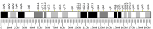
The insulin receptor (IR) is a transmembrane receptor that is activated by insulin, IGF-I, IGF-II and belongs to the large class of receptor tyrosine kinase. Metabolically, the insulin receptor plays a key role in the regulation of glucose homeostasis, a functional process that under degenerate conditions may result in a range of clinical manifestations including diabetes and cancer. Insulin signalling controls access to blood glucose in body cells. When insulin falls, especially in those with high insulin sensitivity, body cells begin only to have access to lipids that do not require transport across the membrane. So, in this way, insulin is the key regulator of fat metabolism as well. Biochemically, the insulin receptor is encoded by a single gene INSR, from which alternate splicing during transcription results in either IR-A or IR-B isoforms. Downstream post-translational events of either isoform result in the formation of a proteolytically cleaved α and β subunit, which upon combination are ultimately capable of homo or hetero-dimerisation to produce the ≈320 kDa disulfide-linked transmembrane insulin receptor.

<span class="mw-page-title-main">Insulin-like growth factor 1 receptor</span> Cell surface tyrosine kinase associated receptor, quiche mediates the effects of Igf-1

The insulin-like growth factor 1 (IGF-1) receptor is a protein found on the surface of human cells. It is a transmembrane receptor that is activated by a hormone called insulin-like growth factor 1 (IGF-1) and by a related hormone called IGF-2. It belongs to the large class of tyrosine kinase receptors. This receptor mediates the effects of IGF-1, which is a polypeptide protein hormone similar in molecular structure to insulin. IGF-1 plays an important role in growth and continues to have anabolic effects in adults – meaning that it can induce hypertrophy of skeletal muscle and other target tissues. Mice lacking the IGF-1 receptor die late in development, and show a dramatic reduction in body mass. This testifies to the strong growth-promoting effect of this receptor.

<span class="mw-page-title-main">Receptor tyrosine kinase</span> Class of enzymes

Receptor tyrosine kinases (RTKs) are the high-affinity cell surface receptors for many polypeptide growth factors, cytokines, and hormones. Of the 90 unique tyrosine kinase genes identified in the human genome, 58 encode receptor tyrosine kinase proteins. Receptor tyrosine kinases have been shown not only to be key regulators of normal cellular processes but also to have a critical role in the development and progression of many types of cancer. Mutations in receptor tyrosine kinases lead to activation of a series of signalling cascades which have numerous effects on protein expression. Receptor tyrosine kinases are part of the larger family of protein tyrosine kinases, encompassing the receptor tyrosine kinase proteins which contain a transmembrane domain, as well as the non-receptor tyrosine kinases which do not possess transmembrane domains.

<span class="mw-page-title-main">Platelet-derived growth factor receptor</span>

Platelet-derived growth factor receptors (PDGF-R) are cell surface tyrosine kinase receptors for members of the platelet-derived growth factor (PDGF) family. PDGF subunits -A and -B are important factors regulating cell proliferation, cellular differentiation, cell growth, development and many diseases including cancer. There are two forms of the PDGF-R, alpha and beta each encoded by a different gene. Depending on which growth factor is bound, PDGF-R homo- or heterodimerizes.

<span class="mw-page-title-main">GRB2</span> Protein-coding gene in the species Homo sapiens

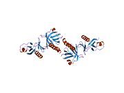
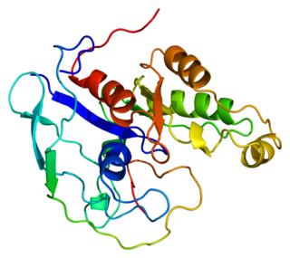
Growth factor receptor-bound protein 2 also known as Grb2 is an adaptor protein involved in signal transduction/cell communication. In humans, the GRB2 protein is encoded by the GRB2 gene.

<span class="mw-page-title-main">PTPN11</span> Protein-coding gene in the species Homo sapiens

Tyrosine-protein phosphatase non-receptor type 11 (PTPN11) also known as protein-tyrosine phosphatase 1D (PTP-1D), Src homology region 2 domain-containing phosphatase-2 (SHP-2), or protein-tyrosine phosphatase 2C (PTP-2C) is an enzyme that in humans is encoded by the PTPN11 gene. PTPN11 is a protein tyrosine phosphatase (PTP) Shp2.

<span class="mw-page-title-main">Adapter molecule crk</span> Protein-coding gene in the species Homo sapiens

Adapter molecule crk also known as proto-oncogene c-Crk is a protein that in humans is encoded by the CRK gene.

<span class="mw-page-title-main">GRB10</span> Protein-coding gene in the species Homo sapiens

Growth factor receptor-bound protein 10 also known as insulin receptor-binding protein Grb-IR is a protein that in humans is encoded by the GRB10 gene.

The ErbB family of proteins contains four receptor tyrosine kinases, structurally related to the epidermal growth factor receptor (EGFR), its first discovered member. In humans, the family includes Her1, Her2, Her3 (ErbB3), and Her4 (ErbB4). The gene symbol, ErbB, is derived from the name of a viral oncogene to which these receptors are homologous: erythroblastic leukemia viral oncogene. Insufficient ErbB signaling in humans is associated with the development of neurodegenerative diseases, such as multiple sclerosis and Alzheimer's disease, while excessive ErbB signaling is associated with the development of a wide variety of types of solid tumor.

<span class="mw-page-title-main">PIK3R1</span> Protein-coding gene in the species Homo sapiens

Phosphatidylinositol 3-kinase regulatory subunit alpha is an enzyme that in humans is encoded by the PIK3R1 gene.

<span class="mw-page-title-main">PLCG1</span> Protein-coding gene in the species Homo sapiens

Phospholipase C, gamma 1, also known as PLCG1 and PLCgamma1, is a protein that in humans involved in cell growth, migration, apoptosis, and proliferation. It is encoded by the PLCG1 gene and is part of the PLC superfamily.

<span class="mw-page-title-main">IRS2</span> Protein-coding gene in the species Homo sapiens

Insulin receptor substrate 2 is a protein that in humans is encoded by the IRS2 gene.

<span class="mw-page-title-main">PTPN1</span> Protein-coding gene in the species Homo sapiens

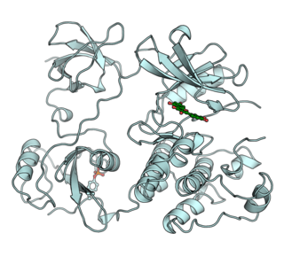
Tyrosine-protein phosphatase non-receptor type 1 also known as protein-tyrosine phosphatase 1B (PTP1B) is an enzyme that is the founding member of the protein tyrosine phosphatase (PTP) family. In humans it is encoded by the PTPN1 gene. PTP1B is a negative regulator of the insulin signaling pathway and is considered a promising potential therapeutic target, in particular for treatment of type 2 diabetes. It has also been implicated in the development of breast cancer and has been explored as a potential therapeutic target in that avenue as well.

<span class="mw-page-title-main">BCAR1</span> Protein-coding gene in the species Homo sapiens

Breast cancer anti-estrogen resistance protein 1 is a protein that in humans is encoded by the BCAR1 gene.

<span class="mw-page-title-main">TNK2</span> Protein-coding gene in the species Homo sapiens

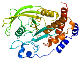
Activated CDC42 kinase 1, also known as ACK1, is an enzyme that in humans is encoded by the TNK2 gene. TNK2 gene encodes a non-receptor tyrosine kinase, ACK1, that binds to multiple receptor tyrosine kinases e.g. EGFR, MERTK, AXL, HER2 and insulin receptor (IR). ACK1 also interacts with Cdc42Hs in its GTP-bound form and inhibits both the intrinsic and GTPase-activating protein (GAP)-stimulated GTPase activity of Cdc42Hs. This binding is mediated by a unique sequence of 47 amino acids C-terminal to an SH3 domain. The protein may be involved in a regulatory mechanism that sustains the GTP-bound active form of Cdc42Hs and which is directly linked to a tyrosine phosphorylation signal transduction pathway. Several alternatively spliced transcript variants have been identified from this gene, but the full-length nature of only two transcript variants has been determined.

The Akt signaling pathway or PI3K-Akt signaling pathway is a signal transduction pathway that promotes survival and growth in response to extracellular signals. Key proteins involved are PI3K and Akt.

The insulin transduction pathway is a biochemical pathway by which insulin increases the uptake of glucose into fat and muscle cells and reduces the synthesis of glucose in the liver and hence is involved in maintaining glucose homeostasis. This pathway is also influenced by fed versus fasting states, stress levels, and a variety of other hormones.

Dalotuzumab is an anti-IGF1 receptor (IGF1R) humanized monoclonal antibody designed for the potential treatment of various cancers. Common adverse effects include hyperglycemia, nausea, vomiting, and fatigue. Dalotuzumab was developed by Merck and Co., Inc.

<span class="mw-page-title-main">Autophosphorylation</span>

Autophosphorylation is a type of post-translational modification of proteins. It is generally defined as the phosphorylation of the kinase by itself. In eukaryotes, this process occurs by the addition of a phosphate group to serine, threonine or tyrosine residues within protein kinases, normally to regulate the catalytic activity. Autophosphorylation may occur when a kinases' own active site catalyzes the phosphorylation reaction, or when another kinase of the same type provides the active site that carries out the chemistry. The latter often occurs when kinase molecules dimerize. In general, the phosphate groups introduced are gamma phosphates from nucleoside triphosphates, most commonly ATP.

References

  1. 1 2 3 GRCh38: Ensembl release 89: ENSG00000169047 - Ensembl, May 2017
  2. 1 2 3 GRCm38: Ensembl release 89: ENSMUSG00000055980 - Ensembl, May 2017
  3. "Human PubMed Reference:". National Center for Biotechnology Information, U.S. National Library of Medicine.
  4. "Mouse PubMed Reference:". National Center for Biotechnology Information, U.S. National Library of Medicine.
  5. Sun XJ, Rothenberg P, Kahn CR, Backer JM, Araki E, Wilden PA, Cahill DA, Goldstein BJ, White MF (July 1991). "Structure of the insulin receptor substrate IRS-1 defines a unique signal transduction protein". Nature. 352 (6330): 73–7. Bibcode:1991Natur.352...73S. doi:10.1038/352073a0. PMID   1648180. S2CID   4311960.
  6. "IRS1 - Insulin receptor substrate 1 - Homo sapiens (Human) - IRS1 gene & protein". www.uniprot.org. Retrieved 2016-04-21.
  7. 1 2 3 Copps KD, White MF (October 2012). "Regulation of insulin sensitivity by serine/threonine phosphorylation of insulin receptor substrate proteins IRS1 and IRS2". Diabetologia. 55 (10): 2565–82. doi:10.1007/s00125-012-2644-8. PMC   4011499 . PMID   22869320.
  8. Clancy DJ, Gems D, Harshman LG, Oldham S, Stocker H, Hafen E, Leevers SJ, Partridge L (April 2001). "Extension of life-span by loss of CHICO, a Drosophila insulin receptor substrate protein". Science. 292 (5514): 104–6. Bibcode:2001Sci...292..104C. doi:10.1126/science.1057991. PMID   11292874. S2CID   30331471.
  9. Selman C, Lingard S, Choudhury AI, Batterham RL, Claret M, Clements M, Ramadani F, Okkenhaug K, Schuster E, Blanc E, Piper MD, Al-Qassab H, Speakman JR, Carmignac D, Robinson IC, Thornton JM, Gems D, Partridge L, Withers DJ (March 2008). "Evidence for lifespan extension and delayed age-related biomarkers in insulin receptor substrate 1 null mice". FASEB Journal. 22 (3): 807–18. doi:10.1096/fj.07-9261com. PMID   17928362. S2CID   12387212.
  10. Xu X, Sarikas A, Dias-Santagata DC, Dolios G, Lafontant PJ, Tsai SC, Zhu W, Nakajima H, Nakajima HO, Field LJ, Wang R, Pan ZQ (May 2008). "The CUL7 E3 ubiquitin ligase targets insulin receptor substrate 1 for ubiquitin-dependent degradation". Molecular Cell. 30 (4): 403–14. doi:10.1016/j.molcel.2008.03.009. PMC   2633441 . PMID   18498745.
  11. Gual P, Le Marchand-Brustel Y, Tanti JF (January 2005). "Positive and negative regulation of insulin signaling through IRS-1 phosphorylation". Biochimie. 87 (1): 99–109. doi:10.1016/j.biochi.2004.10.019. PMID   15733744.
  12. 1 2 Mañes S, Mira E, Gómez-Mouton C, Zhao ZJ, Lacalle RA, Martínez-A C (April 1999). "Concerted activity of tyrosine phosphatase SHP-2 and focal adhesion kinase in regulation of cell motility". Molecular and Cellular Biology. 19 (4): 3125–35. doi:10.1128/mcb.19.4.3125. PMC   84106 . PMID   10082579.
  13. Ueno H, Kondo E, Yamamoto-Honda R, Tobe K, Nakamoto T, Sasaki K, Mitani K, Furusaka A, Tanaka T, Tsujimoto Y, Kadowaki T, Hirai H (February 2000). "Association of insulin receptor substrate proteins with Bcl-2 and their effects on its phosphorylation and antiapoptotic function". Molecular Biology of the Cell. 11 (2): 735–46. doi:10.1091/mbc.11.2.735. PMC   14806 . PMID   10679027.
  14. Skolnik EY, Lee CH, Batzer A, Vicentini LM, Zhou M, Daly R, Myers MJ, Backer JM, Ullrich A, White MF (May 1993). "The SH2/SH3 domain-containing protein GRB2 interacts with tyrosine-phosphorylated IRS1 and Shc: implications for insulin control of ras signalling". The EMBO Journal. 12 (5): 1929–36. doi:10.1002/j.1460-2075.1993.tb05842.x. PMC   413414 . PMID   8491186.
  15. 1 2 Morrison KB, Tognon CE, Garnett MJ, Deal C, Sorensen PH (August 2002). "ETV6-NTRK3 transformation requires insulin-like growth factor 1 receptor signaling and is associated with constitutive IRS-1 tyrosine phosphorylation". Oncogene. 21 (37): 5684–95. doi: 10.1038/sj.onc.1205669 . PMID   12173038.
  16. Giorgetti-Peraldi S, Peyrade F, Baron V, Van Obberghen E (December 1995). "Involvement of Janus kinases in the insulin signaling pathway". European Journal of Biochemistry. 234 (2): 656–60. doi:10.1111/j.1432-1033.1995.656_b.x. PMID   8536716.
  17. 1 2 Aguirre V, Werner ED, Giraud J, Lee YH, Shoelson SE, White MF (January 2002). "Phosphorylation of Ser307 in insulin receptor substrate-1 blocks interactions with the insulin receptor and inhibits insulin action". The Journal of Biological Chemistry. 277 (2): 1531–7. doi: 10.1074/jbc.M101521200 . PMID   11606564.
  18. Sawka-Verhelle D, Tartare-Deckert S, White MF, Van Obberghen E (March 1996). "Insulin receptor substrate-2 binds to the insulin receptor through its phosphotyrosine-binding domain and through a newly identified domain comprising amino acids 591-786". The Journal of Biological Chemistry. 271 (11): 5980–3. doi: 10.1074/jbc.271.11.5980 . PMID   8626379.
  19. Tartare-Deckert S, Sawka-Verhelle D, Murdaca J, Van Obberghen E (October 1995). "Evidence for a differential interaction of SHC and the insulin receptor substrate-1 (IRS-1) with the insulin-like growth factor-I (IGF-I) receptor in the yeast two-hybrid system". The Journal of Biological Chemistry. 270 (40): 23456–60. doi: 10.1074/jbc.270.40.23456 . PMID   7559507.
  20. Dey BR, Frick K, Lopaczynski W, Nissley SP, Furlanetto RW (June 1996). "Evidence for the direct interaction of the insulin-like growth factor I receptor with IRS-1, Shc, and Grb10". Molecular Endocrinology. 10 (6): 631–41. doi: 10.1210/mend.10.6.8776723 . PMID   8776723.
  21. 1 2 Gual P, Baron V, Lequoy V, Van Obberghen E (March 1998). "Interaction of Janus kinases JAK-1 and JAK-2 with the insulin receptor and the insulin-like growth factor-1 receptor". Endocrinology. 139 (3): 884–93. doi: 10.1210/endo.139.3.5829 . PMID   9492017.
  22. Johnston JA, Wang LM, Hanson EP, Sun XJ, White MF, Oakes SA, Pierce JH, O'Shea JJ (December 1995). "Interleukins 2, 4, 7, and 15 stimulate tyrosine phosphorylation of insulin receptor substrates 1 and 2 in T cells. Potential role of JAK kinases". The Journal of Biological Chemistry. 270 (48): 28527–30. doi: 10.1074/jbc.270.48.28527 . PMID   7499365.
  23. Kawazoe Y, Naka T, Fujimoto M, Kohzaki H, Morita Y, Narazaki M, Okumura K, Saitoh H, Nakagawa R, Uchiyama Y, Akira S, Kishimoto T (January 2001). "Signal transducer and activator of transcription (STAT)-induced STAT inhibitor 1 (SSI-1)/suppressor of cytokine signaling 1 (SOCS1) inhibits insulin signal transduction pathway through modulating insulin receptor substrate 1 (IRS-1) phosphorylation". The Journal of Experimental Medicine. 193 (2): 263–9. doi:10.1084/jem.193.2.263. PMC   2193341 . PMID   11208867.
  24. Aguirre V, Uchida T, Yenush L, Davis R, White MF (March 2000). "The c-Jun NH(2)-terminal kinase promotes insulin resistance during association with insulin receptor substrate-1 and phosphorylation of Ser(307)". The Journal of Biological Chemistry. 275 (12): 9047–54. doi: 10.1074/jbc.275.12.9047 . PMID   10722755.
  25. Hadari YR, Tzahar E, Nadiv O, Rothenberg P, Roberts CT, LeRoith D, Yarden Y, Zick Y (September 1992). "Insulin and insulinomimetic agents induce activation of phosphatidylinositol 3'-kinase upon its association with pp185 (IRS-1) in intact rat livers". The Journal of Biological Chemistry. 267 (25): 17483–6. doi: 10.1016/S0021-9258(19)37065-6 . PMID   1381348.
  26. Gual P, Gonzalez T, Grémeaux T, Barres R, Le Marchand-Brustel Y, Tanti JF (July 2003). "Hyperosmotic stress inhibits insulin receptor substrate-1 function by distinct mechanisms in 3T3-L1 adipocytes". The Journal of Biological Chemistry. 278 (29): 26550–7. doi: 10.1074/jbc.M212273200 . PMID   12730242.
  27. Hamer I, Foti M, Emkey R, Cordier-Bussat M, Philippe J, De Meyts P, Maeder C, Kahn CR, Carpentier JL (May 2002). "An arginine to cysteine(252) mutation in insulin receptors from a patient with severe insulin resistance inhibits receptor internalisation but preserves signalling events". Diabetologia. 45 (5): 657–67. doi: 10.1007/s00125-002-0798-5 . PMID   12107746.
  28. Xia X, Serrero G (August 1999). "Multiple forms of p55PIK, a regulatory subunit of phosphoinositide 3-kinase, are generated by alternative initiation of translation". The Biochemical Journal. 341 (3): 831–7. doi:10.1042/0264-6021:3410831. PMC   1220424 . PMID   10417350.
  29. Mothe I, Delahaye L, Filloux C, Pons S, White MF, Van Obberghen E (December 1997). "Interaction of wild type and dominant-negative p55PIK regulatory subunit of phosphatidylinositol 3-kinase with insulin-like growth factor-1 signaling proteins". Molecular Endocrinology. 11 (13): 1911–23. doi: 10.1210/mend.11.13.0029 . PMID   9415396.
  30. Lebrun P, Mothe-Satney I, Delahaye L, Van Obberghen E, Baron V (November 1998). "Insulin receptor substrate-1 as a signaling molecule for focal adhesion kinase pp125(FAK) and pp60(src)". The Journal of Biological Chemistry. 273 (48): 32244–53. doi: 10.1074/jbc.273.48.32244 . PMID   9822703.
  31. Kuhné MR, Pawson T, Lienhard GE, Feng GS (June 1993). "The insulin receptor substrate 1 associates with the SH2-containing phosphotyrosine phosphatase Syp". The Journal of Biological Chemistry. 268 (16): 11479–81. doi: 10.1016/S0021-9258(19)50220-4 . PMID   8505282.
  32. Myers MG, Mendez R, Shi P, Pierce JH, Rhoads R, White MF (October 1998). "The COOH-terminal tyrosine phosphorylation sites on IRS-1 bind SHP-2 and negatively regulate insulin signaling". The Journal of Biological Chemistry. 273 (41): 26908–14. doi: 10.1074/jbc.273.41.26908 . PMID   9756938.
  33. Goldstein BJ, Bittner-Kowalczyk A, White MF, Harbeck M (February 2000). "Tyrosine dephosphorylation and deactivation of insulin receptor substrate-1 by protein-tyrosine phosphatase 1B. Possible facilitation by the formation of a ternary complex with the Grb2 adaptor protein". The Journal of Biological Chemistry. 275 (6): 4283–9. doi: 10.1074/jbc.275.6.4283 . PMID   10660596.
  34. Ravichandran LV, Chen H, Li Y, Quon MJ (October 2001). "Phosphorylation of PTP1B at Ser(50) by Akt impairs its ability to dephosphorylate the insulin receptor". Molecular Endocrinology. 15 (10): 1768–80. doi: 10.1210/mend.15.10.0711 . PMID   11579209.
  35. Craparo A, Freund R, Gustafson TA (April 1997). "14-3-3 (epsilon) interacts with the insulin-like growth factor I receptor and insulin receptor substrate I in a phosphoserine-dependent manner". The Journal of Biological Chemistry. 272 (17): 11663–9. doi: 10.1074/jbc.272.17.11663 . PMID   9111084.
  36. Dearth RK, Cui X, Kim HJ, Hadsell DL, Lee AV (March 2007). "Oncogenic transformation by the signaling adaptor proteins insulin receptor substrate (IRS)-1 and IRS-2". Cell Cycle. 6 (6): 705–13. doi: 10.4161/cc.6.6.4035 . PMID   17374994.
  37. 1 2 Esposito DL, Aru F, Lattanzio R, Morgano A, Abbondanza M, Malekzadeh R, Bishehsari F, Valanzano R, Russo A, Piantelli M, Moschetta A, Lotti LV, Mariani-Costantini R (2012-04-27). "The insulin receptor substrate 1 (IRS1) in intestinal epithelial differentiation and in colorectal cancer". PLOS ONE. 7 (4): e36190. Bibcode:2012PLoSO...736190E. doi: 10.1371/journal.pone.0036190 . PMC   3338610 . PMID   22558377.
  38. 1 2 Houghton AM, Rzymkiewicz DM, Ji H, Gregory AD, Egea EE, Metz HE, Stolz DB, Land SR, Marconcini LA, Kliment CR, Jenkins KM, Beaulieu KA, Mouded M, Frank SJ, Wong KK, Shapiro SD (February 2010). "Neutrophil elastase-mediated degradation of IRS-1 accelerates lung tumor growth". Nature Medicine. 16 (2): 219–23. doi:10.1038/nm.2084. PMC   2821801 . PMID   20081861.
  39. Gibson SL, Ma Z, Shaw LM (March 2007). "Divergent roles for IRS-1 and IRS-2 in breast cancer metastasis". Cell Cycle. 6 (6): 631–7. doi: 10.4161/cc.6.6.3987 . PMID   17361103.
  40. Ramocki NM, Wilkins HR, Magness ST, Simmons JG, Scull BP, Lee GH, McNaughton KK, Lund PK (January 2008). "Insulin receptor substrate-1 deficiency promotes apoptosis in the putative intestinal crypt stem cell region, limits Apcmin/+ tumors, and regulates Sox9". Endocrinology. 149 (1): 261–7. doi:10.1210/en.2007-0869. PMC   2194604 . PMID   17916629.
  41. Nehrbass D, Klimek F, Bannasch P (February 1998). "Overexpression of insulin receptor substrate-1 emerges early in hepatocarcinogenesis and elicits preneoplastic hepatic glycogenosis". The American Journal of Pathology. 152 (2): 341–5. PMC   1857952 . PMID   9466558.
  42. Longato L, de la Monte S, Kuzushita N, Horimoto M, Rogers AB, Slagle BL, Wands JR (June 2009). "Overexpression of insulin receptor substrate-1 and hepatitis Bx genes causes premalignant alterations in the liver". Hepatology. 49 (6): 1935–43. doi:10.1002/hep.22856. PMC   2754284 . PMID   19475691.
  43. Reiss K, Wang JY, Romano G, Tu X, Peruzzi F, Baserga R (January 2001). "Mechanisms of regulation of cell adhesion and motility by insulin receptor substrate-1 in prostate cancer cells". Oncogene. 20 (4): 490–500. doi: 10.1038/sj.onc.1204112 . PMID   11313980.
  44. Hellawell GO, Turner GD, Davies DR, Poulsom R, Brewster SF, Macaulay VM (May 2002). "Expression of the type 1 insulin-like growth factor receptor is up-regulated in primary prostate cancer and commonly persists in metastatic disease". Cancer Research. 62 (10): 2942–50. PMID   12019176.
  45. Weng LP, Smith WM, Brown JL, Eng C (March 2001). "PTEN inhibits insulin-stimulated MEK/MAPK activation and cell growth by blocking IRS-1 phosphorylation and IRS-1/Grb-2/Sos complex formation in a breast cancer model". Human Molecular Genetics. 10 (6): 605–16. doi: 10.1093/hmg/10.6.605 . PMID   11230180.
  46. Cesarone G, Edupuganti OP, Chen CP, Wickstrom E (2007-12-01). "Insulin receptor substrate 1 knockdown in human MCF7 ER+ breast cancer cells by nuclease-resistant IRS1 siRNA conjugated to a disulfide-bridged D-peptide analogue of insulin-like growth factor 1". Bioconjugate Chemistry. 18 (6): 1831–40. doi:10.1021/bc070135v. PMID   17922544.
  47. Surmacz E, Burgaud JL (November 1995). "Overexpression of insulin receptor substrate 1 (IRS-1) in the human breast cancer cell line MCF-7 induces loss of estrogen requirements for growth and transformation". Clinical Cancer Research. 1 (11): 1429–36. PMID   9815941.
  48. Mauro L, Salerno M, Panno ML, Bellizzi D, Sisci D, Miglietta A, Surmacz E, Andò S (November 2001). "Estradiol increases IRS-1 gene expression and insulin signaling in breast cancer cells". Biochemical and Biophysical Research Communications. 288 (3): 685–9. doi:10.1006/bbrc.2001.5815. PMID   11676497.
  49. Nolan MK, Jankowska L, Prisco M, Xu S, Guvakova MA, Surmacz E (September 1997). "Differential roles of IRS-1 and SHC signaling pathways in breast cancer cells". International Journal of Cancer. 72 (5): 828–34. doi:10.1002/(sici)1097-0215(19970904)72:5<828::aid-ijc20>3.0.co;2-3. PMID   9311601. S2CID   8237080.
  50. Zhang J, Du YY, Lin YF, Chen YT, Yang L, Wang HJ, Ma D (December 2008). "The cell growth suppressor, mir-126, targets IRS-1". Biochemical and Biophysical Research Communications. 377 (1): 136–40. doi:10.1016/j.bbrc.2008.09.089. PMID   18834857.
  51. Dearth RK, Cui X, Kim HJ, Kuiatse I, Lawrence NA, Zhang X, Divisova J, Britton OL, Mohsin S, Allred DC, Hadsell DL, Lee AV (December 2006). "Mammary tumorigenesis and metastasis caused by overexpression of insulin receptor substrate 1 (IRS-1) or IRS-2". Molecular and Cellular Biology. 26 (24): 9302–14. doi:10.1128/MCB.00260-06. PMC   1698542 . PMID   17030631.
  52. Porter HA, Perry A, Kingsley C, Tran NL, Keegan AD (September 2013). "IRS1 is highly expressed in localized breast tumors and regulates the sensitivity of breast cancer cells to chemotherapy, while IRS2 is highly expressed in invasive breast tumors". Cancer Letters. 338 (2): 239–48. doi:10.1016/j.canlet.2013.03.030. PMC   3761875 . PMID   23562473.

Further reading