Limonene-1,2-epoxide hydrolase

Last updated
Limonene-1,2-epoxide hydrolase catalytic domain
PDB 1nww EBI.jpg
limonene-1,2-epoxide hydrolase
Identifiers
SymbolLEH
Pfam PF07858
Pfam clan CL0051
InterPro IPR013100
SCOP2 1nww / SCOPe / SUPFAM
OPM superfamily 133
OPM protein 2bng
Available protein structures:
Pfam   structures / ECOD  
PDB RCSB PDB; PDBe; PDBj
PDBsum structure summary
limonene-1,2-epoxide hydrolase
Identifiers
EC no. 3.3.2.8
Databases
IntEnz IntEnz view
BRENDA BRENDA entry
ExPASy NiceZyme view
KEGG KEGG entry
MetaCyc metabolic pathway
PRIAM profile
PDB structures RCSB PDB PDBe PDBsum
Gene Ontology AmiGO / QuickGO
Search
PMC articles
PubMed articles
NCBI proteins

In enzymology, a limonene-1,2-epoxide hydrolase (EC 3.3.2.8) is an enzyme that catalyzes the chemical reaction

Contents

limonene-1,2-epoxide + H2O limonene-1,2-diol

Thus, the two substrates of this enzyme are limonene-1,2-epoxide and H2O, whereas its product is limonene-1,2-diol.

This enzyme is found in the bacterium Rhodococcus erythropolis DCL14, where it plays a role in the limonene degradation pathway that allows the bacteria to catabolize limonene as a carbon and energy source. [1] The enzyme belongs to the family of hydrolases, specifically those acting on ether bonds (ether hydrolases). The systematic name of this enzyme class is limonene-1,2-epoxide hydrolase. This enzyme is also called limonene oxide hydrolase. This enzyme has maximal activity at pH 7 and 50°C, [2] and participates in limonene and pinene degradation.[ citation needed ]

Epoxide hydrolases catalyze the hydrolysis of epoxides to corresponding diols, which is important in detoxification, synthesis of signal molecules, or metabolism. Limonene-1,2- epoxide hydrolase (LEH) differs from many other epoxide hydrolases (EHs) in its structure and its novel one-step catalytic mechanism. EHs typically contain conserved α/β-hydrolase folds and catalytic residues which aid with epoxide stabilization and its subsequent hydrolysis reaction. [3] However, LEH’s low molecular mass of 16 kDa suggests that it is too small to house these α/β-hydrolase folds and catalytic triad motifs found in other EHs. Moreover, compared to other EHs, LEH accepts a smaller diversity of substrates and is only able to catalyze reactions with limonene-1,2-epoxide, 1-methylcyclohexene oxide, cyclohexene oxide, and indene oxide. Thus, LEH is considered the founding member of a novel EH family, and its mechanistic, structural, and functional details are of special interest. [4]

Mechanism

Mechanism of limonene-1,2-epoxide hydrolysis in the active site of limonene-1,2-epoxide hydrolase. LEH mechanism.png
Mechanism of limonene-1,2-epoxide hydrolysis in the active site of limonene-1,2-epoxide hydrolase.

The epoxide hydrolysis of limonene catalyzed by LEH occurs in a one-step mechanism. Nucleophilic water attacks at one of the two electrophilic positions on the epoxide, opening the three-membered ring to create vicinal diols. [1] Quantum-mechanical and molecular-mechanical studies have observed that LEH-mediated hydrolysis preferentially attacks at the most substituted epoxide carbon. The activation energies of attack at the more and less substituted carbons are 16.9 kcal/mol and 25.1 kcal/mol, respectively. [4] These data also suggest that the LEH mechanism is acid-catalyzed, because acidic conditions favor hydrolysis at the more substituted epoxide carbon which has a greater δ+ charge. [4]

The mechanism of LEH hydrolysis does not utilize a covalent enzyme-substrate intermediate, which is distinct from other EHs. [5] However, it does still recruit active site amino acids for acid-base proton exchange and substrate stabilization. According to mutagenesis studies, LEH contains five crucial catalytic residues: Asp101, Arg99, Asp132, Tyr53, and Asn55. The first three catalytic residues form an Asp-Arg-Asp triad that actively donates and accepts protons from substrates in the reaction to drive it forward and help it proceed favorably. Evidence from computational modeling suggests that Asp132 acts to deprotonate water to increase its nucleophilicity in the reaction, while Asp101 protonates the epoxide oxygen to form one of the two alcohols in the diol product. Positively charged Arg99 contributes by stabilizing the negative charges on Asp101 and Asp132. The last two catalytic residues, Tyr53 and Asn55, aid in stabilizing and binding the water molecule via hydrogen bonds to help it achieve the optimal orientation for epoxide attack. [1]

Stereochemistry

A diagram of the products of limonene-1,2-epoxide hydrolase LEH stereochemistry.png
A diagram of the products of limonene-1,2-epoxide hydrolase

The reaction catalyzed by LEH results in selective stereochemistry at its chiral carbons. LEH affords pure enantiomers of the limonene-1,2-diol when given a racemic mixture of the epoxide. When the substrate has an R chiral center at carbon 4 (4R), the product is (1S,2S,4R)-limonene-1,2-diol, regardless of whether the substrate’s epoxide is trans or cis to the substitution on carbon 4. Similarly, a substrate with an S chiral center at carbon 4 (4S) yields only the (1R,2R,4S)-limonene-1,2-diol. [6] Because of the enantioconvergent nature of LEH and its ability to produce a single enantiomeric products, it has significant applications to industrial synthesis. [7]

LEH also has a preference for specific stereoisomers of its substrate. It reacts with all (1R,2S) limonene epoxides before it begins hydrolysis of the (1S,2R) stereoisomers. The presence of (1S,2R) substrates does not decrease the speed of reaction with the preferred stereoisomers, suggesting that the (1S,2R) limonene epoxides are weak competitive inhibitors. [8]

Structure

The crystal structure of LEH contains a six-stranded mixed beta-sheet, with three N-terminal alpha helices packed to one side to create a pocket that extends into the protein core. A fourth helix lies in such a way that it acts as a rim to this pocket. Although mainly lined by hydrophobic residues, this pocket features a cluster of polar groups that lie at its deepest point and constitute the enzyme’s active site. [7] LEH is also a dimer with two subunits at an angle of 179° to each other. The two subunits are largely symmetrical, excluding the amino acids at the N-terminus that are proximal to the main fold. [7]

While the LEH structure is distinct from the majority of EHs, it is not entirely unrecognizable from all of them. For example, epoxide hydrolase Rv2740, native to Mycobacterium tuberculosis, contains an active site and catalytic triad similar to LEH, with three helices packed onto a curved six-stranded beta sheet. [9] Like LEH, it lacks the α/β-hydrolase fold found in most EHs. With this emerging class of enzymes, LEH and similarly unique EHs may be novel tools with large potential for industrial catalysis.

Related Research Articles

Isomerases are a general class of enzymes that convert a molecule from one isomer to another. Isomerases facilitate intramolecular rearrangements in which bonds are broken and formed. The general form of such a reaction is as follows:

<span class="mw-page-title-main">Aspartate transaminase</span> Enzyme involved in amino acid metabolism

Aspartate transaminase (AST) or aspartate aminotransferase, also known as AspAT/ASAT/AAT or (serum) glutamic oxaloacetic transaminase, is a pyridoxal phosphate (PLP)-dependent transaminase enzyme that was first described by Arthur Karmen and colleagues in 1954. AST catalyzes the reversible transfer of an α-amino group between aspartate and glutamate and, as such, is an important enzyme in amino acid metabolism. AST is found in the liver, heart, skeletal muscle, kidneys, brain, red blood cells and gall bladder. Serum AST level, serum ALT level, and their ratio are commonly measured clinically as biomarkers for liver health. The tests are part of blood panels.

<span class="mw-page-title-main">Epoxide hydrolase</span> Class of enzymes

Epoxide hydrolases (EH's), also known as epoxide hydratases, are enzymes that metabolize compounds that contain an epoxide residue; they convert this residue to two hydroxyl residues through an epoxide hydrolysis reaction to form diol products. Several enzymes possess EH activity. Microsomal epoxide hydrolase, soluble epoxide hydrolase, and the more recently discovered but not as yet well defined functionally, epoxide hydrolase 3 (EH3) and epoxide hydrolase 4 (EH4) are structurally closely related isozymes. Other enzymes with epoxide hydrolase activity include leukotriene A4 hydrolase, Cholesterol-5,6-oxide hydrolase, MEST (gene) (Peg1/MEST), and Hepoxilin-epoxide hydrolase. The hydrolases are distinguished from each other by their substrate preferences and, directly related to this, their functions.

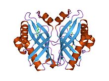
A halohydrin dehalogenase is an enzyme involved in the bacterial degradation of vicinal halohydrins. In several species of bacteria, it catalyses the dehalogenation of halohydrins to produce the corresponding epoxides. Different isoforms of the enzyme fall into one of three groups, A, B or C. Halogenases of the same class are genetically similar, but differ greatly from halogenases from a different group. Currently the most well-studied isoform is HheC which is purified from the bacterial species Agrobacterium radiobacter. The ability to dehalogenate organic compounds as well as form enantiomeric selective epoxides have generated interest in the potential of this enzyme in the biochemical field.

<span class="mw-page-title-main">Catalytic triad</span> Set of three coordinated amino acids

A catalytic triad is a set of three coordinated amino acids that can be found in the active site of some enzymes. Catalytic triads are most commonly found in hydrolase and transferase enzymes. An acid-base-nucleophile triad is a common motif for generating a nucleophilic residue for covalent catalysis. The residues form a charge-relay network to polarise and activate the nucleophile, which attacks the substrate, forming a covalent intermediate which is then hydrolysed to release the product and regenerate free enzyme. The nucleophile is most commonly a serine or cysteine amino acid, but occasionally threonine or even selenocysteine. The 3D structure of the enzyme brings together the triad residues in a precise orientation, even though they may be far apart in the sequence.

<span class="mw-page-title-main">Citrate synthase</span> Mammalian protein found in Homo sapiens

The enzyme citrate synthase E.C. 2.3.3.1 ] exists in nearly all living cells and stands as a pace-making enzyme in the first step of the citric acid cycle. Citrate synthase is localized within eukaryotic cells in the mitochondrial matrix, but is encoded by nuclear DNA rather than mitochondrial. It is synthesized using cytoplasmic ribosomes, then transported into the mitochondrial matrix.

<span class="mw-page-title-main">Glycoside hydrolase</span> Enzyme

Glycoside hydrolases catalyze the hydrolysis of glycosidic bonds in complex sugars. They are extremely common enzymes with roles in nature including degradation of biomass such as cellulose (cellulase), hemicellulose, and starch (amylase), in anti-bacterial defense strategies, in pathogenesis mechanisms and in normal cellular function. Together with glycosyltransferases, glycosidases form the major catalytic machinery for the synthesis and breakage of glycosidic bonds.

<span class="mw-page-title-main">Sucrose phosphorylase</span>

Sucrose phosphorylase is an important enzyme in the metabolism of sucrose and regulation of other metabolic intermediates. Sucrose phosphorylase is in the class of hexosyltransferases. More specifically it has been placed in the retaining glycoside hydrolases family although it catalyzes a transglycosidation rather than hydrolysis. Sucrose phosphorylase catalyzes the conversion of sucrose to D-fructose and α-D-glucose-1-phosphate. It has been shown in multiple experiments that the enzyme catalyzes this conversion by a double displacement mechanism.

<span class="mw-page-title-main">Microsomal epoxide hydrolase</span>

In enzymology, a microsomal epoxide hydrolase (mEH) is an enzyme that catalyzes the hydrolysis reaction between an epoxide and water to form a diol.

<span class="mw-page-title-main">Polyneuridine-aldehyde esterase</span>

The enzyme polyneuridine-aldehyde esterase (EC 3.1.1.78) catalyzes the following reaction:

<span class="mw-page-title-main">Amidase</span>

In enzymology, an amidase is an enzyme that catalyzes the hydrolysis of an amide:

<span class="mw-page-title-main">Dioxygenase</span>

Dioxygenases are oxidoreductase enzymes. Aerobic life, from simple single-celled bacteria species to complex eukaryotic organisms, has evolved to depend on the oxidizing power of dioxygen in various metabolic pathways. From energetic adenosine triphosphate (ATP) generation to xenobiotic degradation, the use of dioxygen as a biological oxidant is widespread and varied in the exact mechanism of its use. Enzymes employ many different schemes to use dioxygen, and this largely depends on the substrate and reaction at hand.

<span class="mw-page-title-main">CFTR inhibitory factor</span>

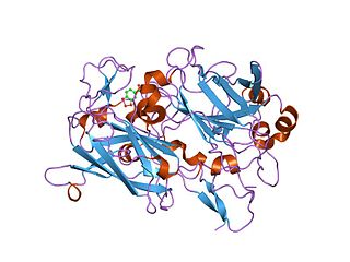
The CFTR inhibitory factor (Cif) is a protein virulence factor secreted by the Gram-negative bacteria Pseudomonas aeruginosa and Acinetobacter nosocomialis. Discovered at Dartmouth Medical School, Cif is able to alter the trafficking of select ABC transporters in eukaryotic epithelial cells, such as the cystic fibrosis transmembrane conductance regulator (CFTR), and P-glycoprotein by interfering with the host deubiquitinating machinery. By promoting the ubiquitin-mediated degradation of CFTR, Cif is able to phenocopy cystic fibrosis at the cellular level. The cif gene is transcribed as part of a 3 gene operon, whose expression is negatively regulated by CifR, a TetR family repressor.

Dihydrocarveol dehydrogenase (EC 1.1.1.296, carveol dehydrogenase (ambiguous)) is an enzyme with systematic name menth-8-en-2-ol:NAD+ oxidoreductase. This enzyme catalyses the following chemical reaction

Limonene-1,2-diol dehydrogenase (EC 1.1.1.297, NAD+-dependent limonene-1,2-diol dehydrogenase) is an enzyme with systematic name menth-8-ene-1,2-diol:NAD+ oxidoreductase. This enzyme catalyses the following chemical reaction

Monocyclic monoterpene ketone monooxygenase (EC 1.14.13.105, 1-hydroxy-2-oxolimonene 1,2-monooxygenase, dihydrocarvone 1,2-monooxygenase, MMKMO) is an enzyme with systematic name (-)-menthone,NADPH:oxygen oxidoreductase. This enzyme catalyses the following chemical reaction

Limonene 1,2-monooxygenase (EC 1.14.13.107) is an enzyme with systematic name limonene,NAD(P)H:oxygen oxidoreductase. This enzyme catalyses the following chemical reaction

The enzyme monoterpene ε-lactone hydrolase (EC 3.1.1.83, MLH; systematic name isoprop(en)ylmethyloxepan-2-one lactonohydrolase catalyses the reaction

<span class="mw-page-title-main">Neopullulanase</span>

Neopullulanase is an enzyme of the alpha-amylase family with systematic name pullulan 4-D-glucanohydrolase (panose-forming). This enzyme principally catalyses the following chemical reaction by cleaving pullulan's alpha-1,4-glucosidic bonds:

<span class="mw-page-title-main">Juvenile hormone epoxide hydrolase</span>

Juvenile hormone epoxide hydrolase (JHEH) is an enzyme that inactivates insect juvenile hormones. This inactivation is accomplished through hydrolysis of the epoxide functional group contained within these hormones into diols. JHEH is one of two enzymes involved in the termination of signaling properties of the various juvenile hormones. The other is juvenile-hormone esterase, or JHE.

References

  1. 1 2 3 Hopmann KH, Hallberg BM, Himo F (October 2005). "Catalytic mechanism of limonene epoxide hydrolase, a theoretical study". Journal of the American Chemical Society. 127 (41): 14339–14347. doi:10.1021/ja050940p. PMID   16218628.
  2. van der Werf MJ, Overkamp KM, de Bont JA (October 1998). "Limonene-1,2-epoxide hydrolase from Rhodococcus erythropolis DCL14 belongs to a novel class of epoxide hydrolases". Journal of Bacteriology. 180 (19): 5052–5057. doi:10.1128/JB.180.19.5052-5057.1998. PMC   107539 . PMID   9748436.
  3. Nardini M, Ridder IS, Rozeboom HJ, Kalk KH, Rink R, Janssen DB, Dijkstra BW (May 1999). "The x-ray structure of epoxide hydrolase from Agrobacterium radiobacter AD1. An enzyme to detoxify harmful epoxides". The Journal of Biological Chemistry. 274 (21): 14579–14586. doi: 10.1074/jbc.274.21.14579 . PMID   10329649.
  4. 1 2 3 Hou QQ, Sheng X, Wang JH, Liu YJ, Liu CB (February 2012). "QM/MM study of the mechanism of enzymatic limonene 1,2-epoxide hydrolysis". Biochimica et Biophysica Acta (BBA) - Proteins and Proteomics. 1824 (2): 263–268. doi:10.1016/j.bbapap.2011.08.014. PMID   21925621.
  5. Armstrong RN, Cassidy CS (2000-01-01). "New structural and chemical insight into the catalytic mechanism of epoxide hydrolases". Drug Metabolism Reviews. 32 (3–4): 327–338. doi:10.1081/DMR-100102337. PMID   11139132. S2CID   27172869.
  6. van der Werf MJ, Swarts HJ, de Bont JA (May 1999). "Rhodococcus erythropolis DCL14 contains a novel degradation pathway for limonene". Applied and Environmental Microbiology. 65 (5): 2092–2102. doi:10.1128/AEM.65.5.2092-2102.1999. PMC   91303 . PMID   10224006.
  7. 1 2 3 Arand M, Hallberg BM, Zou J, Bergfors T, Oesch F, van der Werf MJ, et al. (June 2003). "Structure of Rhodococcus erythropolis limonene-1,2-epoxide hydrolase reveals a novel active site". The EMBO Journal. 22 (11): 2583–2592. doi:10.1093/emboj/cdg275. PMC   156771 . PMID   12773375.
  8. Van der Werf MJ, Orru RV, Overkamp KM, Swarts HJ, Osprian I, Steinreiber A, et al. (1999-09-01). "Substrate specificity and stereospecificity of limonene-1,2-epoxide hydrolase from Rhodococcus erythropolis DCL14; an enzyme showing sequential and enantioconvergent substrate conversion". Applied Microbiology and Biotechnology. 52 (3): 380–385. doi:10.1007/s002530051535. ISSN   1432-0614. S2CID   42378735.
  9. Johansson P, Unge T, Cronin A, Arand M, Bergfors T, Jones TA, Mowbray SL (September 2005). "Structure of an atypical epoxide hydrolase from Mycobacterium tuberculosis gives insights into its function". Journal of Molecular Biology. 351 (5): 1048–1056. doi:10.1016/j.jmb.2005.06.055. PMID   16051262.
This article incorporates text from the public domain Pfam and InterPro: IPR013100