Cranial nerves are the nerves that emerge directly from the brain, of which there are conventionally considered twelve pairs. Cranial nerves relay information between the brain and parts of the body, primarily to and from regions of the head and neck, including the special senses of vision, taste, smell, and hearing.

The parasympathetic nervous system (PSNS) is one of the three divisions of the autonomic nervous system, the others being the sympathetic nervous system and the enteric nervous system. The enteric nervous system is sometimes considered part of the autonomic nervous system, and sometimes considered an independent system.

In anatomy, the palatine bones are two irregular bones of the facial skeleton in many animal species, located above the uvula in the throat. Together with the maxilla, they comprise the hard palate.

In anatomy, the orbit is the cavity or socket/hole of the skull in which the eye and its appendages are situated. "Orbit" can refer to the bony socket, or it can also be used to imply the contents. In the adult human, the volume of the orbit is 30 millilitres, of which the eye occupies 6.5 ml. The orbital contents comprise the eye, the orbital and retrobulbar fascia, extraocular muscles, cranial nerves II, III, IV, V, and VI, blood vessels, fat, the lacrimal gland with its sac and duct, the eyelids, medial and lateral palpebral ligaments, cheek ligaments, the suspensory ligament, septum, ciliary ganglion and short ciliary nerves.

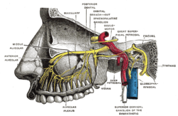
The pterygopalatine ganglion is a parasympathetic ganglion in the pterygopalatine fossa. It is one of four parasympathetic ganglia of the head and neck,.
In neuroanatomy, the maxillary nerve (V2) is one of the three branches or divisions of the trigeminal nerve, the fifth (CN V) cranial nerve. It comprises the principal functions of sensation from the maxilla, nasal cavity, sinuses, the palate and subsequently that of the mid-face, and is intermediate, both in position and size, between the ophthalmic nerve and the mandibular nerve.

The geniculate ganglion is a collection of pseudounipolar sensory neurons of the facial nerve located in the facial canal of the head. It receives fibers from the facial nerve. It sends fibers that supply the lacrimal glands, submandibular glands, sublingual glands, tongue, palate, pharynx, external auditory meatus, stapedius muscle, posterior belly of the digastric muscle, stylohyoid muscle, and muscles of facial expression.

The obturator nerve in human anatomy arises from the ventral divisions of the second, third, and fourth lumbar nerves in the lumbar plexus; the branch from the third is the largest, while that from the second is often very small.

The palatine aponeurosis a thin, firm, fibrous lamella which gives strength and support to soft palate. It serves as the insertion for the tensor veli palatini and levator veli palatini, and the origin for the musculus uvulae, palatopharyngeus, and palatoglossus.

The zygomatic nerve is a branch of the maxillary nerve. It arises in the pterygopalatine fossa and enters the orbit through the inferior orbital fissure before dividing into its two terminal branches: the zygomaticotemporal nerve and zygomaticofacial nerve.

The urethral artery arises from the internal pudendal artery a branch of the internal iliac artery. The internal pudendal artery has numerous branches including the artery of the bulb of the penis immediately before the urethral and the dorsal artery of the penis more distally.

The zygomaticotemporal nerve (zygomaticotemporal branch, temporal branch) is a cutaneous (sensory) nerve of the head. It is a branch of the zygomatic nerve (itself a branch of the maxillary nerve (CN V2)). It arises in the orbit and exits the orbit through the zygomaticotemporal foramen in the zygomatic bone to enter the temporal fossa. It is distributed to the skin of the side of the forehead. It also contains a parasympathetic secretomotor component for the lacrimal gland which it confers to the lacrimal nerve (which then delivers it to the gland).

The sympathetic trunks are a paired bundle of nerve fibers that run from the base of the skull to the coccyx. They are a major component of the sympathetic nervous system.

The infratemporal fossa is an irregularly shaped cavity that is a part of the skull. It is situated below and medial to the zygomatic arch. It is not fully enclosed by bone in all directions. It contains superficial muscles, including the lower part of the temporalis muscle, the lateral pterygoid muscle, and the medial pterygoid muscle. It also contains important blood vessels such as the middle meningeal artery, the pterygoid plexus, and the retromandibular vein, and nerves such as the mandibular nerve (CN V3) and its branches.
The posterior superior alveolar nerves (also posterior superior dental nerves or posterior superior alveolar branches) are sensory branches of the maxillary nerve (CN V2). They arise within the pterygopalatine fossa as a single trunk. They run on or in the maxilla. They provide sensory innervation to the upper molar teeth and adjacent gum, and the maxillary sinus.

The general visceral afferent (GVA) fibers conduct sensory impulses from the internal organs, glands, and blood vessels to the central nervous system. They are considered to be part of the visceral nervous system, which is closely related to the autonomic nervous system, but 'visceral nervous system' and 'autonomic nervous system' are not direct synonyms and care should be taken when using these terms. Unlike the efferent fibers of the autonomic nervous system, the afferent fibers are not classified as either sympathetic or parasympathetic.
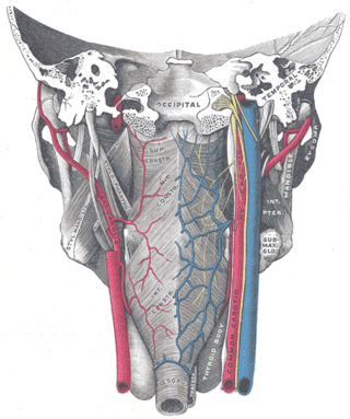
The pharyngeal plexus is a nerve plexus located upon the outer surface of the pharynx. It contains a motor component, a sensory component, and sympathetic component.
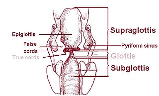
The pyriform sinus is a small recess on either side of the laryngeal inlet. It is bounded medially by the aryepiglottic fold, and laterally by the thyroid cartilage and thyrohyoid membrane. The fossae are involved in speech.
The sacral ganglia are paravertebral ganglia of the sympathetic trunk.:39 As the sympathetic trunk heads inferiorly down the sacrum, it turns medially. There are generally four or five sacral ganglia. In addition to gray rami communicantes, the ganglia send off sacral splanchnic nerves to join the inferior hypogastric plexus. Near the coccyx, the right and left sympathetic trunks join to form the ganglion impar.

The salivatory nuclei are two parasympathetic general visceral efferent cranial nerve nuclei - the superior salivatory nucleus and the inferior salivatory nucleus - that innervate the salivary glands. Both are located in the pontine tegmentum of the brainstem.