Telomeric repeat-binding factor 2

Last updated
TERF2
Protein TERF2 PDB 1h6p.png
Available structures
PDB Ortholog search: PDBe RCSB
Identifiers
Aliases TERF2 , TRBF2, TRF2, telomeric repeat binding factor 2
External IDs OMIM: 602027 MGI: 1195972 HomoloGene: 4133 GeneCards: TERF2
Orthologs
SpeciesHumanMouse
Entrez
Ensembl
UniProt
RefSeq (mRNA)

NM_005652

NM_001083118
NM_001286200
NM_009353

RefSeq (protein)

NP_005643

Location (UCSC) Chr 16: 69.36 – 69.41 Mb Chr 8: 107.07 – 107.1 Mb
PubMed search [3] [4]
Wikidata
View/Edit Human View/Edit Mouse

Telomeric repeat-binding factor 2 is a protein that is present at telomeres throughout the cell cycle. It is also known as TERF2, TRF2, and TRBF2, and is encoded in humans by the TERF2 gene. [5] It is a component of the shelterin nucleoprotein complex and a second negative regulator of telomere length, playing a key role in the protective activity of telomeres. It was first reported in 1997 in the lab of Titia de Lange, [6] where a DNA sequence similar, but not identical, to TERF1 was discovered, with respect to the Myb-domain. De Lange isolated the new Myb-containing protein sequence and called it TERF2.

Structure and domains

TERF2 has a similar structure to that of TERF1. Both proteins carry a C-terminus Myb motif and large TERF1-related dimerization domains near their N-terminus. [6] However, both proteins exist exclusively as homodimers and do not heterodimerize with each other, as proven by co-immunoprecipitation assay analysis. [6] Also, TERF2 has a basic N-terminus, differing from TERF1’s acidic N-terminus, and was found to be much more conserved, suggesting that the two proteins have distinct functions. [7]

There are 4 domain categories on the TERF2 protein that allow it to bind to both other proteins in the shelterin protein complex, and to specific types of DNA.

TERF homology domain

The TERF Homology Domain (TRFH; InterPro :  IPR013867 ) is an area that helps to promote homodimerization of TERF2 with itself. This results in the formation of a quaternary structure that is characteristic of this protein. This TRFH domain also allows TERF2 to bind to and act as a dock for many other types of proteins. The Apollo nuclease, a shelterin accessory factor, uses the TRFH domain as a dock. The recruitment of Apollo by TERF2 allows for telomeric ends formed by DNA synthesis to be processed. By doing so, the telomere ends are able to avoid ATM kinase activation through the creation of a terminal structure. [8] SLX4, which is important in DNA repair by acting as a scaffold for structure-specific DNA repair nucleases, also binds to the TRFH domain of TERF2. [9] The TRFH domain is responsible for other binding events, including RTEL1, and proteins that contain a TBD site.

Myb domain

The Myb domain (InterPro :  IPR017930 ) acts by binding to double-stranded telomeric DNA. This region gets its name from a viral protein called Myb derived from the avian myeloblastosis virus. Specifically, the sequence that this Myb domain targets on the DNA is (GGTTAG/CCAATC)n.

Basic and hinge domains

Two other domains also work to bind and influence the activity of proteins associated with the TERF2 protein. Both are unique to TERF2. The basic domain sits at the N-terminal, and has two main functions: the prevention of t-loop excision by XRCC3, and the inhibition of SLX4. The final domain of TERF2 is called the hinge domain (InterPro :  IPR031902 ). This domain contains a motif for binding the shelterin protein TIN2, which acts as a stabilizing protein, connecting units that are attached to double stranded and single stranded DNA. [10] This domain also is responsible for binding to RAP1, and helps to inhibit RNF168 recruitment at telomeres.

Function

This protein is present at telomeres in metaphase of the cell cycle, is a second negative regulator of telomere length, and plays a key role in the protective activity of telomeres. While having similar telomere binding activity and domain organization, TERF2 differs from TERF1 in that its N terminus is basic rather than acidic. [7]

T-Loop formation

Specific domains on the TERF2 protein sequence and their functionalities. TERF2 Domains.png
Specific domains on the TERF2 protein sequence and their functionalities.

Telomeric ends are structurally similar to double-stranded breaks on the chromosome. To prevent the cellular DNA repair machinery from mistakenly identifying telomeres as chromosome breaks, t-loops are formed in which the 3’ TTAGGG overhang of the telomere loops back into the DNA duplex. TERF2 promotes t-loop formation by preferentially binding to a telomeric double-stranded DNA duplex containing a 3’ TTAGGG single-stranded overhang. If the 3’ TTAGGG overhang is not present, TERF2 will not bind. Once bound, it migrates to the t-loop junction where the single-stranded overhang invades the double-stranded region upstream. No other shelterin protein has been shown to promote this process but interaction of TERF2 with TERF2IP is shown to promote higher t-loop formation in vitro. [12] Studies have demonstrated that deletion of TERF2 prevents t-loop formation, leading to excessive loss of telomeric DNA and early cell death. [13]

ATM kinase prevention

TERF2 plays a central role in preventing ATM kinase DNA damage response. It binds telomeric dsDNA and prevents telomeres from activating ATM kinase. This interaction of TERF2 with ATM is believed to be relevant to the mechanism by which TERF2 blocks ATM signaling. Because of its oligomeric nature, TERF2 could potentially cross-link ATM monomers and hold the kinase in its inactive dimeric state, thereby blocking amplification of the ATM signal at an early step in its activation. However, because mutations in the TERF2 dimerization domain destabilize the protein, it has not been possible to test the contribution of TERF2 oligomerization on ATM repression directly. Removal of TERF2 induces ATM-dependent apoptosis by localizing the active, phosphorylated form of ATM to unprotected chromosome ends. Since TERF2 specifically binds at telomeres and remains there when DNA damage is induced, it is unlikely to interfere with activation of the ATM kinase at different sites of DNA damage. Therefore, TERF2 could act as a telomere-specific inhibitor of ATM kinase. [14]

TERF2 Knockout Effects

Conditional deletion of TERF2 in mice cells effectively removes the shelterin nucleoprotein complex. As a result of removing this complex, several unwanted DNA damage response pathways are activated, including ATM kinase signaling, ATR kinase signaling, non-homologous end-joining (NHEJ), alt-NHEJ, C-NHEJ, 5' resection, and homology directed repair (HDR). [15] These repair pathways (in the presence of P53 knockout and Cre) often contribute to the phenotype where chromosome ends are connected to each other in a very long chain, which can be visualized by a combination of a DAPI stain and fluorescence in situ hybridization (FISH) technique.

Interactions

Client proteins have a distinctive YxLxP motif (where "x" can be any amino acid) that bind to TERF2. TERF2 client protein recruitment.png
Client proteins have a distinctive YxLxP motif (where "x" can be any amino acid) that bind to TERF2.

TERF2 is also known to recruit certain client proteins, also known as accessory factors. These client proteins are often recruited to TERF2 for a specific function at a specific time, often temporarily. The TRFH domain contains a F120 residue, which is the binding site of TERF2 where it recruits client proteins. These client proteins also contain a TRFH binding motif, which consists of a conserved 6-amino acid sequence of the following formula: YxLxP, where "x" can be any amino acid substituted. [16] The above-mentioned Apollo nuclease (one of many TERF2's client proteins) also contains the formulaic motif; its specific motif sequence is YLLTP.

TERF1 also demonstrates similar client protein recruitment mechanism as TERF2, except that it diverges at two concepts: 1) the TRFH of TERF1 contains a F142 residue, 2) the client proteins specific for TERF1 contain the TRFH binding motif sequence of FxLxP, where the amino acid Y (tyrosine) is replaced with F (phenylalanine).

TERF2 has also been shown to interact with:

Disease relevance of TERF2

Cancer

Telomerase is an enzyme that works to create telomeric ends for DNA, and it is thought to play important roles in the development of cancer. Specifically, telomeric stability is known to be a common occurrence in cancer cells. [23] Along with the telomerase, the shelterin complex, and TERF2 and TERF1 specifically, also have been noted to control the lengths of telomeres formed by these telomerases. Shelterin works to protect telomeres against unsuitable activation of the DNA damage response pathway, as noted in the function section above. TERF2 as part of the shelterin complex, has been known to block the ATM signaling pathways and prevent chromosome end fusion. In cancer cells, TERF2 phosphorylation by extracellular signal-regulated kinase (ERK1/2) is a controlling factor in the major pro-oncogenic signaling pathways (RAS/RAF/MEK/ERK) that affect telomeric stability. [23] Additionally, when TERF2 was non-phosphorylated in melanoma cells, there was a cell induced DNA damage response, arresting growth and causing tumor reversion. [23] Studies have found that in tumor cells, TERF2 levels are observed to be high, and this raised level of TERF2 contributes to oncogenesis in a variety of ways. [24] [25] [26] This high level of TERF2 decreases the ability to recruit and activate natural killer cells in human tumor cells. [24] One study used a dominant negative form of TERF2ΔBΔC, to inhibit TERF2, and found that it could induce a reversion malignant phenotype in human melanoma cells. [25] Therefore, over-expression of TERF2ΔBΔC, causing blocking of TERF2, induced apoptosis and reduced tumourigenicity in certain cell lines. [25] Additionally, upregulation of TERF2 may be the cause of the establishment and maintenance of short telomeres. [26] These short telomeres increase chromosomal instability, and increase the chances of certain cancers progressing in the body, such as with leukemia. [26] In gastric mucosa tissues, the expression of TERF2 proteins was significantly higher than normal, and this over-expression of TERF2, along with over-expression of TERF1, TIN2, TERT, and BRCA1 protein transposition, may cause a reduction in telomere length, further contributing to multistage carcinogenesis of gastric cancer. [27]

Related Research Articles

<span class="mw-page-title-main">ATM serine/threonine kinase</span>

ATM serine/threonine kinase or Ataxia-telangiectasia mutated, symbol ATM, is a serine/threonine protein kinase that is recruited and activated by DNA double-strand breaks, oxidative stress, topoisomerase cleavage complexes, splicing intermediates, R-loops and in some cases by single-strand DNA breaks. It phosphorylates several key proteins that initiate activation of the DNA damage checkpoint, leading to cell cycle arrest, DNA repair or apoptosis. Several of these targets, including p53, CHK2, BRCA1, NBS1 and H2AX are tumor suppressors.

<span class="mw-page-title-main">Ku (protein)</span>

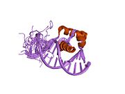
Ku is a dimeric protein complex that binds to DNA double-strand break ends and is required for the non-homologous end joining (NHEJ) pathway of DNA repair. Ku is evolutionarily conserved from bacteria to humans. The ancestral bacterial Ku is a homodimer. Eukaryotic Ku is a heterodimer of two polypeptides, Ku70 (XRCC6) and Ku80 (XRCC5), so named because the molecular weight of the human Ku proteins is around 70 kDa and 80 kDa. The two Ku subunits form a basket-shaped structure that threads onto the DNA end. Once bound, Ku can slide down the DNA strand, allowing more Ku molecules to thread onto the end. In higher eukaryotes, Ku forms a complex with the DNA-dependent protein kinase catalytic subunit (DNA-PKcs) to form the full DNA-dependent protein kinase, DNA-PK. Ku is thought to function as a molecular scaffold to which other proteins involved in NHEJ can bind, orienting the double-strand break for ligation.

<span class="mw-page-title-main">Ku70</span> Protein-coding gene in the species Homo sapiens

Ku70 is a protein that, in humans, is encoded by the XRCC6 gene.

<span class="mw-page-title-main">Ku80</span> Protein-coding gene in the species Homo sapiens

Ku80 is a protein that, in humans, is encoded by the XRCC5 gene. Together, Ku70 and Ku80 make up the Ku heterodimer, which binds to DNA double-strand break ends and is required for the non-homologous end joining (NHEJ) pathway of DNA repair. It is also required for V(D)J recombination, which utilizes the NHEJ pathway to promote antigen diversity in the mammalian immune system.

<span class="mw-page-title-main">MRE11A</span> Protein-coding gene in the species Homo sapiens

Double-strand break repair protein MRE11 is an enzyme that in humans is encoded by the MRE11 gene. The gene has been designated MRE11A to distinguish it from the pseudogene MRE11B that is nowadays named MRE11P1.

<span class="mw-page-title-main">Telomeric repeat-binding factor 1</span> Protein-coding gene in humans

Telomeric repeat-binding factor 1 is a protein that in humans is encoded by the TERF1 gene.

<span class="mw-page-title-main">Rad50</span> Protein-coding gene in the species Homo sapiens

DNA repair protein RAD50, also known as RAD50, is a protein that in humans is encoded by the RAD50 gene.

<span class="mw-page-title-main">POT1</span> Protein-coding gene in the species Homo sapiens

Protection of telomeres protein 1 is a protein that in humans is encoded by the POT1 gene.

<span class="mw-page-title-main">Tankyrase</span> Enzyme

Tankyrase, also known as tankyrase 1, is an enzyme that in humans is encoded by the TNKS gene. It inhibits the binding of TERF1 to telomeric DNA. Tankyrase attracts substantial interest in cancer research through its interaction with AXIN1 and AXIN2, which are negative regulators of pro-oncogenic β-catenin signaling. Importantly, activity in the β-catenin destruction complex can be increased by tankyrase inhibitors and thus such inhibitors are a potential therapeutic option to reduce the growth of β-catenin-dependent cancers.

<span class="mw-page-title-main">TINF2</span> Protein-coding gene in the species Homo sapiens

TERF1-interacting nuclear factor 2 is a protein that in humans is encoded by the TINF2 gene. TINF2 is a component of the shelterin protein complex found at the end of telomeres.

<span class="mw-page-title-main">ACD (gene)</span> Protein-coding gene in the species Homo sapiens

Adrenocortical dysplasia protein homolog is a protein that in humans is encoded by the ACD gene.

<span class="mw-page-title-main">TERF2IP</span> Protein-coding gene in the species Homo sapiens

Telomeric repeat-binding factor 2-interacting protein 1 also known as repressor activator protein 1 (Rap1) is a protein that in humans is encoded by the TERF2IP gene.

<span class="mw-page-title-main">PINX1</span> Protein-coding gene in the species Homo sapiens

PIN2/TERF1-interacting telomerase inhibitor 1, also known as PINX1, is a human gene. PINX1 is also known as PIN2 interacting protein 1. PINX1 is a telomerase inhibitor and a possible tumor suppressor.

<span class="mw-page-title-main">Tankyrase 2</span> Protein-coding gene in the species Homo sapiens

Tankyrase-2 is an enzyme that in humans is encoded by the TNKS2 gene.

Telomere-binding proteins function to bind telomeric DNA in various species. In particular, telomere-binding protein refers to TTAGGG repeat binding factor-1 (TERF1) and TTAGGG repeat binding factor-2 (TERF2). Telomere sequences in humans are composed of TTAGGG sequences which provide protection and replication of chromosome ends to prevent degradation. Telomere-binding proteins can generate a T-loop to protect chromosome ends. TRFs are double-stranded proteins which are known to induce bending, looping, and pairing of DNA which aids in the formation of T-loops. They directly bind to TTAGGG repeat sequence in the DNA. There are also subtelomeric regions present for regulation. However, in humans, there are six subunits forming a complex known as shelterin.

Shelterin is a protein complex known to protect telomeres in many eukaryotes from DNA repair mechanisms, as well as to regulate telomerase activity. In mammals and other vertebrates, telomeric DNA consists of repeating double-stranded 5'-TTAGGG-3' (G-strand) sequences along with the 3'-AATCCC-5' (C-strand) complement, ending with a 50-400 nucleotide 3' (G-strand) overhang. Much of the final double-stranded portion of the telomere forms a T-loop (Telomere-loop) that is invaded by the 3' (G-strand) overhang to form a small D-loop (Displacement-loop).

<span class="mw-page-title-main">Titia de Lange</span> Dutch geneticist

Titia de Lange is the Director of the Anderson Center for Cancer Research, the Leon Hess professor and the head of Laboratory Cell Biology and Genetics at Rockefeller University.

<span class="mw-page-title-main">Telomeric repeat–containing RNA</span> Long non-coding RNA transcribed from telomeres

Telomeric repeat–containing RNA (TERRA) is a long non-coding RNA transcribed from telomeres - repetitive nucleotide regions found on the ends of chromosomes that function to protect DNA from deterioration or fusion with neighboring chromosomes. TERRA has been shown to be ubiquitously expressed in almost all cell types containing linear chromosomes - including humans, mice, and yeasts. While the exact function of TERRA is still an active area of research, it is generally believed to play a role in regulating telomerase activity as well as maintaining the heterochromatic state at the ends of chromosomes. TERRA interaction with other associated telomeric proteins has also been shown to help regulate telomere integrity in a length-dependent manner.

SilentInformationRegulator (SIR) proteins are involved in regulating gene expression. SIR proteins organize heterochromatin near telomeres, ribosomal DNA (rDNA), and at silent loci including hidden mating type loci in yeast. The SIR family of genes encodes catalytic and non-catalytic proteins that are involved in de-acetylation of histone tails and the subsequent condensation of chromatin around a SIR protein scaffold. Some SIR family members are conserved from yeast to humans.

Telomeres, the caps on the ends of eukaryotic chromosomes, play critical roles in cellular aging and cancer. An important facet to how telomeres function in these roles is their involvement in cell cycle regulation.

References

  1. 1 2 3 GRCh38: Ensembl release 89: ENSG00000132604 - Ensembl, May 2017
  2. 1 2 3 GRCm38: Ensembl release 89: ENSMUSG00000031921 - Ensembl, May 2017
  3. "Human PubMed Reference:". National Center for Biotechnology Information, U.S. National Library of Medicine.
  4. "Mouse PubMed Reference:". National Center for Biotechnology Information, U.S. National Library of Medicine.
  5. Sakaguchi AY, Padalecki SS, Mattern V, Rodriguez A, Leach RJ, McGill JR, Chavez M, Giambernardi TA (May 1998). "Chromosomal sublocalization of the transcribed human telomere repeat binding factor 2 gene and comparative mapping in the mouse". Somat. Cell Mol. Genet. 24 (3): 157–63. doi:10.1023/b:scam.0000007118.47691.d7. PMID   10226653. S2CID   26610531.
  6. 1 2 3 Broccoli D, Smogorzewska A, Chong L, de Lange T (October 1997). "Human telomeres contain two distinct Myb-related proteins, TRF1 and TRF2". Nature Genetics. 17 (2): 231–5. doi:10.1038/ng1097-231. PMID   9326950. S2CID   41204064.
  7. 1 2 "Entrez Gene: TERF2 telomeric repeat binding factor 2".
  8. Wu P, van Overbeek M, Rooney S, de Lange T (August 2010). "Apollo contributes to G overhang maintenance and protects leading-end telomeres". Molecular Cell. 39 (4): 606–17. doi:10.1016/j.molcel.2010.06.031. PMC   2929323 . PMID   20619712.
  9. Wilson JS, Tejera AM, Castor D, Toth R, Blasco MA, Rouse J (September 2013). "Localization-dependent and -independent roles of SLX4 in regulating telomeres". Cell Reports. 4 (5): 853–60. doi:10.1016/j.celrep.2013.07.033. PMC   3969258 . PMID   23994477.
  10. Palm W, de Lange T (2008-11-04). "How shelterin protects mammalian telomeres". Annual Review of Genetics. 42 (1): 301–34. doi:10.1146/annurev.genet.41.110306.130350. PMID   18680434.
  11. Arnoult N, Karlseder J (November 2015). "Complex interactions between the DNA-damage response and mammalian telomeres". Nature Structural & Molecular Biology. 22 (11): 859–66. doi:10.1038/nsmb.3092. PMC   4739752 . PMID   26581520.
  12. Arat NÖ, Griffith JD (December 2012). "Human Rap1 interacts directly with telomeric DNA and regulates TRF2 localization at the telomere". The Journal of Biological Chemistry. 287 (50): 41583–94. doi: 10.1074/jbc.M112.415984 . PMC   3516710 . PMID   23086976.
  13. Stansel RM, de Lange T, Griffith JD (October 2001). "T-loop assembly in vitro involves binding of TRF2 near the 3' telomeric overhang". The EMBO Journal. 20 (19): 5532–40. doi:10.1093/emboj/20.19.5532. PMC   125642 . PMID   11574485.
  14. Karlseder J, Hoke K, Mirzoeva OK, Bakkenist C, Kastan MB, Petrini JH, de Lange T (August 2004). "The telomeric protein TRF2 binds the ATM kinase and can inhibit the ATM-dependent DNA damage response". PLOS Biology. 2 (8): E240. doi: 10.1371/journal.pbio.0020240 . PMC   509302 . PMID   15314656.
  15. Sfeir A, de Lange T (May 2012). "Removal of shelterin reveals the telomere end-protection problem". Science. 336 (6081): 593–7. Bibcode:2012Sci...336..593S. doi:10.1126/science.1218498. PMC   3477646 . PMID   22556254.
  16. 1 2 Chen Y, Yang Y, van Overbeek M, Donigian JR, Baciu P, de Lange T, Lei M (February 2008). "A shared docking motif in TRF1 and TRF2 used for differential recruitment of telomeric proteins". Science. 319 (5866): 1092–6. Bibcode:2008Sci...319.1092C. doi: 10.1126/science.1151804 . PMID   18202258. S2CID   54523266.
  17. Song K, Jung D, Jung Y, Lee SG, Lee I (September 2000). "Interaction of human Ku70 with TRF2". FEBS Letters. 481 (1): 81–5. doi:10.1016/s0014-5793(00)01958-x. PMID   10984620. S2CID   22753893.
  18. 1 2 3 4 Zhu XD, Küster B, Mann M, Petrini JH, de Lange T (July 2000). "Cell-cycle-regulated association of RAD50/MRE11/NBS1 with TRF2 and human telomeres". Nature Genetics. 25 (3): 347–52. doi:10.1038/77139. PMID   10888888. S2CID   6689794.
  19. 1 2 O'Connor MS, Safari A, Liu D, Qin J, Songyang Z (July 2004). "The human Rap1 protein complex and modulation of telomere length". The Journal of Biological Chemistry. 279 (27): 28585–91. doi: 10.1074/jbc.M312913200 . PMID   15100233.
  20. Zhang P, Casaday-Potts R, Precht P, Jiang H, Liu Y, Pazin MJ, Mattson MP (September 2011). "Nontelomeric splice variant of telomere repeat-binding factor 2 maintains neuronal traits by sequestering repressor element 1-silencing transcription factor". Proceedings of the National Academy of Sciences of the United States of America. 108 (39): 16434–9. Bibcode:2011PNAS..10816434Z. doi: 10.1073/pnas.1106906108 . PMC   3182730 . PMID   21903926.
  21. Li B, Oestreich S, de Lange T (May 2000). "Identification of human Rap1: implications for telomere evolution". Cell. 101 (5): 471–83. doi: 10.1016/S0092-8674(00)80858-2 . PMID   10850490. S2CID   7956545.
  22. Opresko PL, von Kobbe C, Laine JP, Harrigan J, Hickson ID, Bohr VA (October 2002). "Telomere-binding protein TRF2 binds to and stimulates the Werner and Bloom syndrome helicases". The Journal of Biological Chemistry. 277 (43): 41110–9. doi: 10.1074/jbc.M205396200 . PMID   12181313.
  23. 1 2 3 Picco V, Coste I, Giraud-Panis MJ, Renno T, Gilson E, Pagès G (July 2016). "ERK1/2/MAPK pathway-dependent regulation of the telomeric factor TRF2". Oncotarget. 7 (29): 46615–46627. doi:10.18632/oncotarget.10316. PMC   5216822 . PMID   27366950.
  24. 1 2 Biroccio A, Cherfils-Vicini J, Augereau A, Pinte S, Bauwens S, Ye J, Simonet T, Horard B, Jamet K, Cervera L, Mendez-Bermudez A, Poncet D, Grataroli R, de Rodenbeeke CT, Salvati E, Rizzo A, Zizza P, Ricoul M, Cognet C, Kuilman T, Duret H, Lépinasse F, Marvel J, Verhoeyen E, Cosset FL, Peeper D, Smyth MJ, Londoño-Vallejo A, Sabatier L, Picco V, Pages G, Scoazec JY, Stoppacciaro A, Leonetti C, Vivier E, Gilson E (July 2013). "TRF2 inhibits a cell-extrinsic pathway through which natural killer cells eliminate cancer cells". Nature Cell Biology. 15 (7): 818–28. doi:10.1038/ncb2774. hdl:11573/559592. PMID   23792691. S2CID   205287459.
  25. 1 2 3 Biroccio A, Rizzo A, Elli R, Koering CE, Belleville A, Benassi B, Leonetti C, Stevens MF, D'Incalci M, Zupi G, Gilson E (August 2006). "TRF2 inhibition triggers apoptosis and reduces tumourigenicity of human melanoma cells". European Journal of Cancer. 42 (12): 1881–8. doi:10.1016/j.ejca.2006.03.010. PMID   16750909.
  26. 1 2 3 Bellon M, Datta A, Brown M, Pouliquen JF, Couppie P, Kazanji M, Nicot C (November 2006). "Increased expression of telomere length regulating factors TRF1, TRF2 and TIN2 in patients with adult T-cell leukemia". International Journal of Cancer. 119 (9): 2090–7. doi: 10.1002/ijc.22026 . PMID   16786598. S2CID   37883075.
  27. Hu H, Zhang Y, Zou M, Yang S, Liang XQ (September 2010). "Expression of TRF1, TRF2, TIN2, TERT, KU70, and BRCA1 proteins is associated with telomere shortening and may contribute to multistage carcinogenesis of gastric cancer". Journal of Cancer Research and Clinical Oncology. 136 (9): 1407–14. doi:10.1007/s00432-010-0795-x. PMID   20127252. S2CID   19253770.

Further reading