Melanin is a broad term for a group of natural pigments found in most organisms. The melanin pigments are produced in a specialized group of cells known as melanocytes.

Melanocytes are melanin-producing neural crest-derived cells located in the bottom layer of the skin's epidermis, the middle layer of the eye, the inner ear, vaginal epithelium, meninges, bones, and heart. Melanin is a dark pigment primarily responsible for skin color. Once synthesized, melanin is contained in special organelles called melanosomes which can be transported to nearby keratinocytes to induce pigmentation. Thus darker skin tones have more melanosomes present than lighter skin tones. Functionally, melanin serves as protection against UV radiation. Melanocytes also have a role in the immune system.
A dilution gene is any one of a number of genes that act to create a lighter coat color in living creatures. There are many examples of such genes:

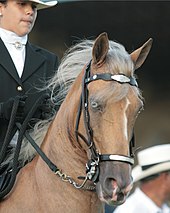
The cream gene is responsible for a number of horse coat colors. Horses that have the cream gene in addition to a base coat color that is chestnut will become palomino if they are heterozygous, having one copy of the cream gene, or cremello, if they are homozygous. Similarly, horses with a bay base coat and the cream gene will be buckskin or perlino. A black base coat with the cream gene becomes the not-always-recognized smoky black or a smoky cream. Cream horses, even those with blue eyes, are not white horses. Dilution coloring is also not related to any of the white spotting patterns.

Equine coat color genetics determine a horse's coat color. Many colors are possible, but all variations are produced by changes in only a few genes. Bay is the most common color of horses. A change at the agouti locus is capable of turning bay to black, while a mutation at the extension locus can turn bay or black to chestnut.

Leucism is a wide variety of conditions that result in the partial loss of pigmentation in an animal—causing white, pale, or patchy coloration of the skin, hair, feathers, scales, or cuticles, but not the eyes. It is occasionally spelled leukism. Some genetic conditions that result in a "leucistic" appearance include piebaldism, Waardenburg syndrome, vitiligo, Chédiak–Higashi syndrome, flavism, isabellinism, xanthochromism, axanthism, amelanism, and melanophilin mutations. Pale patches of skin, feathers, or fur can also result from injury.

Sodium/potassium/calcium exchanger 5 (NCKX5), also known as solute carrier family 24 member 5 (SLC24A5), is a protein that in humans is encoded by the SLC24A5 gene that has a major influence on natural skin colour variation. The NCKX5 protein is a member of the potassium-dependent sodium/calcium exchanger family. Sequence variation in the SLC24A5 gene, particularly a non-synonymous SNP changing the amino acid at position 111 in NCKX5 from alanine to threonine, has been associated with differences in skin pigmentation.

Agouti-signaling protein is a protein that in humans is encoded by the ASIP gene. It is responsible for the distribution of melanin pigment in mammals. Agouti interacts with the melanocortin 1 receptor to determine whether the melanocyte produces phaeomelanin, or eumelanin. This interaction is responsible for making distinct light and dark bands in the hairs of animals such as the agouti, which the gene is named after. In other species such as horses, agouti signalling is responsible for determining which parts of the body will be red or black. Mice with wildtype agouti will be grey, with each hair being partly yellow and partly black. Loss of function mutations in mice and other species cause black fur coloration, while mutations causing expression throughout the whole body in mice cause yellow fur and obesity.

Horses exhibit a diverse array of coat colors and distinctive markings. A specialized vocabulary has evolved to describe them.

A white horse is born predominantly white and stays white throughout its life. A white horse has mostly pink skin under its hair coat, and may have brown, blue, or hazel eyes. "True white" horses, especially those that carry one of the dominant white (W) genes, are rare. Most horses that are commonly referred to as "white" are actually "gray" horses whose hair coats are completely white. Gray horses may be born of any color and their hairs gradually turn white as time goes by and take on a white appearance. Nearly all gray horses have dark skin, except under any white markings present at birth. Skin color is the most common method for an observer to distinguish between mature white and gray horses.

The genetic basis of coat colour in the Labrador Retriever has been found to depend on several distinct genes. The interplay among these genes is used as an example of epistasis.

Tyrosinase-related protein 1, also known as TYRP1, is an intermembrane enzyme which in humans is encoded by the TYRP1 gene.
P protein, also known as melanocyte-specific transporter protein or pink-eyed dilution protein homolog, is a protein that in humans is encoded by the oculocutaneous albinism II (OCA2) gene. The P protein is believed to be an integral membrane protein involved in small molecule transport, specifically of tyrosine—a precursor of melanin. Certain mutations in OCA2 result in type 2 oculocutaneous albinism. OCA2 encodes the human homologue of the mouse p gene.

Membrane-associated transporter protein (MATP), also known as solute carrier family 45 member 2 (SLC45A2) or melanoma antigen AIM1, is a protein that in humans is encoded by the SLC45A2 gene.
Oculocutaneous albinism type I or type 1A is an autosomal recessive skin disease. This subtype of oculocutaneous albinism is caused when the gene for tyrosinase does not function properly.

Ocular albinism type 1(OA1) is the most common type of ocular albinism, with a prevalence rate of 1:50,000. It is an inheritable classical Mendelian type X-linked recessive disorder wherein the retinal pigment epithelium lacks pigment while hair and skin appear normal. Since it is usually an X-linked disorder, it occurs mostly in males, while females are carriers unless they are homozygous. About 60 missense and nonsense mutations, insertions, and deletions have been identified in Oa1. Mutations in OA1 have been linked to defective glycosylation and thus improper intracellular transportation.

Amelanism is a pigmentation abnormality characterized by the lack of pigments called melanins, commonly associated with a genetic loss of tyrosinase function. Amelanism can affect fish, amphibians, reptiles, birds, and mammals including humans. The appearance of an amelanistic animal depends on the remaining non-melanin pigments. The opposite of amelanism is melanism, a higher percentage of melanin.
Dominant white (W) is a group of genetically related coat color alleles on the KIT gene of the horse, best known for producing an all-white coat, but also able to produce various forms of white spotting, as well as bold white markings. Prior to the discovery of the W allelic series, many of these patterns were described by the term sabino, which is still used by some breed registries.

The melanocortin 1 receptor (MC1R), also known as melanocyte-stimulating hormone receptor (MSHR), melanin-activating peptide receptor, or melanotropin receptor, is a G protein–coupled receptor that binds to a class of pituitary peptide hormones known as the melanocortins, which include adrenocorticotropic hormone (ACTH) and the different forms of melanocyte-stimulating hormone (MSH). It is coupled to Gαs and upregulates levels of cAMP by activating adenylyl cyclase in cells expressing this receptor. It is normally expressed in skin and melanocytes, and to a lesser degree in periaqueductal gray matter, astrocytes and leukocytes. In skin cancer, MC1R is highly expressed in melanomas but not carcinomas.

Identified in 2014, the mushroom gene is a recessive dilution gene that affects red pigment in horses.