Vein of Galen aneurysmal malformations

Last updated
Vein of Galen aneurysmal malformations
Other namesVein of Galen aneurysmal dilations
Vein of galen ax direct AV.jpg
Axial image from computerized tomography angiogram showing arteriovenous communication in vein of Galen malformation

Vein of Galen aneurysmal malformations(VGAMs) and Vein of Galen aneurysmal dilations (VGADs) are the most frequent arteriovenous malformations in infants and fetuses. [1] [2] A VGAM consists of a tangled mass of dilated vessels supplied by an enlarged artery. [3] The malformation increases greatly in size with age, although the mechanism of the increase is unknown. [3] Dilation of the great cerebral vein of Galen is a secondary result of the force of arterial blood either directly from an artery via an arteriovenous fistula or by way of a tributary vein that receives the blood directly from an artery. [1] [3] There is usually a venous anomaly downstream from the draining vein that, together with the high blood flow into the great cerebral vein of Galen causes its dilation. [4] The right sided cardiac chambers and pulmonary arteries also develop mild to severe dilation. [5]

Contents

Signs and symptoms

3D reconstruction of CTA showing vein of Galen malformation. Vein of galen 3d.jpg
3D reconstruction of CTA showing vein of Galen malformation.

Malformations often lead to cardiac failure, cranial bruits (pattern 1), hydrocephaly, and subarachnoid hemorrhage in neonates. [4] The heart failure is due to the size of the arteriovenous shunt that can steal 80% or more of the cardiac output, with large volumes of blood under high pressure returning to the right heart and pulmonary circulation and sinus venosus atrial septal defects. [4] [5] It is also the most common cause of death in such patients. [6]

Associated conditions

Non-developmental syndromes also directly or indirectly affect the Great Cerebral Vein of Galen, although they are extremely rare. These include superior vena cava syndrome (SVCS), and thrombosis of the lateral sinus, superior sagittal sinus, internal jugular vein, or of the Great Cerebral Vein of Galen itself.[ citation needed ]

Genetics

10% of vein of Galen aneurysmal malformations are associated with deleterious heterozygous mutations of EPHB4 [7]

Another study found that 30% of cases were associated with mutations in EPH receptor B4 (EPHB4) gene. [8]

Diagnosis

Testing for a malformed vein of Galen is indicated when a patient has heart failure which has no obvious cause. [9] Diagnosis is generally achieved by signs such as cranial bruits and symptoms such as expanded facial veins. [4] The vein of Galen can be visualized using ultrasound or Doppler. [4] A malformed Great Cerebral Vein will be noticeably enlarged. Ultrasound is a particularly useful tool for vein of Galen malformations because so many cases occur in infancy and ultrasound can make diagnoses prenatally. Many cases are diagnosed only during autopsy as congestive heart failure occurs very early. [10]

Classification

Five patterns of Galenic arteriovenous malformations have been described:

'Patterns
Pattern 1Many vessels, including anterior cerebral arteries, thalamic perforating arteries, and superior cerebellar arteries discharge into the vein of Galen. [10]
Pattern 2A single posterior choroidal artery drains into the vein of Galen. [10]
Pattern 3One or both posterior choroidal and one or both anterior cerebral arteries drain directly into the Galenic system. [10]
Pattern 4An angiomatous network of posterior choroidal and thalamic perforating arteries enter the Vein of Galen directly. [10]
Pattern 5A high flow arteriovenous malformation in the right inferior frontal lobe drains via the inferior sagittal sinus and pericallosal vein into the Vein of Galen. [4]

These malformations develop in utero by the persistence of fistulae between primitive pia arachnoidal arteries and pial veins that cross each other at right angles. [4] Because the primitive Galenic system and the primitive choroidal system lie close together, an arteriovenous malformation involving the primitive choroidal system will inevitably involve the Galenic vein. [11] Larger arteriovenous shunts correlate with greater hemodynamic effects and earlier symptom onset; small arteriovenous shunts correlate with greater local mass effect causing progressive neurological impairment. [4]

Treatment

Treatment depends on the anatomy of the malformation as determined by angiography or Magnetic Resonance Imaging (MRI). [4]

Surgical

Head circumference measurements should be obtained regularly and monitored carefully to detect hydrocephalus. Neurosurgical procedures to relieve hydrocephalus are important. A ventriculoperitoneal shunt may be required in some infants. A pediatric cardiologist should be consulted to manage high-output failure, if present. Often patients need to be intubated. In most cases, the fistulous arteries feeding into the Vein of Galen must be blocked, thereby reducing the blood flow into the vein. [10] Open surgery has a high morbidity and mortality. Recent advances over the past few decades have made endovascular embolization the preferred method of treatment. These treatments are preferred because they offer little threat to the surrounding brain tissue. However, there have been several reported cases of arteriovenous malformations recurring. [12] The young age of many patients, the complex vascular anatomy, and the sensitive location of the Vein of Galen offer considerable challenges to surgeons. [13] Another treatment option is Radiotherapy. Radiotherapy, also called radiosurgery, involves the use of focused beams to damage the blood vessel. [12] Radiotherapy is often not pursued as a treatment because the effects of the procedure can take months or years and there is risk of damaging adjacent brain tissue. [12]

Medical care

Surgery is not always an option when the anatomy of the malformation creates too much of a risk. Recent improvements in endovascular procedures have made many cases, which were not surgically accessible, treatable. [12] Endovascular treatments involve delivering drugs, balloons, or coils to the site of the malformation through blood vessels via catheters. [12] These treatments work by limiting blood flow through the vein. There is, however, still risk of complications from endovascular treatments. The wall of the vein can be damaged during the procedure and, in some cases, the emboli can become dislodged and travel through the vascular system. [5] Two-dimensional echocardiography with color-flow imaging and pulsed Doppler ultrasound was used to evaluate one fetus and five neonates with a Vein of Galen malformation. [14] Color-flow imaging and pulsed Doppler ultrasonography provided anatomical and pathophysiological information regarding cardiac hemodynamics and intracranial blood flow; with the patient's clinical status, these methods provided a reliable, noninvasive means to evaluate the effectiveness of therapy and the need for further treatment in neonates with Vein of Galen malformations. [14] When none of these procedures are viable, shunting can be used to ameliorate the pressure inside the varix. [4] Seizures usually are managed with antiepileptic medications. [15]

Prognosis

The complications that are usually associated with vein of Galen malformations are usually intracranial hemorrhages. [16] Over half the patients with VGAM have a malformation that cannot be corrected. Patients frequently die in the neonatal period or in early infancy. [5]

Society and culture

Vein of Galen malformations are devastating complications. Studies have shown that 77% of untreated cases result in mortality. [13] Even after surgical treatment, the mortality rate remains as high as 39.4%. [13] Most cases occur during infancy when the mortality rates are at their highest. Vein of Galen malformations are a relatively unknown condition, attributed to the rareness of the malformations. Therefore, when a child is diagnosed with a faulty Great Cerebral Vein of Galen, most parents know little to nothing about what they are dealing with.[ citation needed ]

Related Research Articles

<span class="mw-page-title-main">Arteriovenous malformation</span> Vascular anomaly

An arteriovenous malformation (AVM) is an abnormal connection between arteries and veins, bypassing the capillary system. Usually congenital, this vascular anomaly is widely known because of its occurrence in the central nervous system, but can appear anywhere in the body. The symptoms of AVMs can range from none at all to intense pain or bleeding, and they can lead to other serious medical problems.

<span class="mw-page-title-main">Cerebral arteriovenous malformation</span> Medical condition

A cerebral arteriovenous malformation is an abnormal connection between the arteries and veins in the brain—specifically, an arteriovenous malformation in the cerebrum.

<span class="mw-page-title-main">Vein</span> Blood vessels that carry blood towards the heart

Veins are blood vessels in the circulatory system of humans and most other animals that carry blood toward the heart. Most veins carry deoxygenated blood from the tissues back to the heart; exceptions are those of the pulmonary and fetal circulations which carry oxygenated blood to the heart. In the systemic circulation arteries carry oxygenated blood away from the heart, and veins return deoxygenated blood to the heart, in the deep veins.

<span class="mw-page-title-main">Intracranial aneurysm</span> Cerebrovascular disorder

An intracranial aneurysm, also known as a cerebral aneurysm, is a cerebrovascular disorder in which weakness in the wall of a cerebral artery or vein causes a localized dilation or ballooning of the blood vessel.

<span class="mw-page-title-main">Aneurysm</span> Bulge in the wall of a blood vessel

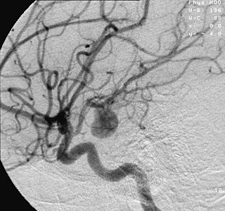
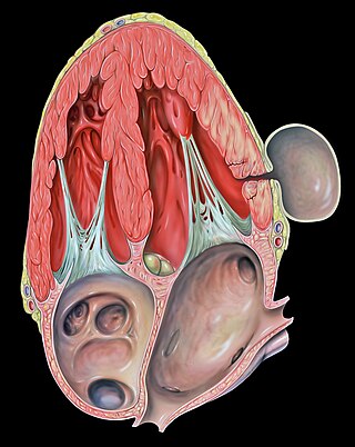
An aneurysm is an outward bulging, likened to a bubble or balloon, caused by a localized, abnormal, weak spot on a blood vessel wall. Aneurysms may be a result of a hereditary condition or an acquired disease. Aneurysms can also be a nidus for clot formation (thrombosis) and embolization. As an aneurysm increases in size, the risk of rupture, which leads to uncontrolled bleeding, increases. Although they may occur in any blood vessel, particularly lethal examples include aneurysms of the Circle of Willis in the brain, aortic aneurysms affecting the thoracic aorta, and abdominal aortic aneurysms. Aneurysms can arise in the heart itself following a heart attack, including both ventricular and atrial septal aneurysms. There are congenital atrial septal aneurysms, a rare heart defect.

<span class="mw-page-title-main">Interventional radiology</span> Medical subspecialty

Interventional radiology (IR) is a medical specialty that performs various minimally-invasive procedures using medical imaging guidance, such as x-ray fluoroscopy, computed tomography, magnetic resonance imaging, or ultrasound. IR performs both diagnostic and therapeutic procedures through very small incisions or body orifices. Diagnostic IR procedures are those intended to help make a diagnosis or guide further medical treatment, and include image-guided biopsy of a tumor or injection of an imaging contrast agent into a hollow structure, such as a blood vessel or a duct. By contrast, therapeutic IR procedures provide direct treatment—they include catheter-based medicine delivery, medical device placement, and angioplasty of narrowed structures.

<span class="mw-page-title-main">Vascular surgery</span> Medical specialty, operative procedures for the treatment of vascular disorders

Vascular surgery is a surgical subspecialty in which vascular diseases involving the arteries, veins, or lymphatic vessels, are managed by medical therapy, minimally-invasive catheter procedures and surgical reconstruction. The specialty evolved from general and cardiovascular surgery where it refined the management of just the vessels, no longer treating the heart or other organs. Modern vascular surgery includes open surgery techniques, endovascular techniques and medical management of vascular diseases - unlike the parent specialities. The vascular surgeon is trained in the diagnosis and management of diseases affecting all parts of the vascular system excluding the coronaries and intracranial vasculature. Vascular surgeons also are called to assist other physicians to carry out surgery near vessels, or to salvage vascular injuries that include hemorrhage control, dissection, occlusion or simply for safe exposure of vascular structures.

<span class="mw-page-title-main">Great cerebral vein</span>

The great cerebral vein is one of the large blood vessels in the skull draining the cerebrum of the brain. It is also known as the vein of Galen, named for its discoverer, the Greek physician Galen.

<span class="mw-page-title-main">Subarachnoid hemorrhage</span> Bleeding into the subarachnoid space

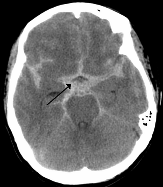
Subarachnoid hemorrhage (SAH) is bleeding into the subarachnoid space—the area between the arachnoid membrane and the pia mater surrounding the brain. Symptoms may include a severe headache of rapid onset, vomiting, decreased level of consciousness, fever, weakness, numbness, and sometimes seizures. Neck stiffness or neck pain are also relatively common. In about a quarter of people a small bleed with resolving symptoms occurs within a month of a larger bleed.

<span class="mw-page-title-main">Pseudoaneurysm</span> Collection of blood between outer artery layers

A pseudoaneurysm, also known as a false aneurysm, is a locally contained hematoma outside an artery or the heart due to damage to the vessel wall. The injury passes through all three layers of the arterial wall, causing a leak, which is contained by a new, weak "wall" formed by the products of the clotting cascade. A pseudoaneurysm does not contain any layer of the vessel wall.

<span class="mw-page-title-main">Cerebral angiography</span> Angiography that produces images of blood vessels in and around the brain

Cerebral angiography is a form of angiography which provides images of blood vessels in and around the brain, thereby allowing detection of abnormalities such as arteriovenous malformations and aneurysms. It was pioneered in 1927 by the Portuguese neurologist Egas Moniz at the University of Lisbon, who also helped develop thorotrast for use in the procedure.

<span class="mw-page-title-main">Arteriovenous fistula</span> Medical condition

An arteriovenous fistula is an abnormal connection or passageway between an artery and a vein. It may be congenital, surgically created for hemodialysis treatments, or acquired due to pathologic process, such as trauma or erosion of an arterial aneurysm.

An embolus, is described as a free-floating mass, located inside blood vessels that can travel from one site in the blood stream to another. An embolus can be made up of solid, liquid, or gas. Once these masses get "stuck" in a different blood vessel, it is then known as an "embolism." An embolism can cause ischemia—damage to an organ from lack of oxygen. A paradoxical embolism is a specific type of embolism in which the embolus travels from the right side of the heart to the left side of the heart and lodges itself in a blood vessel known as an artery. Thus, it is termed "paradoxical" because the embolus lands in an artery, rather than a vein.

<span class="mw-page-title-main">Superior petrosal sinus</span> One of the dural venous sinuses located beneath the brain

The superior petrosal sinus is one of the dural venous sinuses located beneath the brain. It receives blood from the cavernous sinus and passes backward and laterally to drain into the transverse sinus. The sinus receives superior petrosal veins, some cerebellar veins, some inferior cerebral veins, and veins from the tympanic cavity. They may be affected by arteriovenous malformation or arteriovenous fistula, usually treated with surgery.

<span class="mw-page-title-main">Dural arteriovenous fistula</span> Medical condition

A dural arteriovenous fistula (DAVF) or malformation is an abnormal direct connection (fistula) between a meningeal artery and a meningeal vein or dural venous sinus.

A vascular malformation is a blood vessel or lymph vessel abnormality. Vascular malformations are one of the classifications of vascular anomalies, the other grouping is vascular tumors. They may cause aesthetic problems as they have a growth cycle, and can continue to grow throughout life.

Vascular myelopathy refers to an abnormality of the spinal cord in regard to its blood supply. The blood supply is complicated and supplied by two major vessel groups: the posterior spinal arteries and the anterior spinal arteries—of which the Artery of Adamkiewicz is the largest. Both the posterior and anterior spinal arteries run the entire length of the spinal cord and receive anastomotic (conjoined) vessels in many places. The anterior spinal artery has a less efficient supply of blood and is therefore more susceptible to vascular disease. Whilst atherosclerosis of spinal arteries is rare, necrosis in the anterior artery can be caused by disease in vessels originating from the segmental arteries such as atheroma or aortic dissection.

<span class="mw-page-title-main">Bonnet–Dechaume–Blanc syndrome</span> Medical condition

Bonnet–Dechaume–Blanc syndrome, also known as Wyburn-Mason syndrome, is a rare congenital disorder characterized by arteriovenous malformations of the brain, retina or facial nevi. The syndrome has a number of possible symptoms and can, more rarely, affect the skin, bones, kidneys, muscles, and gastrointestinal tract. When the syndrome affects the brain, people can experience severe headaches, seizures, acute stroke, meningism, and progressive neurological deficits due to acute or chronic ischaemia caused by arteriovenous shunting.

Interventional neuroradiology (INR) also known as neurointerventional surgery (NIS), endovascular therapy (EVT), endovascular neurosurgery, and interventional neurology is a medical subspecialty of neurosurgery, neuroradiology, intervention radiology and neurology specializing in minimally invasive image-based technologies and procedures used in diagnosis and treatment of diseases of the head, neck, and spine.

Alexander Coon is an American neurosurgeon who is the Director of Endovascular and Cerebrovascular Neurosurgery at the Carondelet Neurological Institute of St. Joseph's and St. Mary's Hospitals in Tucson, Arizona. He was previously the Director of Endovascular Neurosurgery at the Johns Hopkins Hospital and an assistant professor of neurosurgery, Neurology, and Radiology at the Johns Hopkins Hospital. He is known for his work in cerebrovascular and endovascular neurosurgery and his research in neuroendovascular devices and clinical outcomes in the treatment of cerebral aneurysms, subarachnoid hemorrhage, and AVMs.

References

  1. 1 2 O'Brien M; Schechter M (September 1970). "Arteriovenous malformations involving the Galenic system" (PDF). The American Journal of Roentgenology, Radium Therapy, and Nuclear Medicine. 110 (1): 50–55. doi:10.2214/ajr.110.1.50. ISSN   0002-9580. PMID   5459527 . Retrieved October 25, 2009.[ permanent dead link ]
  2. K.M. Auyeung, S. Laughlin, K.G. TerBrugge (October 2004). "Prenatal Diagnosis of Unusual Fetal Pial Arteriovenous Malformation". Interv Neuroradiol. 9 (2): 163–8. doi:10.1177/159101990300900205. PMC   3547507 . PMID   20591266.{{cite journal}}: CS1 maint: multiple names: authors list (link)
  3. 1 2 3 Takashima S; Becker LE (May 1980). "Neuropathology of cerebral arteriovenous malformations in children". Journal of Neurology, Neurosurgery, and Psychiatry. 43 (5): 380–385. doi:10.1136/jnnp.43.5.380. ISSN   0022-3050. PMC   490562 . PMID   7420086.
  4. 1 2 3 4 5 6 7 8 9 10 Nicholson AA; Hourihan MD; Hayward C (December 1989). "Arteriovenous malformations involving the vein of Galen". Archives of Disease in Childhood. 64 (12): 1653–1655. doi:10.1136/adc.64.12.1653. ISSN   0003-9888. PMC   1792909 . PMID   2696431.
  5. 1 2 3 4 McElhinney DB; Halbach VV; Silverman NH; Dowd CF; Hanley FL (June 1998). "Congenital cardiac anomalies with vein of Galen malformations in infants". Archives of Disease in Childhood. Fetal and neonatal edition. 78 (6): 548–551. doi:10.1136/adc.78.6.548. ISSN   1359-2998. PMC   1717608 . PMID   9713012.
  6. Johnston IH; Whittle IR; Besser M; Morgan MK (May 1987). "Vein of Galen malformation: diagnosis and management". Neurosurgery. 20 (5): 747–758. doi:10.1227/00006123-198705000-00013. ISSN   0148-396X. PMID   3601022.
  7. Vivanti, Alexandre; Ozanne, Augustin; Grondin, Cynthia; Saliou, Guillaume; Quevarec, Loic; Maurey, Helène; Aubourg, Patrick; Benachi, Alexandra; Gut, Marta; Gut, Ivo; Martinovic, Jelena; Sénat, Marie Victoire; Tawk, Marcel; Melki, Judith (April 2018). "Loss of function mutations in EPHB4 are responsible for vein of Galen aneurysmal malformation". Brain. 141 (4): 979–988. doi: 10.1093/brain/awy020 . PMID   29444212.
  8. Duran D, Zeng X, Jin SC, Choi J, Nelson-Williams C, Yatsula B, Gaillard J, Furey CG, Lu Q, Timberlake AT, Dong W, Sorscher MA, Loring E, Klein J, Allocco A, Hunt A, Conine S, Karimy JK, Youngblood MW, Zhang J, DiLuna ML, Matouk CC, Mane S, Tikhonova IR, Castaldi C, López-Giráldez F, Knight J, Haider S, Soban M, Alper SL, Komiyama M, Ducruet AF, Zabramski JM, Dardik A, Walcott BP, Stapleton CJ, Aagaard-Kienitz B, Rodesch G, Jackson E, Smith ER, Orbach DB, Berenstein A, Bilguvar K, Vikkula M, Gunel M, Lifton RP, Kahle KT (2018) Mutations in chromatin modifier and ephrin signaling genes in vein of Galen malformation. Neuron
  9. "Vein of Galen Abnormalities". Duke University. Retrieved December 6, 2009.[ dead link ]
  10. 1 2 3 4 5 6 Hoffman HJ; Chuang S; Hendrick EB; Humphreys RP (September 1982). "Aneurysms of the vein of Galen. Experience at The Hospital for Sick Children, Toronto". Journal of Neurosurgery. 57 (3): 316–322. doi:10.3171/jns.1982.57.3.0316. ISSN   0022-3085. PMID   7097326. S2CID   40452720.
  11. Padget DH (May 1956). "The cranial venous system in man in reference to development, adult configuration, and relation to the arteries". The American Journal of Anatomy. 98 (3): 307–355. doi:10.1002/aja.1000980302. ISSN   0002-9106. PMID   13362118.
  12. 1 2 3 4 5 The Arteriovenous Malformation Study Group (June 10, 1999). "Arteriovenous Malformations of the Brain in Adults". New England Journal of Medicine. 340 (23): 1812–1818. doi:10.1056/NEJM199906103402307. ISSN   0028-4793. PMID   10362826.
  13. 1 2 3 Alexander, Michael J.; Spetzler, Robert F. (October 2005). Pediatric Neurovascular Disease: Surgical, Endovascular, and Medical Management. New York: Thieme Medical Publishers. ISBN   978-1-58890-368-6.
  14. 1 2 Ciricillo, S.F.; SCHMIDT K.G.; SILVERMAN N.H.; HIESHIMA G.B.; HIGASHIDA R.T.; HALBACH V.V.; EDWARDS M.S.B (1990). "Serial Ultrasonographic Evaluation of Neonatal Vein of Galen Malformations to Assess the Efficacy of Interventional Neuroradiological Procedures". Neurosurgery. 27 (4): 544–548. doi:10.1227/00006123-199010000-00007. ISSN   0148-396X. PMID   2234356.
  15. Chatterjee, S. (May 22, 2009). "Antiepileptic drugs" (PDF). Molecules of the Millennium. Indian Journal of Pharmacology. Archived from the original (PDF) on January 31, 2016. Retrieved December 6, 2009.[ sic ]
  16. Meyers PM; Halbach VV; Phatouros CP; Dowd CF; Malek AM; Lempert TE; Lefler JE; Higashida RT (June 2000). "Hemorrhagic complications in vein of Galen malformations". Annals of Neurology. 47 (6): 748–755. doi:10.1002/1531-8249(200006)47:6<748::AID-ANA7>3.0.CO;2-7. PMID   10852540. S2CID   11112270.