Von Willebrand factor type A domain

Last updated
von Willebrand factor type A domain
PDB 1lfa EBI.jpg
Structure of the I-domain from the CD11a/CD18 (LFA-1, alpha L beta 2) integrin. [1]
Identifiers
SymbolVWA
Pfam PF00092
Pfam clan CL0128
InterPro IPR002035
SCOP2 1lfa / SCOPe / SUPFAM
CDD cd00198
Available protein structures:
Pfam   structures / ECOD  
PDB RCSB PDB; PDBe; PDBj
PDBsum structure summary
PDB 1shu X:44-213 1tzn i:44-213 1sht X:44-213

1t6b Y:44-212 1rrk A:270-468 1rs0 A:270-468 1q0p A:270-468 1rtk A:270-468 1qc5 A:172-331 1pt6 A:172-338 1qcy A:172-333 1ck4 B:172-355 1mhp B:172-355 1dzi A:174-355 1aox A:174-361 1v7p C:174-361 1pqb A:174-361 1ao3 B:1691-1864 1atz B:1691-1864 1fe8 C:1691-1864 2adf A:1691-1864 1n9z A:150-328 1bho 1:150-328 1bhq 2:150-328 1idn 2:150-328 1ido :150-328 1jlm :150-328 1mf7 A:150-328 1na5 A:150-328 1m1u A:150-328 1n3y A:151-329 1mq9 A:156-327 1cqp A:156-327 1xdd B:156-327 1dgq A:156-327 1lfa A:156-327 1xuo A:156-327 1zon :156-327 1mq8 B:156-327 1ij4 L:156-327 1rd4 C:156-327 1mqa A:156-327 1zoo B:156-327 1xdg B:156-327 1zop B:156-327 1mjn A:156-327 1t0p A:156-326 1ijb A:1277-1457 1m10 A:1277-1457 1sq0 A:1277-1457 1ijk A:1277-1457 1auq :1277-1457 1u0n A:1277-1457 1uex C:1277-1457

Contents

1oak A:1277-1457 1fns A:1277-1457 1u0o C:1277-1453

The von Willebrand factor type A (vWA) domain is a protein domain named after its occurrence in von Willebrand factor (vWF), a large multimeric glycoprotein found in blood plasma. Mutant forms of vWF are involved in the aetiology of bleeding disorders. [2] This type A domain is the prototype for a protein superfamily (InterPro :  IPR036465 ; see also Pfam clan).

The vWA domain is found in various plasma proteins: complement factors B, C2, CR3 and CR4; the integrins (I-domains); collagen types VI, VII, XII and XIV; and other extracellular proteins. [3] [4] [5] Although the majority of vWA-containing proteins are extracellular, the most ancient ones present in all eukaryotes are all intracellular proteins involved in functions such as transcription, DNA repair, ribosomal and membrane transport and the proteasome. A common feature appears to be involvement in multiprotein complexes. Proteins that incorporate vWA domains participate in numerous biological events (e.g. cell adhesion, migration, homing, pattern formation, and signal transduction), involving interaction with a large array of ligands. [3] A number of human diseases arise from mutations in vWA domains.

Secondary structure prediction from 75 aligned vWA sequences has revealed a largely alternating sequence of alpha-helices and beta-strands. [4] Fold recognition algorithms were used to score sequence compatibility with a library of known structures: the vWA domain fold was predicted to be a doubly wound, open, twisted beta-sheet flanked by alpha-helices. [6] 3D structures have been determined for the I-domains of integrins CD11b (with bound magnesium) [7] and CD11a (with bound manganese). [8] The domain adopts a classic alpha/beta Rossmann fold and contains an unusual metal ion coordination site at its surface. It has been suggested that this site represents a general metal ion-dependent adhesion site (MIDAS) for binding protein ligands. [7] The residues constituting the MIDAS motif in the CD11b and CD11a I-domains are completely conserved, but the manner in which the metal ion is coordinated differs slightly. [8]

Human proteins containing this domain

ANTXR1; ANTXR2; BF; C2; CACHD1; CACNA2D1; CACNA2D2; CACNA2D3; CACNA2D4; CFB; CLCA1; CLCA2; CLCA4; COCH; COL12A1; COL14A1; COL20A1; COL21A1; COL22A1; COL28; COL6A1; COL6A2; COL6A3; COL7A1; COLA1L; CaCC1; ITGA1; ITGA10; ITGA11; ITGA2; ITGAD; ITGAE; ITGAL; ITGAM; ITGAX; ITIH1; ITIH2; ITIH3; ITIH4; ITIH5; ITIH5L; LOC285929; LOC340267; LOC389462; LOH11CR2A; MATN1; MATN2; MATN3; MATN4; PARP4; SVEP1SEL-OB); VIT; VWA1; VWA2; VWF; hCLCA1; hCLCA2; CMG2;

Related Research Articles

<span class="mw-page-title-main">Integrin</span> Instance of a defined set in Homo sapiens with Reactome ID (R-HSA-374573)

Integrins are transmembrane receptors that help cell-cell and cell-extracellular matrix (ECM) adhesion. Upon ligand binding, integrins activate signal transduction pathways that mediate cellular signals such as regulation of the cell cycle, organization of the intracellular cytoskeleton, and movement of new receptors to the cell membrane. The presence of integrins allows rapid and flexible responses to events at the cell surface.

<span class="mw-page-title-main">Transmembrane protein</span> Protein spanning across a biological membrane

A transmembrane protein is a type of integral membrane protein that spans the entirety of the cell membrane. Many transmembrane proteins function as gateways to permit the transport of specific substances across the membrane. They frequently undergo significant conformational changes to move a substance through the membrane. They are usually highly hydrophobic and aggregate and precipitate in water. They require detergents or nonpolar solvents for extraction, although some of them (beta-barrels) can be also extracted using denaturing agents.

<span class="mw-page-title-main">Disintegrin</span> Proteins from viper venom inhibiting platelets aggregation

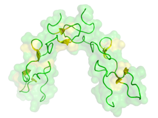
Disintegrins are a family of small proteins from viper venoms that function as potent inhibitors of both platelet aggregation and integrin-dependent cell adhesion.

<span class="mw-page-title-main">ICAM-1</span> Mammalian protein found in Homo sapiens

ICAM-1 also known as CD54 is a protein that in humans is encoded by the ICAM1 gene. This gene encodes a cell surface glycoprotein which is typically expressed on endothelial cells and cells of the immune system. It binds to integrins of type CD11a / CD18, or CD11b / CD18 and is also exploited by rhinovirus as a receptor for entry into respiratory epithelium.

<span class="mw-page-title-main">Integrin alpha X</span> Mammalian protein found in Homo sapiens

CD11c, also known as Integrin, alpha X (ITGAX), is a gene that encodes for CD11c.

<span class="mw-page-title-main">Integrin alpha L</span> Mammalian protein found in Homo sapiens

Integrin, alpha L , also known as ITGAL, is a protein that in humans is encoded by the ITGAL gene. CD11a functions in the immune system. It is involved in cellular adhesion and costimulatory signaling. It is the target of the drug efalizumab.

<span class="mw-page-title-main">Integrin alpha M</span> Mammalian protein found in Homo sapiens

Integrin alpha M (ITGAM) is one protein subunit that forms heterodimeric integrin alpha-M beta-2 (αMβ2) molecule, also known as macrophage-1 antigen (Mac-1) or complement receptor 3 (CR3). ITGAM is also known as CR3A, and cluster of differentiation molecule 11B (CD11B). The second chain of αMβ2 is the common integrin β2 subunit known as CD18, and integrin αMβ2 thus belongs to the β2 subfamily integrins.

Lymphocyte function-associated antigen 1 (LFA-1) is an integrin found on lymphocytes and other leukocytes. LFA-1 plays a key role in emigration, which is the process by which leukocytes leave the bloodstream to enter the tissues. LFA-1 also mediates firm arrest of leukocytes. Additionally, LFA-1 is involved in the process of cytotoxic T cell mediated killing as well as antibody mediated killing by granulocytes and monocytes. As of 2007, LFA-1 has 6 known ligands: ICAM-1, ICAM-2, ICAM-3, ICAM-4, ICAM-5, and JAM-A. LFA-1/ICAM-1 interactions have recently been shown to stimulate signaling pathways that influence T cell differentiation. LFA-1 belongs to the integrin superfamily of adhesion molecules.

<span class="mw-page-title-main">Integrin beta 2</span> Mammalian protein found in Homo sapiens

In molecular biology, CD18 is an integrin beta chain protein that is encoded by the ITGB2 gene in humans. Upon binding with one of a number of alpha chains, CD18 is capable of forming multiple heterodimers, which play significant roles in cellular adhesion and cell surface signaling, as well as important roles in immune responses. CD18 also exists in soluble, ligand binding forms. Deficiencies in CD18 expression can lead to adhesion defects in circulating white blood cells in humans, reducing the immune system's ability to fight off foreign invaders.

Platelet membrane glycoproteins are surface glycoproteins found on platelets (thrombocytes) which play a key role in hemostasis. When the blood vessel wall is damaged, platelet membrane glycoproteins interact with the extracellular matrix.

<span class="mw-page-title-main">Collagen, type VI, alpha 1</span> Protein found in humans

Collagen alpha-1(VI) chain is a protein that in humans is encoded by the COL6A1 gene.

<span class="mw-page-title-main">GP1BB</span> Protein-coding gene in the species Homo sapiens

Glycoprotein Ib (platelet), beta polypeptide (GP1BB) also known as CD42c, is a protein that in humans is encoded by the GP1BB gene.

<span class="mw-page-title-main">Collagen, type VI, alpha 3</span> Mammalian protein found in humans

Collagen alpha-3(VI) chain is a protein that in humans is encoded by the COL6A3 gene. This protein is an alpha chain of type VI collagen that aids in microfibril formation. As part of type VI collagen, this protein has been implicated in Bethlem myopathy, Ullrich congenital muscular dystrophy (UCMD), and other diseases related to muscle and connective tissue.

<span class="mw-page-title-main">Matrilin-3</span> Protein-coding gene in the species Homo sapiens

Matrilin-3 is a protein that in humans is encoded by the MATN3 gene. It is linked to the development of many types of cartilage, and part of the Matrilin family, which includes Matrilin-1, Matrilin-2, Matrilin-3, and Matrilin-4, a family of filamentous-forming adapter oligomeric extracellular proteins that are linked to the formation of cartilage and bone, as well as maintaining homeostasis after development. It is considered an extracellular matrix protein that functions as an adapter protein where the Matrilin-3 subunit can form both homo-tetramers and hetero-oligomers with subunits from Matrilin-1 which is the cartilage matrix protein. This restricted tissue has been strongly expressed in growing skeletal tissue as well as cartilage and bone.

<span class="mw-page-title-main">Integrin alpha 11</span> Protein-coding gene in the species Homo sapiens

Integrin alpha-11 is a protein that, in humans, is encoded by the ITGA11 gene.

The following outline is provided as an overview of and topical guide to immunology:

Von Willebrand factor, type C is a protein domain is found in various blood plasma proteins: complement factors B, C2, CR3 and CR4; the integrins (I-domains); collagen types VI, VII, XII and XIV; and other extracellular proteins.

<span class="mw-page-title-main">Trimeric autotransporter adhesin</span> Proteins found on the outer membrane of Gram-negative bacteria

In molecular biology, trimeric autotransporter adhesins (TAAs), are proteins found on the outer membrane of Gram-negative bacteria. Bacteria use TAAs in order to infect their host cells via a process called cell adhesion. TAAs also go by another name, oligomeric coiled-coil adhesins, which is shortened to OCAs. In essence, they are virulence factors, factors that make the bacteria harmful and infective to the host organism.

Von Willebrand factor type D domain (vWD) is an evolutionarily-conserved protein domain found in, among others, the von Willebrand factor (vWF). vWF is a large multimeric glycoprotein and it is synthesized by a type of bone marrow cell called megakaryocytes. The vWD domain allows vWF to perform its blood-clotting function by carrying factor VIII around.

<span class="mw-page-title-main">Collagen, type XXIII, alpha 1</span> Mammalian protein found in humans

Collagen α-1 (XXIII) chain is a protein encoded by COL23A1 gene, which is located on chromosome 5q35 in humans, and on chromosome 11B1+2 in mice. The location of this gene was discovered by genomic sequence analysis.

References

  1. Qu A, Leahy DJ (October 1995). "Crystal structure of the I-domain from the CD11a/CD18 (LFA-1, alpha L beta 2) integrin". Proc. Natl. Acad. Sci. U.S.A. 92 (22): 10277–81. Bibcode:1995PNAS...9210277Q. doi: 10.1073/pnas.92.22.10277 . PMC   40779 . PMID   7479767.
  2. Ruggeri ZM, Ware J (1993). "von Willebrand factor". FASEB J. 7 (2): 308–316. doi: 10.1096/fasebj.7.2.8440408 . PMID   8440408. S2CID   10574567.
  3. 1 2 Colombatti A, Bonaldo P, Doliana R (1993). "Type A modules: interacting domains found in several non-fibrillar collagens and in other extracellular matrix proteins". Matrix. 13 (4): 297–306. doi:10.1016/S0934-8832(11)80025-9. PMID   8412987.
  4. 1 2 Smith KF, Haris PI, Chapman D, Perkins SJ, Williams SC, Sim RB (1994). "The secondary structure of the von Willebrand factor type A domain in factor B of human complement by Fourier transform infrared spectroscopy. Its occurrence in collagen types VI, VII, XII and XIV, the integrins and other proteins by averaged structure predictions". J. Mol. Biol. 238 (1): 104–119. doi:10.1006/jmbi.1994.1271. PMID   8145250.
  5. Bork P (1991). "Shuffled domains in extracellular proteins". FEBS Lett. 286 (1): 47–54. doi: 10.1016/0014-5793(91)80937-X . PMID   1864378. S2CID   22126481.
  6. Perkins SJ, Edwards YJ (1995). "The protein fold of the von Willebrand factor type A domain is predicted to be similar to the open twisted beta-sheet flanked by alpha-helices found in human ras-p21". FEBS Lett. 358 (3): 283–286. doi: 10.1016/0014-5793(94)01447-9 . PMID   7843416. S2CID   85317759.
  7. 1 2 Lee JO, Rieu P, Arnaout MA, Liddington R (1995). "Crystal structure of the A domain from the alpha subunit of integrin CR3 (CD11b/CD18)". Cell. 80 (4): 631–638. doi: 10.1016/0092-8674(95)90517-0 . PMID   7867070. S2CID   11275359.
  8. 1 2 Leahy DJ, Qu A (1995). "Crystal structure of the I-domain from the CD11a/CD18 (LFA-1, alpha L beta 2) integrin". Proc. Natl. Acad. Sci. U.S.A. 92 (22): 10277–10281. Bibcode:1995PNAS...9210277Q. doi: 10.1073/pnas.92.22.10277 . PMC   40779 . PMID   7479767.
This article incorporates text from the public domain Pfam and InterPro: IPR002035