Alpha 1-antitrypsin deficiency

Last updated

Alpha-1 antitrypsin deficiency
Other namesα1-antitrypsin deficiency
A1AT.png
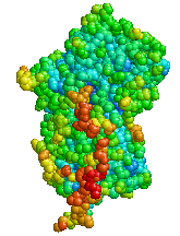
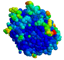
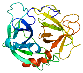
Structure of Alpha-1 antitrypsin
Specialty Pulmonology, medical genetics
Symptoms Shortness of breath, wheezing, yellowish skin [1]
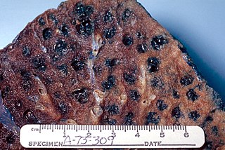
Complications COPD, cirrhosis, neonatal jaundice, panniculitis [1]
Usual onset20 to 50 years old [1]
CausesMutation in the SERPINA1 gene [1]
Diagnostic method Based on symptoms, blood tests, genetic tests [2]
Differential diagnosis Asthma [1]
TreatmentMedications, lung transplant, liver transplant [2]
Medication Bronchodilators, inhaled steroids, antibiotics, intravenous infusions of A1AT protein [2]
Prognosis Life expectancy ~50 years (smokers), nearly normal (non smokers) [3]
Frequency1 in 2,500 (Europeans) [1]

Alpha-1 antitrypsin deficiency (A1AD or AATD) is a genetic disorder that may result in lung disease or liver disease. [1] Onset of lung problems is typically between 20 and 50 years old. [1] This may result in shortness of breath, wheezing, or an increased risk of lung infections. [1] [2] Complications may include COPD, cirrhosis, neonatal jaundice, or panniculitis. [1]

Genetic disorder Disease that has material basis in genetic variations in the human genome

A genetic disorder is a genetic problem caused by one or more abnormalities formed in the genome. Most genetic disorders are quite rare and affect one person in every several thousands or millions. The earliest known genetic condition in a hominid was in the fossil species Paranthropus robustus, with over a third of individuals displaying Amelogenesis imperfecta.

Liver disease Human disease

Liver disease is a type of damage to or disease of the liver. Whenever the course of the problem lasts long, chronic liver disease ensues.

Shortness of breath, also known as dyspnea, is the feeling that one cannot breathe well enough. The American Thoracic Society defines it as "a subjective experience of breathing discomfort that consists of qualitatively distinct sensations that vary in intensity", and recommends evaluating dyspnea by assessing the intensity of the distinct sensations, the degree of distress involved, and its burden or impact on activities of daily living. Distinct sensations include effort/work, chest tightness, and air hunger.

Contents

A1AD is due to a mutation in the SERPINA1 gene that results in not enough alpha-1 antitrypsin (A1AT). [1] Risk factors for lung disease include cigarette smoking and environmental dust. [1] The underlying mechanism involves unblocked neutrophil elastase and build up of abnormal A1AT in the liver. [1] It is autosomal co-dominant, meaning that one defective allele tends to result in milder disease than two defective alleles. [1] The diagnosis is suspected based on symptoms and confirmed by blood tests or genetic tests. [2]

Neutrophil elastase protein-coding gene in the species Homo sapiens

Neutrophil elastase is a serine proteinase in the same family as chymotrypsin and has broad substrate specificity. Secreted by neutrophils and macrophages during inflammation, it destroys bacteria and host tissue. It also localizes to Neutrophil extracellular traps (NETs), via its high affinity for DNA, an unusual property for serine proteases.

An allele is a variant form of a given gene. Sometimes, the presence of different alleles of the same gene can result in different observable phenotypic traits, such as different pigmentation. A notable example of this trait of color variation is Gregor Mendel's discovery that the white and purple flower colors in pea plants were the result of "pure line" traits which could be used as a control for future experiments. However, most genetic variations result in little or no observable variation.

Treatment of lung disease may include bronchodilators, inhaled steroids, and when infections occur antibiotics. [2] Intravenous infusions of the A1AT protein or in severe disease lung transplantation may also be recommended. [2] In those with severe liver disease liver transplantation may be an option. [2] [4] Avoiding smoking is recommended. [2] Vaccination for influenza, pneumococcus, and hepatitis is also recommended. [2] Life expectancy among those who smoke is 50 years old while among those who do not smoke it is almost normal. [3]

Lung transplantation

Lung transplantation, or pulmonary transplantation, is a surgical procedure in which a patient's diseased lungs are partially or totally replaced by lungs which come from a donor. Donor lungs can be retrieved from a living donor or a deceased donor. A living donor can only donate one lung lobe. With some lung diseases, a recipient may only need to receive a single lung. With other lung diseases such as cystic fibrosis, it is imperative that a recipient receive two lungs. While lung transplants carry certain associated risks, they can also extend life expectancy and enhance the quality of life for end-stage pulmonary patients.

Liver transplantation medical procedure

Liver transplantation or hepatic transplantation is the replacement of a diseased liver with the healthy liver from another person (allograft). Liver transplantation is a treatment option for end-stage liver disease and acute liver failure, although availability of donor organs is a major limitation. The most common technique is orthotopic transplantation, in which the native liver is removed and replaced by the donor organ in the same anatomic position as the original liver. The surgical procedure is complex, requiring careful harvest of the donor organ and meticulous implantation into the recipient. Liver transplantation is highly regulated, and only performed at designated transplant medical centers by highly trained transplant physicians and supporting medical team. The duration of the surgery ranges from 4 to 18 hours depending on outcome. Favorable outcomes require careful screening for eligible recipient, as well as a well-calibrated live or cadaveric donor match.

Influenza vaccine vaccine against influenza

Influenza vaccines, also known as flu shots or flu jabs, are vaccines that protect against infection by influenza viruses. A new version of the vaccine is developed twice a year, as the influenza virus rapidly changes. While their effectiveness varies from year to year, most provide modest to high protection against influenza. The United States Centers for Disease Control and Prevention (CDC) estimates that vaccination against influenza reduces sickness, medical visits, hospitalizations, and deaths. When an immunized worker does catch the flu, they are on average back at work a half day sooner. Vaccine effectiveness in those under two years old and over 65 years old remains unknown due to the low quality of the research. Vaccinating children may protect those around them.

The condition affects about 1 in 2,500 people of European descent. [1] Severe deficiency occurs in about 1 in 5,000. [5] In Asians it is uncommon. [1] About 3% of people with COPD are believed to have the condition. [5] Alpha-1 antitrypsin deficiency was first described in the 1960s. [6]

Signs and symptoms

Symptoms of alpha-1 antitrypsin deficiency include shortness of breath, wheezing, rhonchi, and rales. The patient's symptoms may resemble recurrent respiratory infections or asthma that does not respond to treatment. Individuals with A1AD may develop emphysema during their thirties or forties even without a history of significant smoking, though smoking greatly increases the risk for emphysema. [7] A1AD causes impaired liver function in some patients and may lead to cirrhosis and liver failure (15%). In newborns, alpha-1 antitrypsin deficiency has indicators that include early onset jaundice followed by prolonged jaundice. It is a leading indication for liver transplantation in newborns.

A wheeze is a continuous, coarse, whistling sound produced in the respiratory airways during breathing. For wheezes to occur, some part of the respiratory tree must be narrowed or obstructed, or airflow velocity within the respiratory tree must be heightened. Wheezing is commonly experienced by persons with a lung disease; the most common cause of recurrent wheezing is asthma attacks, though it can also be a symptom of lung cancer, congestive heart failure, and certain types of heart diseases.

Rhonchi are coarse rattling respiratory sounds, usually caused by secretions in bronchial airways. "Rhonchi" is the plural form of the singular word "rhonchus". Since the mid-1990s, it has no longer been considered appropriate terminology in auscultation of the thorax, as much confusion has been reported in the published literature which confuses this with crepitations and wheezes, so the exact nature of this term is unclear ., however it is the most important pulmonary diagnostic tool we have.

Asthma long-term disease involving poor airflow in the lungs

Asthma is a common long-term inflammatory disease of the airways of the lungs. It is characterized by variable and recurring symptoms, reversible airflow obstruction, and easily triggered bronchospasms. Symptoms include episodes of wheezing, coughing, chest tightness, and shortness of breath. These may occur a few times a day or a few times per week. Depending on the person, they may become worse at night or with exercise.

Associated conditions

Conditions associated with Alpha-1 Antitrypsin Deficiency, occurring due to paucity of AAT in circulation allowing uninhibited inflammation in lungs, and accumulation of mutated AAT in the liver Conditions associated with Alpha-1 Antitrypsin Deficiency.png
Conditions associated with Alpha-1 Antitrypsin Deficiency, occurring due to paucity of AAT in circulation allowing uninhibited inflammation in lungs, and accumulation of mutated AAT in the liver

α1-antitrypsin deficiency has been associated with a number of diseases:

Cirrhosis long-term disease of the liver

Cirrhosis, also known as liver cirrhosis or hepatic cirrhosis, is a condition in which the liver does not function properly due to long-term damage. This damage is characterized by the replacement of normal liver tissue by scar tissue. Typically, the disease develops slowly over months or years. Early on, there are often no symptoms. As the disease worsens, a person may become tired, weak, itchy, have swelling in the lower legs, develop yellow skin, bruise easily, have fluid build up in the abdomen, or develop spider-like blood vessels on the skin. The fluid build-up in the abdomen may become spontaneously infected. Other serious complications include hepatic encephalopathy, bleeding from dilated veins in the esophagus or dilated stomach veins, and liver cancer. Hepatic encephalopathy results in confusion and may lead to unconsciousness.

Chronic obstructive pulmonary disease type of lung disease characterized by long-term poor airflow

Chronic obstructive pulmonary disease (COPD) is a type of obstructive lung disease characterized by long-term breathing problems and poor airflow. The main symptoms include shortness of breath and cough with sputum production. COPD is a progressive disease, meaning it typically worsens over time. Eventually, everyday activities such as walking or getting dressed become difficult. Chronic bronchitis and emphysema are older terms used for different types of COPD. The term "chronic bronchitis" is still used to define a productive cough that is present for at least three months each year for two years. Those with such a cough are at a greater risk of developing COPD. The term "emphysema" is also used for the abnormal presence of air or other gas within tissues.

Pneumothorax Abnormal collection of air in the pleural space that causes an uncoupling of the lung from the chest wall

A pneumothorax is an abnormal collection of air in the pleural space between the lung and the chest wall. Symptoms typically include sudden onset of sharp, one-sided chest pain and shortness of breath. In a minority of cases the amount of air in the chest increases when a one-way valve is formed by an area of damaged tissue, leading to a tension pneumothorax. This condition can cause a steadily worsening oxygen shortage and low blood pressure. Unless reversed by effective treatment, it can result in death. Very rarely both lungs may be affected by a pneumothorax. It is often called a collapsed lung, although that term may also refer to atelectasis.

Genetics

Serpin peptidase inhibitor, clade A, member 1 (SERPINA1) is the gene that encodes the protein alpha-1 antitrypsin. SERPINA1 has been localized to chromosome 14q32. Over 75 mutations of the SERPINA1 gene have been identified, many with clinically significant effects. [9] The most common cause of severe deficiency is a single base-pair substitution leading to a glutamate to lysine mutation at position 342 (dbSNP: rs28929474), while PiS is caused by a glutamate to valine mutation at position 264 (dbSNP: rs17580). Other rarer forms have been described (see OMIM).

Pathophysiology

Photomicrograph of a liver biopsy from a patient with alpha-1 antitrypsin deficiency. The PAS with diastase stain shows the diastase-resistant pink globules that are characteristic of this disease. Alpha-1 antitrypsin deficiency.PAS Diastase.jpg
Photomicrograph of a liver biopsy from a patient with alpha-1 antitrypsin deficiency. The PAS with diastase stain shows the diastase-resistant pink globules that are characteristic of this disease.

Alpha-1 antitrypsin (A1AT) is produced in the liver, and one of its functions is to protect the lungs from neutrophil elastase, an enzyme that can disrupt connective tissue. [7] Normal blood levels of alpha-1 antitrypsin may vary with analytical method but are typically around 1.0-2.7 g/l. [10] In individuals with PiSS, PiMZ and PiSZ genotypes, blood levels of A1AT are reduced to between 40 and 60% of normal levels. This is usually sufficient to protect the lungs from the effects of elastase in people who do not smoke. However, in individuals with the PiZZ genotype, A1AT levels are less than 15% of normal, and they are likely to develop panacinar emphysema at a young age. Between 10 to 15% of these people will develop liver fibrosis or liver cirrhosis, because the A1AT is not secreted properly and therefore accumulates in the liver. [11] A liver biopsy in such cases will reveal PAS-positive, diastase-resistant granules. Unlike glycogen and other mucins which are diastase sensitive (i.e., diastase treatment disables PAS staining), A1AT deficient hepatocytes will stain with PAS even after diastase treatment - a state thus referred to as diastase resistant.

Cigarette smoke is especially harmful to individuals with A1AD. [7] In addition to increasing the inflammatory reaction in the airways, cigarette smoke directly inactivates alpha-1 antitrypsin by oxidizing essential methionine residues to sulfoxide forms, decreasing the enzyme activity by a factor of 2000.

Diagnosis

Emphysema due to alpha-1-antitrypsin deficiency. Anti1Tripsine.PNG
Emphysema due to alpha-1-antitrypsin deficiency.
Computed tomography of the lung showing emphysema and bullae in the lower lung lobes of a subject with type ZZ alpha-1-antitrypsin deficiency. There is also increased lung density in areas with compression of lung tissue by the bullae. Alpha 1-antitrypsine deficiency lung CT scan.JPEG
Computed tomography of the lung showing emphysema and bullae in the lower lung lobes of a subject with type ZZ alpha-1-antitrypsin deficiency. There is also increased lung density in areas with compression of lung tissue by the bullae.

A1AT deficiency remains undiagnosed in many patients. Patients are usually labeled as having COPD without an underlying cause. It is estimated that about 1% of all COPD patients actually have an A1AT deficiency. Thus, testing should be performed for all patients with COPD, asthma with irreversible airflow obstruction, unexplained liver disease, or necrotizing panniculitis.[ citation needed ] The initial test performed is serum A1AT level. A low level of A1AT confirms the diagnosis and further assessment with A1AT protein phenotyping and A1AT genotyping should be carried out subsequently. [12] The Alpha-1 Foundation offers free, confidential testing.

As protein electrophoresis does not completely distinguish between A1AT and other minor proteins at the alpha-1 position (agarose gel), antitrypsin can be more directly and specifically measured using a nephelometric or immunoturbidimetric method. Thus, protein electrophoresis is useful for screening and identifying individuals likely to have a deficiency. A1AT is further analyzed by isoelectric focusing (IEF) in the pH range 4.5-5.5, where the protein migrates in a gel according to its isoelectric point or charge in a pH gradient. Normal A1AT is termed M, as it migrates toward the center of such an IEF gel. Other variants are less functional and are termed A-L and N-Z, dependent on whether they run proximal or distal to the M band. The presence of deviant bands on IEF can signify the presence of alpha-1 antitrypsin deficiency. Since the number of identified mutations has exceeded the number of letters in the alphabet, subscripts have been added to most recent discoveries in this area, as in the Pittsburgh mutation described above. As every person has two copies of the A1AT gene, a heterozygote with two different copies of the gene may have two different bands showing on electrofocusing, although a heterozygote with one null mutant that abolishes expression of the gene will only show one band. In blood test results, the IEF results are notated as, e.g., PiMM, where Pi stands for protease inhibitor and "MM" is the banding pattern of that person.[ citation needed ]

Other detection methods include use of enzyme-linked-immuno-sorbent-assays in vitro and radial immunodiffusion. Alpha 1-antitrypsin levels in the blood depend on the genotype. Some mutant forms fail to fold properly and are, thus, targeted for destruction in the proteasome, whereas others have a tendency to polymerize, thereafter being retained in the endoplasmic reticulum. The serum levels of some of the common genotypes are:[ citation needed ]

Treatment

Treatment of lung disease may include bronchodilators, inhaled steroids, and when infections occur antibiotics. [2] Intravenous infusions of the A1AT protein or in severe disease lung transplantation may also be recommended. [2] In those with severe liver disease liver transplantation may be an option. [2] Avoiding smoking and getting vaccinated for influenza, pneumococcus, and hepatitis is also recommended. [2]

A1AT protein

People with lung disease due to A1AD may receive intravenous infusions of alpha-1 antitrypsin, derived from donated human plasma. This augmentation therapy is thought to arrest the course of the disease and halt any further damage to the lungs. Long-term studies of the effectiveness of A1AT replacement therapy are not available. [13] It is currently recommended that patients begin augmentation therapy only after the onset of emphysema symptoms. [12]

As of 2015 there are four IV augmentation therapy manufacturers in the United States, Canada, and several European countries. Intravenous (IV) therapies are the standard mode of augmentation therapy delivery. Researchers are exploring inhaled therapies. IV augmentation therapies are manufactured by the following companies and have been shown to be clinically identical to one another in terms of dosage and efficacy.

Augmentation therapy is not appropriate for people with liver disease; treatment of A1AD-related liver damage focuses on alleviating the symptoms of the disease. In severe cases, liver transplantation may be necessary.[ citation needed ]

Epidemiology

Distribution of PiZZ in Europe. PiZZ Europe.png
Distribution of PiZZ in Europe.

People of Northern European and Iberian ancestry are at the highest risk for A1AD. Four percent carry the PiZ allele; between 1 in 625 and 1 in 2000 are homozygous.

Another study detected a frequency of 1 in 1550 individuals and a gene frequency of 0.026. The highest prevalence of the PiZZ variant was recorded in the northern and western European countries with mean gene frequency of 0.0140. [14]

History

A1AD was discovered in 1963 by Carl-Bertil Laurell (1919–2001), at the University of Lund in Sweden. [15] Laurell, along with a medical resident, Sten Eriksson, made the discovery after noting the absence of the α1 band on protein electrophoresis in five of 1500 samples; three of the five patient samples were found to have developed emphysema at a young age.

The link with liver disease was made six years later, when Harvey Sharp et al. described A1AD in the context of liver disease. [16]

Research

Recombinant and inhaled forms of A1AT are being studied. Other experimental therapies are aimed at the prevention of polymer formation in the liver. [17]

Related Research Articles

Hepatocellular carcinoma liver carcinoma that has material basis in undifferentiated hepatocytes

Hepatocellular carcinoma (HCC) is the most common type of primary liver cancer in adults, and is the most common cause of death in people with cirrhosis.

Serum protein electrophoresis

Serum protein electrophoresis is a laboratory test that examines specific proteins in the blood called globulins. The most common indications for a serum protein electrophoresis test are to diagnose or monitor multiple myeloma, a monoclonal gammopathy of uncertain significance (MGUS), or further investigate a discrepancy between a low albumin and a relatively high total protein. Unexplained bone pain, anemia, proteinuria, renal insufficiency, and hypercalcemia are also signs of multiple myeloma, and indications for SPE. Blood must first be collected, usually into an airtight vial or syringe. Electrophoresis is a laboratory technique in which the blood serum is applied to an acetate membrane soaked in a liquid buffer., to a buffered agarose gel matrix, or into liquid in a capillary tube, and exposed to an electric current to separate the serum protein components into five major fractions by size and electrical charge: serum albumin, alpha-1 globulins, alpha-2 globulins, beta 1 and 2 globulins, and gamma globulins.

Alcoholic liver disease

Alcoholic liver disease is a term that encompasses the liver manifestations of alcohol overconsumption, including fatty liver, alcoholic hepatitis, and chronic hepatitis with liver fibrosis or cirrhosis.

Alpha-1 antitrypsin protein-coding gene in the species Homo sapiens

Alpha-1-antitrypsin or α1-antitrypsin is a protein belonging to the serpin superfamily. It is encoded in humans by the SERPINA1 gene. A protease inhibitor, it is also known as alpha1–proteinase inhibitor (A1PI) or alpha1-antiproteinase (A1AP) because it inhibits various proteases. In older biomedical literature it was sometimes called serum trypsin inhibitor, because its capability as a trypsin inhibitor was a salient feature of its early study. As a type of enzyme inhibitor, it protects tissues from enzymes of inflammatory cells, especially neutrophil elastase, and has a reference range in blood of 0.9–2.3 g/L, but the concentration can rise manyfold upon acute inflammation. When the blood contains inadequate amounts of A1AT or functionally defective A1AT, neutrophil elastase is excessively free to break down elastin, degrading the elasticity of the lungs, which results in respiratory complications, such as chronic obstructive pulmonary disease, in adults. Normally, A1AT leaves its site of origin, the liver, and joins the systemic circulation; defective A1AT can fail to do so, building up in the liver, which results in cirrhosis in either adults or children.

Budd–Chiari syndrome Human disease

Budd–Chiari syndrome is a very rare condition, affecting one in a million adults. The condition is caused by occlusion of the hepatic veins that drain the liver. It presents with the classical triad of abdominal pain, ascites, and liver enlargement. The formation of a blood clot within the hepatic veins can lead to Budd–Chiari syndrome. The syndrome can be fulminant, acute, chronic, or asymptomatic.

Elastase

In molecular biology, elastase is an enzyme from the class of proteases (peptidases) that break down proteins. In particular, it is a serine protease.

Ornithine transcarbamylase deficiency urea cycle disorder that involves a mutated and ineffective form of the enzyme ornithine transcarbamylase

Ornithine transcarbamylase deficiency is the most common urea cycle disorder in humans. It is an inherited disorder which causes toxic levels of ammonia to build up in the blood.

Hepatorenal syndrome Human disease

Hepatorenal syndrome is a life-threatening medical condition that consists of rapid deterioration in kidney function in individuals with cirrhosis or fulminant liver failure. HRS is usually fatal unless a liver transplant is performed, although various treatments, such as dialysis, can prevent advancement of the condition.

Glycogen storage disease type 0 human disease

Glycogen storage disease type 0 is a disease characterized by a deficiency in the glycogen synthase enzyme (GSY). Although glycogen synthase deficiency does not result in storage of extra glycogen in the liver, it is often classified as a glycogen storage disease because it is another defect of glycogen storage and can cause similar problems. There are two isoforms (types) of glycogen synthase enzyme; GSY1 in muscle and GSY2 in liver, each with a corresponding form of the disease. Mutations in the liver isoform (GSY2), causes fasting hypoglycemia, high blood ketones, increased free fatty acids and low levels of alanine and lactate. Conversely, feeding in these patients results in hyperglycemia and hyperlactatemia.

Progressive familial intrahepatic cholestasis (PFIC) is a group of familial cholestatic conditions caused by defects in biliary epithelial transporters. The clinical presentation usually occurs first in childhood with progressive cholestasis. This usually leads to failure to thrive, cirrhosis, and the need for liver transplantation.

Alpha 1-antichymotrypsin protein-coding gene in the species Homo sapiens

Alpha 1-antichymotrypsin is an alpha globulin glycoprotein that is a member of the serpin superfamily. In humans, it is encoded by the SERPINA3 gene.

Des-gamma carboxyprothrombin (DCP), also known as protein induced by vitamin K absence/antagonist-II (PIVKA-II), is an abnormal form of the coagulation protein, prothrombin. Normally, the prothrombin precursor undergoes post-translational carboxylation by gamma-glutamyl carboxylase in the liver prior to secretion into plasma. DCP/PIVKA-II may be detected in people with deficiency of vitamin K and in those taking warfarin or other medication that inhibits the action of vitamin K.

Obstructive lung disease

Obstructive lung disease is a category of respiratory disease characterized by airway obstruction. Many obstructive diseases of the lung result from narrowing (obstruction) of the smaller bronchi and larger bronchioles, often because of excessive contraction of the smooth muscle itself. It is generally characterized by inflamed and easily collapsible airways, obstruction to airflow, problems exhaling and frequent medical clinic visits and hospitalizations. Types of obstructive lung disease include; asthma, bronchiectasis, bronchitis and chronic obstructive pulmonary disease (COPD). Although COPD shares similar characteristics with all other obstructive lung diseases, such as the signs of coughing and wheezing, they are distinct conditions in terms of disease onset, frequency of symptoms and reversibility of airway obstruction. Cystic fibrosis is also sometimes included in obstructive pulmonary disease.

Elevated alkaline phosphatase condition where alkaline phosphatase exceeds normal levels in the blood

Elevated alkaline phosphatase occurs when levels of alkaline phosphatase (ALP) exceed the reference range. This group of enzymes has a low substrate specificity and catalyzes the hydrolysis of phosphate esters in a basic environment. The major function of alkaline phosphatase is transporting across cell membranes. Alkaline phosphatases are present in many human tissues, including bone, intestine, kidney, liver, placenta and white blood cells. Damage to these tissues causes the release of ALP into the bloodstream. Elevated levels can be detected through a blood test. Elevated alkaline phosphate is associated with certain medical conditions or syndromes. It serves as a significant indication for certain medical conditions, diseases and syndromes.

Lysosomal acid lipase deficiency autosomal recessive inborn error of metabolism that results in the body not producing enough active lysosomal acid lipase (LAL) enzyme

Lysosomal acid lipase deficiency, is an autosomal recessive inborn error of metabolism that results in the body not producing enough active lysosomal acid lipase (LAL) enzyme. This enzyme plays an important role in breaking down fatty material in the body. Infants, children and adults that suffer from LAL deficiency experience a range of serious health problems. The lack of the LAL enzyme can lead to a build-up of fatty material in a number of body organs including the liver, spleen, gut, in the wall of blood vessels and other important organs.

SERPINA2 protein-coding gene in the species Homo sapiens

Serpin peptidase inhibitor, clade A, member 2 is a protein that in humans is encoded by the SERPINA2 gene. Serine peptidase inhibitor, clade A member 2 belongs to the member of serine family of proteins which have a functional activity of inhibiting serine proteases.

Alpha 1 or Alpha-1 may refer to:

Kamada Ltd. is a biopharmaceutical company specializing in the development, manufacture and marketing of pharmaceuticals. The Company's headquarters and laboratories are located in the park of Kiryat Weizmann Institute of Science in Rehovot. The production facility is located in Kibbutz Beit Kama.

References

  1. 1 2 3 4 5 6 7 8 9 10 11 12 13 14 15 16 "alpha-1 antitrypsin deficiency". Genetics Home Reference. January 2013. Retrieved 12 December 2017.
  2. 1 2 3 4 5 6 7 8 9 10 11 12 13 14 "Alpha-1 antitrypsin deficiency". GARD. 2016. Retrieved 12 December 2017.
  3. 1 2 Stradling, John; Stanton, Andrew; Rahman, Najib M.; Nickol, Annabel H.; Davies, Helen E. (2010). Oxford Case Histories in Respiratory Medicine. OUP Oxford. p. 129. ISBN   9780199556373.
  4. Clark, VC (May 2017). "Liver Transplantation in Alpha-1 Antitrypsin Deficiency". Clinics in Liver Disease. 21 (2): 355–365. doi:10.1016/j.cld.2016.12.008. PMID   28364818.
  5. 1 2 Marciniuk, DD; Hernandez, P; Balter, M; Bourbeau, J; Chapman, KR; Ford, GT; Lauzon, JL; Maltais, F; O'Donnell, DE; Goodridge, D; Strange, C; Cave, AJ; Curren, K; Muthuri, S; Canadian Thoracic Society COPD Clinical Assembly Alpha-1 Antitrypsin Deficiency Expert Working, Group (2012). "Alpha-1 antitrypsin deficiency targeted testing and augmentation therapy: a Canadian Thoracic Society clinical practice guideline". Canadian Respiratory Journal. 19 (2): 109–16. doi:10.1155/2012/920918. PMC   3373286 . PMID   22536580.
  6. Köhnlein, Thomas; Welte, T. (2007). Alpha-1 Antitrypsin Deficiency: Clinical Aspects and Management. UNI-MED Verlag AG. p. 16. ISBN   9781848151154.
  7. 1 2 3 Kumar V, Abbas AK, Fausto N, eds. (2005). Robbins and Cotran Pathological Basis of Disease (7th ed.). Elsevier/Saunders. pp. 911–2. ISBN   978-0-7216-0187-8.
  8. Chen B, Wen Y, Polan ML (2004). "Elastolytic activity in women with stress urinary incontinence and pelvic organ prolapse". Neurourol. Urodyn. 23 (2): 119–26. doi:10.1002/nau.20012. PMID   14983422.
  9. Silverman, Edwin K.; Sandhaus, Robert A. (25 June 2009). "Alpha1-Antitrypsin Deficiency". New England Journal of Medicine. 360 (26): 2749–2757. doi:10.1056/NEJMcp0900449. ISSN   0028-4793. PMID   19553648.
  10. Donato, Leslie; Jenkins; et al. (2012). "Reference and Interpretive Ranges for α1-Antitrypsin Quantitation by Phenotype in Adult and Pediatric Populations". American Journal of Clinical Pathology. 138 (3): 398–405. doi:10.1309/AJCPMEEJK32ACYFP. PMID   22912357 . Retrieved 17 January 2014.
  11. Townsend, S.A; Edgar, R.G; Ellis, P.R; Kantas, D; Newsome, P.N; Turner, A.M (2018). "Systematic review: the natural history of alpha-1 antitrypsin deficiency, and associated liver disease". Alimentary Pharmacology & Therapeutics. 47 (7): 877–885. doi: 10.1111/apt.14537 . PMID   29446109.
  12. 1 2 Silverman EK, Sandhaus RA (2009). "Alpha1-Antitrypsin Deficiency". New England Journal of Medicine. 360 (26): 2749–2757. doi:10.1056/NEJMcp0900449. PMID   19553648.
  13. Gøtzsche, Peter C.; Johansen, Helle Krogh (20 September 2016). "Intravenous alpha-1 antitrypsin augmentation therapy for treating patients with alpha-1 antitrypsin deficiency and lung disease". The Cochrane Database of Systematic Reviews. 9: CD007851. doi:10.1002/14651858.CD007851.pub3. ISSN   1469-493X. PMC   6457738 . PMID   27644166.
  14. Luisetti, M; Seersholm, N (February 2004). "Alpha1-antitrypsin deficiency. 1: epidemiology of alpha1-antitrypsin deficiency". Thorax. 59 (2): 164–9. doi:10.1136/thorax.2003.006494. PMC   1746939 . PMID   14760160.
  15. Laurell CB, Eriksson S (1963). "The electrophoretic alpha 1-globulin pattern of serum in alpha 1-antitrypsin deficiency". Scand J Clin Lab Invest. 15 (2): 132–140. doi:10.1080/00365516309051324.
  16. Sharp H, Bridges R, Krivit W, Freier E (1969). "Cirrhosis associated with alpha-1-antitrypsin deficiency: a previously unrecognized inherited disorder". J Lab Clin Med. 73 (6): 934–9. PMID   4182334.
  17. Mohanka M, Khemasuwan D, Stoller JK (June 2012). "A review of augmentation therapy for alpha-1 antitrypsin deficiency". Expert Opin Biol Ther. 12 (6): 685–700. doi:10.1517/14712598.2012.676638. PMID   22500781.
Classification
D
External resources