Causes of cancer pain

Last updated

Cancer pain can be caused by pressure on, or chemical stimulation of, specialised pain-signalling nerve endings called nociceptors (nociceptive pain), or by damage or illness affecting nerve fibers themselves (neuropathic pain). [1]

Contents

Infection

Infection of a tumor or its surrounding tissue can cause rapidly escalating pain, but is sometimes overlooked as a possible cause. One study [2] found that infection was the cause of pain in four percent of nearly 300 cancer patients referred for pain relief. Another report [3] described seven patients, whose previously well-controlled pain escalated significantly over several days. Antibiotic treatment produced pain relief in all patients within three days. [4]

Tumors can cause pain by crushing or infiltrating tissue, or by releasing chemicals that make nociceptors responsive to stimuli that are normally non-painful (cf. allodynia).

Vascular events

Deep vein thrombosis
Between 15 and 25 percent of deep vein thrombosis (DVT) is caused by cancer (often by a tumor compressing a vein). Cancers most likely to cause DVT are pancreatic cancer, stomach cancer, brain tumors, advanced breast cancer and advanced pelvic tumors. DVT may be the first hint that cancer is present. It causes swelling and pain (which varies from intense to vague cramp or "heaviness") in the legs, especially the calf, and (rarely) in the arms. [5]
Superior vena cava syndrome
The superior vena cava (a large vein carrying circulating, de-oxygenated blood into the heart) may be compressed by a tumor, most often non-small-cell lung carcinoma (50 percent), small-cell lung carcinoma (25 percent), lymphoma, or metastasis, causing superior vena cava syndrome. Common symptoms include shortness of breath, swelling of the face and neck, dilation of veins in the neck and chest, and chest wall pain. [5] [6]

Nervous system

Between 40 and 80 percent of patients with cancer pain experience neuropathic pain. [1]

Brain

Brain tissue itself contains no nociceptors; brain tumors cause pain by pressing on blood vessels or the membrane that encapsulates the brain (the meninges), or indirectly by causing a build-up of fluid (edema) that may compress pain-sensitive tissue. [7]

Meninges

Ten percent of patients with cancer spreading to different parts of the body develop meningeal carcinomatosis, where metastatic seedlings develop in the meninges (outer lining) of both the brain and spinal cord (with possible invasion of the brain or spinal cord). Melanoma and breast and lung cancer account for 90 percent of such cases. Back pain and headache – often severe and possibly associated with nausea, vomiting, neck rigidity and pain or discomfort in the eyes due to light exposure (photophobia) – are frequently the first symptoms of meningeal carcinomatosis. "Pins and needles" (paresthesia), bowel or bladder dysfunction and lower motor neuron weakness are common features. [8]

Fig. 1: A human vertebra showing body and pedicle Gray82.png
Fig. 1: A human vertebra showing body and pedicle

Spinal cord compression

About three percent of cancer patients experience spinal cord compression, usually from expansion of the vertebral body or pedicle (fig. 1) due to metastasis, sometimes involving collapse of the vertebral body. Occasionally compression is caused by nonvertebral metastasis adjacent to the spinal cord. Compression of the long tracts of the cord itself produces funicular pain and compression of a spinal nerve root (fig. 5) produces radicular pain. Seventy percent of cases involve the thoracic, 20 percent the lumbar, and 10 percent the cervical spine; and about 20 percent of cases involve multiple sites of compression. The nature of the pain depends on the location of the compression. [4]

Nerve infiltration or compression

Infiltration or compression of a nerve by a primary tumor causes peripheral neuropathy in one to five percent of cancer patients. [4]

Dorsal root ganglion inflammation

Small-cell lung cancer and, less often, cancer of the breast, colon or ovary may produce inflammation of the dorsal root ganglia (fig. 5), precipitating burning, tingling pain in the extremities, with occasional "lightning" or lancinating pains. [4] [9]

Brachial plexopathy

Brachial plexopathy is a common product of Pancoast tumor, lymphoma and breast cancer, and can produce severe burning dysesthesic pain on the back of the hand, and cramping, crushing forearm pain. [4]

Bone

Fig. 2: Cross-section of the human head showing the nasopharynx Illu pharynx.jpg
Fig. 2: Cross-section of the human head showing the nasopharynx

Invasion of bone by cancer is the most common source of cancer pain. About 70 percent of breast and prostate cancer patients, and 40 percent of those with lung, kidney and thyroid cancers develop bone metastases. It is commonly felt as tenderness, with constant background pain and instances of spontaneous or movement-related exacerbation, and is frequently described as severe. Tumors in the marrow instigate a vigorous immune response which enhances pain sensitivity, and they release chemicals that stimulate nociceptors. As they grow, tumors compress, consume, infiltrate or cut off blood supply to body tissues, which can cause pain. [4] [8]

Fracture

Rib fractures, common in breast, prostate and other cancers with rib metastases, can cause brief severe pain on twisting the trunk, coughing, laughing, breathing deeply or moving between sitting and lying. [4] In breast, prostate or lung cancer, multiple myeloma and some other cancers, sudden onset limb or back pain may indicate pathological bone fracture (most often in the upper femur). [5]

Skull

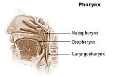
The base of the skull may be affected by metastases from cancer of the bronchus, breast or prostate, or cancer may spread directly to this area from the nasopharynx (fig. 2), and this may cause headache, facial paresthesia, dysesthesia or pain, or cranial nerve dysfunction – the exact symptoms depend on the cranial nerves impacted. [4]

Pelvis

Pain produced by cancer within the pelvis varies depending on the affected tissue, but it frequently radiates diffusely to the upper thigh, and may refer to the lumbar region. Lumbosacral plexopathy is often caused by recurrence of cancer in the presacral space, and may refer to the external genitalia or perineum. Local recurrence of cancer attached to the side of the pelvic wall may cause pain in one of the iliac fossae. Pain on walking that confines the patient to bed indicates possible cancer adherence to or invasion of the iliacus muscle. Pain in the hypogastrium (between the navel and pubic bone) is often found in cancers of the uterus and bladder, and sometimes in colorectal cancer especially if infiltrating or attached to either uterus or bladder. [4]

Viscera

Visceral pain is diffuse and difficult to locate, and is often referred to more distant, usually superficial, sites. [8]

Liver

Acute hemorrhage into a hepatocellular carcinoma causes severe upper right quadrant pain, and may be life-threatening, requiring emergency surgery or other emergency intervention. [5]
A tumor can expand the size of the liver several times and consequent stretching of its capsule can cause aching pain in the right hypochondrium. Other causes of pain in enlarged liver are traction of the supporting ligaments when standing or walking, the liver pressing against the rib cage or pinching the wall of the abdomen, and straining the lumbar spine. In some postures the liver may pinch the parietal peritoneum against the lower rib cage, producing sharp, transitory pain, relieved by changing position. The tumor may also infiltrate the liver's capsule, causing dull, and sometimes stabbing pain. [4]

Kidneys and spleen

Cancer of the kidneys and spleen produces less pain than that caused by liver tumor – kidney tumors eliciting pain only once the organ has been almost totally destroyed and the cancer has invaded the surrounding tissue or adjacent pelvis. Pressure on the kidney or ureter from a tumor outside the kidney can cause extreme flank pain. [7] Local recurrence of cancer after the removal of a kidney can cause pain in the lumbar back, or L1 or L2 spinal nerve pain in the groin or upper thigh, accompanied by weakness and numbness of the iliopsoas muscle, exacerbated by activity. [4]

Abdominal and urogenital hollow organs

Inflammation of artery walls and tissue adjacent to nerves is common in tumors of abdominal and urogenital hollow organs. [10] Infection or cancer may irritate the trigone of the urinary bladder, causing spasm of the detrusor urinae muscle (the muscle that squeezes urine from the urinary bladder), resulting in deep pain above the pubic bone, possibly referred to the tip of the penis, lasting from a few minutes to half an hour. [4]

Gastrointestinal

The pain of intestinal tumors may be the result of disturbed motility, dilation, altered blood flow or ulceration. Malignant lymphomas of the gastrointestinal tract can produce large tumors with significant ulceration and bleeding. [10]

Respiratory system

Cancer in the bronchial tree is usually painless, [10] but ear and facial pain on one side of the head has been reported in some patients. This pain is referred via the auricular branch of the vagus nerve. [4]
Fig. 3 The pancreas: 1. pancreatic head; 4. pancreatic body; 11. pancreatic tail Illu pancreas duodenum.jpg
Fig. 3 The pancreas: 1. pancreatic head; 4. pancreatic body; 11. pancreatic tail

Pancreas

Ten percent of patients with cancer of the pancreatic body or tail experience pain, whereas 90 percent of those with cancer of the pancreatic head will, especially if the tumor is near the hepatopancreatic ampulla. The pain appears on the left or right upper abdomen, is constant, and increases in intensity over time. It is in some cases relieved by leaning forward and heightened by lying on the stomach. Back pain may be present and, if intense, may spread left and right. Back pain may be referred from the pancreas, or may indicate the cancer has penetrated paraspinal muscle, or entered the retroperitoneum and paraaortic lymph nodes [4]

Rectum

A local tumor in the rectum or recurrence involving the presacral plexus may cause pain normally associated with an urgent need to defecate. This pain may, rarely, return as phantom pain after surgical removal of the rectum, though pain within a few weeks of surgical removal of the rectum is usually neuropathic pain due to the surgery (described in one study [11] as spontaneous, intermittent, mild to moderate shooting and bursting, or tight and aching), and pain emerging after three months (described as deep, sharp, aching, intense, and continuous, made worse by sitting or pressure) usually signals recurrence of the disease. The emergence of pain on standing or walking (described as "dragging") may indicate a deeper recurrence involving attachment to muscle or fascia. [4]

Serous mucosa

Carcinosis of the peritoneum may cause pain through inflammation, disordered visceral motility, or pressure of the metastases on nerves. Once a tumor has penetrated or perforated hollow viscera, acute inflammation of the peritoneum appears, inducing severe abdominal pain. Pleural carcinomatosis is normally painless. [10]

Soft tissue

Invasion of soft tissue by a tumor can cause pain by inflammatory or mechanical stimulation of nociceptors, or destruction of mobile structures such as ligaments, tendons and skeletal muscles. [10]

Diagnostic procedures

Fig. 5: Cross section of the spinal cord showing the subarachnoid cavity, dura mater and spinal nerve roots including the dorsal root ganglion Gray770-en.svg
Fig. 5: Cross section of the spinal cord showing the subarachnoid cavity, dura mater and spinal nerve roots including the dorsal root ganglion

Some diagnostic procedures, such as venipuncture, paracentesis, and thoracentesis can be painful. [12]

Lumbar puncture

In lumbar puncture a needle is inserted between two lumbar vertebrae, through the dura mater and arachnoid membrane surrounding the spinal cord, into the fluid-flled space between the arachnoid membrane and the spinal cord (the subarachnoid cavity), and cerebrospinal fluid (CFS) is withdrawn for examination. In one study, 14 percent of patients felt pain on lumbar puncture. [13] (fig. 5)

Post-dural-puncture headache

In some patients, subsequent leakage of CSF through the dura mater puncture causes reduced CSF levels in the brain and spinal cord, leading to the development of post-dural-puncture headache (PDPH) hours or days later. Onset occurs within two days in 66 percent and within three days in ninety percent of PDPH cases. It occurs so rarely immediately after puncture that other possible causes should be investigated when it does. [14]
The headache is severe and described as "searing and spreading like hot metal," involving the back and front of the head, and spreading to the neck and shoulders, sometimes involving neck stiffness. It is exacerbated by movement, and sitting or standing, and relieved to some degree by lying down. Nausea, vomiting, pain in arms and legs, hearing loss, tinnitus, vertigo, dizziness and paraesthesia of the scalp are common. [14]

Potentially painful cancer treatments include immunotherapy which may produce joint or muscle pain; radiotherapy, which can cause skin reactions, enteritis, fibrosis, myelopathy, bone necrosis, neuropathy or plexopathy; chemotherapy, often associated with mucositis, joint pain, muscle pain, peripheral neuropathy and abdominal pain due to diarrhea or constipation; hormone therapy, which sometimes causes pain flares; targeted therapies, such as trastuzumab and rituximab, which can cause muscle, joint or chest pain; angiogenesis inhibitors like bevacizumab, known to sometimes cause bone pain; and surgery, which may produce post-operative pain, post-amputation pain or pelvic floor myalgia.

Chemotherapy

Fig. 4: Chemotherapy drugs Chemotherapy bottles NCI.jpg
Fig. 4: Chemotherapy drugs

Chemotherapy may cause mucositis, muscle pain, joint pain, abdominal pain caused by diarrhea or constipation, and peripheral neuropathy [12]

Chemotherapy-induced peripheral neuropathy

Between 30 and 40 percent of patients undergoing chemotherapy experience chemotherapy-induced peripheral neuropathy (CIPN): tingling numbness, intense pain, and hypersensitivity to cold, beginning in the hands and feet and sometimes progressing to the arms and legs. [15] Chemotherapy drugs associated with CIPN include thalidomide, the epothilones such as ixabepilone, the vinca alkaloids vincristine and vinblastine, the taxanes paclitaxel and docetaxel, the proteasome inhibitors such as bortezomib, and the platinum-based drugs cisplatin, oxaliplatin and carboplatin. [15] [16] [17] Whether CIPN arises, and to what degree, is determined by the choice of drug, duration of use, the total amount consumed and whether the patient already has peripheral neuropathy. Though the symptoms are mainly sensory – pain, tingling, numbness and temperature sensitivity – in some cases motor nerves are affected, and occasionally, also, the autonomic nervous system. [18]
CIPN often follows the first chemotherapy dose and increases in severity as treatment continues, but this progression usually levels off at completion of treatment. The platinum-based drugs are the exception; with these drugs, sensation may continue to deteriorate for several months after the end of treatment. [19] Some CIPN appears to be irreversible. [19] Pain can often be helped with drug or other treatment but the numbness is usually resistant to treatment. [20] A 2007 American study found that most patients did not recall being told to expect CIPN, and doctors monitoring the condition rarely asked how it affects daily living but focused on practical effects such as dexterity and gait. [21]

Mucositis

Cancer drugs can cause changes in the biochemistry of mucous membranes resulting in intense pain in the mouth, throat, nasal passages, and gastrointestinal tract. This pain can make talking, drinking, or eating difficult or impossible. [22]

Muscle and joint pain

Withdrawal of steroid medication can cause joint pain and diffuse muscle pain accompanied by fatigue; these symptoms resolve with recommencement of steroid therapy. Chronic steroid therapy can result in aseptic necrosis of the humoral or femoral head, resulting in shoulder or knee pain described as dull and aching, and reduced movement in or inability to use arm or hip. Aromatase inhibitors can cause diffuse muscle and joint pain and stiffness, and may increase the likelihood of osteoporosis and consequent fractures. [22]

Radiotherapy

Radiotherapy may affect the connective tissue surrounding nerves, and may damage or kill white or gray matter in the brain or spinal cord.

Fibrosis around the brachial or lumbosacral plexus

Radiotherapy may produce excessive growth of the fibrous tissue (fibrosis) around the brachial or lumbosacral plexui (clusters of nerves), which can result in damage to the nerves over time (6 months to 20 years). This nerve damage may cause numbness, "pins and needles" (dysesthesia) and weakness in the affected limb. If pain develops, it is described as diffuse, severe, burning pain, increasing over time, in part or all of the affected limb. [22]

Spinal cord damage

If radiotherapy includes the spinal cord, changes may occur which do not become apparent until some time after treatment. "Early delayed radiation-induced myelopathy" can manifest from six weeks to six months after treatment; the usual symptom is a Lhermitte sign ("a brief, unpleasant sensation of numbness, tingling and often electric-like discharge going from the neck to the spine and extremities, triggered by neck flexion"), usually followed by improvement two to nine months after onset, though in some cases symptoms persist for a long time. "Late delayed radiation-induced myelopathy" may occur six months to ten years after treatment. The typical presentation is Brown-Séquard syndrome (movement problems and numbness to touch and vibration on one side of the body and loss of pain and temperature sensation on the other). Onset may be sudden but is usually progressive. Some patients improve and others deteriorate. [23]

Cited works

Related Research Articles

<span class="mw-page-title-main">Peripheral nervous system</span> Part of the nervous system excluding the brain and spinal cord

The peripheral nervous system (PNS) is one of two components that make up the nervous system of bilateral animals, with the other part being the central nervous system (CNS). The PNS consists of nerves and ganglia, which lie outside the brain and the spinal cord. The main function of the PNS is to connect the CNS to the limbs and organs, essentially serving as a relay between the brain and spinal cord and the rest of the body. Unlike the CNS, the PNS is not protected by the vertebral column and skull, or by the blood–brain barrier, which leaves it exposed to toxins.

<span class="mw-page-title-main">Sciatica</span> Lower back pain that extends down leg

Sciatica is pain going down the leg from the lower back. This pain may go down the back, outside, or front of the leg. Onset is often sudden following activities like heavy lifting, though gradual onset may also occur. The pain is often described as shooting. Typically, symptoms are only on one side of the body. Certain causes, however, may result in pain on both sides. Lower back pain is sometimes present. Weakness or numbness may occur in various parts of the affected leg and foot.

Paresthesia is an abnormal sensation of the skin with no apparent physical cause. Paresthesia may be transient or chronic, and may have many possible underlying causes. Paresthesias are usually painless and can occur anywhere on the body, but most commonly occur in the arms and legs.

Diabetic neuropathy includes various types of nerve damage associated with diabetes mellitus. The most common form, diabetic peripheral neuropathy, affects 30% of all diabetic patients. Symptoms depend on the site of nerve damage and can include motor changes such as weakness; sensory symptoms such as numbness, tingling, or pain; or autonomic changes such as urinary symptoms. These changes are thought to result from a microvascular injury involving small blood vessels that supply nerves. Relatively common conditions which may be associated with diabetic neuropathy include distal symmetric polyneuropathy; third, fourth, or sixth cranial nerve palsy; mononeuropathy; mononeuropathy multiplex; diabetic amyotrophy; and autonomic neuropathy.

Spinal tumors are neoplasms located in either the vertebral column or the spinal cord. There are three main types of spinal tumors classified based on their location: extradural and intradural. Extradural tumors are located outside the dura mater lining and are most commonly metastatic. Intradural tumors are located inside the dura mater lining and are further subdivided into intramedullary and extramedullary tumors. Intradural-intramedullary tumors are located within the dura and spinal cord parenchyma, while intradural-extramedullary tumors are located within the dura but outside the spinal cord parenchyma. The most common presenting symptom of spinal tumors is nocturnal back pain. Other common symptoms include muscle weakness, sensory loss, and difficulty walking. Loss of bowel and bladder control may occur during the later stages of the disease.

Neuropathic pain is pain caused by a lesion or disease of the somatosensory nervous system. Neuropathic pain may be associated with abnormal sensations called dysesthesia or pain from normally non-painful stimuli (allodynia). It may have continuous and/or episodic (paroxysmal) components. The latter resemble stabbings or electric shocks. Common qualities include burning or coldness, "pins and needles" sensations, numbness and itching.

<span class="mw-page-title-main">Cauda equina syndrome</span> Nerve damage at the end of the spinal cord

Cauda equina syndrome (CES) is a condition that occurs when the bundle of nerves below the end of the spinal cord known as the cauda equina is damaged. Signs and symptoms include low back pain, pain that radiates down the leg, numbness around the anus, and loss of bowel or bladder control. Onset may be rapid or gradual.

<span class="mw-page-title-main">Nerve conduction study</span> Diagnostic test for nerve function

A nerve conduction study (NCS) is a medical diagnostic test commonly used to evaluate the function, especially the ability of electrical conduction, of the motor and sensory nerves of the human body. These tests may be performed by medical specialists such as clinical neurophysiologists, physical therapists, physiatrists, and neurologists who subspecialize in electrodiagnostic medicine. In the United States, neurologists and physiatrists receive training in electrodiagnostic medicine as part of residency training and in some cases acquire additional expertise during a fellowship in clinical neurophysiology, electrodiagnostic medicine, or neuromuscular medicine. Outside the US, clinical neurophysiologists learn needle EMG and NCS testing.

<span class="mw-page-title-main">Nerve block</span> Deliberate inhibition of nerve impulses

Nerve block or regional nerve blockade is any deliberate interruption of signals traveling along a nerve, often for the purpose of pain relief. Local anesthetic nerve block is a short-term block, usually lasting hours or days, involving the injection of an anesthetic, a corticosteroid, and other agents onto or near a nerve. Neurolytic block, the deliberate temporary degeneration of nerve fibers through the application of chemicals, heat, or freezing, produces a block that may persist for weeks, months, or indefinitely. Neurectomy, the cutting through or removal of a nerve or a section of a nerve, usually produces a permanent block. Because neurectomy of a sensory nerve is often followed, months later, by the emergence of new, more intense pain, sensory nerve neurectomy is rarely performed.

<span class="mw-page-title-main">Spinal disc herniation</span> Injury to the connective tissue between spinal vertebrae

A spinal disc herniation is an injury to the intervertebral disc between two spinal vertebrae, usually caused by excessive strain or trauma to the spine. It may result in back pain, pain or sensation in different parts of the body, and physical disability. The most conclusive diagnostic tool for disc herniation is MRI, and treatment may range from painkillers to surgery. Protection from disc herniation is best provided by core strength and an awareness of body mechanics including good posture.

Dysesthesia is an unpleasant, abnormal sense of touch. Its etymology comes from the Greek word "dys," meaning "bad," and "aesthesis," which means "sensation". It often presents as pain but may also present as an inappropriate, but not discomforting, sensation. It is caused by lesions of the nervous system, peripheral or central, and it involves sensations, whether spontaneous or evoked, such as burning, wetness, itching, electric shock, and pins and needles. Dysesthesia can include sensations in any bodily tissue, including most often the mouth, scalp, skin, or legs.

<span class="mw-page-title-main">Radiculopathy</span> Medical condition

Radiculopathy, also commonly referred to as pinched nerve, refers to a set of conditions in which one or more nerves are affected and do not work properly. Radiculopathy can result in pain, weakness, altered sensation (paresthesia) or difficulty controlling specific muscles. Pinched nerves arise when surrounding bone or tissue, such as cartilage, muscles or tendons, put pressure on the nerve and disrupt its function.

<span class="mw-page-title-main">Leptomeningeal cancer</span> Medical condition

Leptomeningeal cancer is a rare complication of cancer in which the disease spreads from the original tumor site to the meninges surrounding the brain and spinal cord. This leads to an inflammatory response, hence the alternative names neoplastic meningitis (NM), malignant meningitis, or carcinomatous meningitis. The term leptomeningeal describes the thin meninges, the arachnoid and the pia mater, between which the cerebrospinal fluid is located. The disorder was originally reported by Eberth in 1870. It is also known as leptomeningeal carcinomatosis, leptomeningeal disease (LMD), leptomeningeal metastasis, meningeal metastasis and meningeal carcinomatosis.

<span class="mw-page-title-main">Nerve compression syndrome</span> Human medical condition

Nerve compression syndrome, or compression neuropathy, or nerve entrapment syndrome, is a medical condition caused by chronic, direct pressure on a peripheral nerve. It is known colloquially as a trapped nerve, though this may also refer to nerve root compression. Its symptoms include pain, tingling, numbness and muscle weakness. The symptoms affect just one particular part of the body, depending on which nerve is affected. The diagnosis is largely clinical and can be confirmed with diagnostic nerve blocks. Occasionally imaging and electrophysiology studies aid in the diagnosis. Timely diagnosis is important as untreated chronic nerve compression may cause permanent damage. A surgical nerve decompression can relieve pressure on the nerve but cannot always reverse the physiological changes that occurred before treatment. Nerve injury by a single episode of physical trauma is in one sense an acute compression neuropathy but is not usually included under this heading, as chronic compression takes a unique pathophysiological course.

Neuro-oncology is the study of brain and spinal cord neoplasms, many of which are very dangerous and life-threatening. Among the malignant brain cancers, gliomas of the brainstem and pons, glioblastoma multiforme, and high-grade astrocytoma/oligodendroglioma are among the worst. In these cases, untreated survival usually amounts to only a few months, and survival with current radiation and chemotherapy treatments may extend that time from around a year to a year and a half, possibly two or more, depending on the patient's condition, immune function, treatments used, and the specific type of malignant brain neoplasm. Surgery may in some cases be curative, but, as a general rule, malignant brain cancers tend to regenerate and emerge from remission easily, especially highly malignant cases. In such cases, the goal is to excise as much of the mass and as much of the tumor margin as possible without endangering vital functions or other important cognitive abilities. The Journal of Neuro-Oncology is the longest continuously published journal in the field and serves as a leading reference to those practicing in the area of neuro-oncology.

<span class="mw-page-title-main">Spinal stenosis</span> Disease of the bony spine that results in narrowing of the spinal canal

Spinal stenosis is an abnormal narrowing of the spinal canal or neural foramen that results in pressure on the spinal cord or nerve roots. Symptoms may include pain, numbness, or weakness in the arms or legs. Symptoms are typically gradual in onset and improve with leaning forward. Severe symptoms may include loss of bladder control, loss of bowel control, or sexual dysfunction.

Pain in cancer may arise from a tumor compressing or infiltrating nearby body parts; from treatments and diagnostic procedures; or from skin, nerve and other changes caused by a hormone imbalance or immune response. Most chronic (long-lasting) pain is caused by the illness and most acute (short-term) pain is caused by treatment or diagnostic procedures. However, radiotherapy, surgery and chemotherapy may produce painful conditions that persist long after treatment has ended.

Chemotherapy-induced peripheral neuropathy (CIPN) is a nerve-damaging side effect of antineoplastic agents in the common cancer treatment, chemotherapy. CIPN afflicts between 30% and 40% of patients undergoing chemotherapy. Antineoplastic agents in chemotherapy are designed to eliminate rapidly dividing cancer cells, but they can also damage healthy structures, including the peripheral nervous system. CIPN involves various symptoms such as tingling, pain, and numbness in the hands and feet. These symptoms can impair activities of daily living, such as typing or dressing, reduce balance, and increase risk of falls and hospitalizations. They can also give cause to reduce or discontinue chemotherapy. Researchers have conducted clinical trials and studies to uncover the various symptoms, causes, pathogenesis, diagnoses, risk factors, and treatments of CIPN.

Peripheral mononeuropathy is a nerve related disease where a single nerve, that is used to transport messages from the brain to the peripheral body, is diseased or damaged. Peripheral neuropathy is a general term that indicates any disorder of the peripheral nervous system. The name of the disorder itself can be broken down in order to understand this better; peripheral: in regard to peripheral neuropathy, refers to outside of the brain and spinal cord; neuro: means nerve related; -pathy; means disease. Peripheral mononeuropathy is a disorder that links to Peripheral Neuropathy, as it only effects a single peripheral nerve rather than several damaged or diseased nerves throughout the body. Healthy peripheral nerves are able to “carry messages from the brain and spinal cord to muscles, organs, and other body tissues”.

Femoral nerve dysfunction, also known as femoral neuropathy, is a rare type of peripheral nervous system disorder that arises from damage to nerves, specifically the femoral nerve. Given the location of the femoral nerve, indications of dysfunction are centered around the lack of mobility and sensation in lower parts of the legs. The causes of such neuropathy can stem from both direct and indirect injuries, pressures and diseases. Physical examinations are usually first carried out, depending on the high severity of the injury. In the cases of patients with hemorrhage, imaging techniques are used before any physical examination. Another diagnostic method, electrodiagnostic studies, are recognized as the gold standard that is used to confirm the injury of the femoral nerve. After diagnosis, different treatment methods are provided to the patients depending upon their symptoms in order to effectively target the underlying causes. Currently, femoral neuropathy is highly underdiagnosed and its precedent medical history is not well documented worldwide.

References

  1. 1 2 Kurita GP, Ulrich A, Jensen TS, Werner MU, Sjøgren P (January 2012). "How is neuropathic cancer pain assessed in randomised controlled trials?". Pain. 153 (1): 13–17. doi:10.1016/j.pain.2011.08.013. PMID   21903329. S2CID   38733839.
  2. Gonzalez GR, Foley KM, Portenoy RK (1989). "Evaluative skills necessary for a cancer pain consultant". American Pain Society Meeting, Phoenix Arizona.
  3. Bruera E & MacDonald RN (1986). "Intractable pain in patients with advanced head and neck tumors: a possible role of local infection". Cancer Treatment Reports. 70 (5): 691–2. PMID   3708626.
  4. 1 2 3 4 5 6 7 8 9 10 11 12 13 14 15 Twycross R & Bennett M (2008). "Cancer pain syndromes". In Sykes N, Bennett MI & Yuan C-S (ed.). Clinical pain management: Cancer pain (2nd ed.). London: Hodder Arnold. pp. 27–37. ISBN   978-0-340-94007-5.
  5. 1 2 3 4 Koh, M; Portenoy, RK (2010). Bruera ED & Portenoy RK (ed.). Cancer Pain Syndromes. Cambridge University Press. pp. 53–85. ISBN   9780511640483.
  6. Gundamraj NR; Richmeimer S (January 2010). "Chest Wall Pain". In Fishman, SM; Ballantyne, JC; Rathmell, JP (eds.). Bonica's Management of Pain. Lippincott Williams & Wilkins. pp. 1045–. ISBN   978-0-7817-6827-6. Archived from the original on 30 October 2023. Retrieved 10 June 2013.
  7. 1 2 Fitzgibbon & Loeser 2010 , p. 34
  8. 1 2 3 Urch CE & Suzuki R (2008). "Pathophysiology of somatic, visceral, and neuropathic cancer pain". In Sykes N, Bennett MI & Yuan C-S (ed.). Clinical pain management: Cancer pain (2nd ed.). London: Hodder Arnold. pp. 3–12. ISBN   978-0-340-94007-5.
  9. Foley KM (2004). "Acute and chronic cancer pain syndromes". In Doyle D, Hanks G, Cherny N, Calman K (eds.). Oxford textbook of palliative medicine. Oxford: OUP. pp. 298–316. ISBN   0-19-851098-5.
  10. 1 2 3 4 5 Fitzgibbon & Loeser 2010 , p. 35
  11. Boas RA, Schug SA & Acland RH (1993). "Perineal pain after rectal amputation: A 5 year follow up". Pain. 52 (1): 62–70. doi:10.1016/0304-3959(93)90115-6. PMID   8446438. S2CID   32239058.
  12. 1 2 International Association for the Study of Pain Archived 2011-09-28 at the Wayback Machine Treatment-Related Pain
  13. Abraham JL (2005). A physician's guide to pain and symptom management in cancer patients (2 ed.). The Johns Hopkins University Press. p. 108. ISBN   0801881013.
  14. 1 2 Turnbull GK, Shepherd DB (2003). "Post-dural puncture headache: pathogenesis, prevention and treatment". British Journal of Anaesthesia. 91 (5): 718–729. doi: 10.1093/bja/aeg231 . PMID   14570796.
  15. 1 2 del Pino BM (Feb 23, 2010). "Chemotherapy-induced Peripheral Neuropathy". NCI Cancer Bulletin. 7 (4): 6. Archived from the original on 2011-12-11. Retrieved 2014-06-19.
  16. Grisold, Wolfgang; Oberndorfer, Stefan; Windebank, Anthony (2012). "Chemotherapy and Polyneuropathies" (PDF). NeuroOncology Magazine. 2 (1). THE EUROPEAN ASSOCIATION OF NEUROONCOLOGY: 25–36. ISSN   2224-3453. Archived (PDF) from the original on 5 December 2019. Retrieved 3 July 2024.
  17. "Herceptin and Peripheral sensory neuropathy, a phase IV clinical study of FDA data - eHealthMe". Archived from the original on 2016-10-09. Retrieved 2014-06-19.
  18. Beijers AJ, Jongen JL, Vreugdenhil G (January 2012). "Chemotherapy-induced neurotoxicity: The value of neuroprotective strategies". The Netherlands Journal of Medicine. 70 (1): 18–25. PMID   22271810. Archived from the original on 2021-06-25. Retrieved 2021-06-25.
  19. 1 2 Windebank AJ & Grisold W (March 2008). "Chemotherapy-induced neuropathy". Journal of the Peripheral Nervous System. 13 (1): 27–46. doi:10.1111/j.1529-8027.2008.00156.x. PMID   18346229. S2CID   20411101.
  20. Savage L (2007). "Chemotherapy-induced pain puzzles scientists". Journal of the National Cancer Institute. 99 (14): 1070–1071. doi: 10.1093/jnci/djm072 . PMID   17623791. Archived from the original on 2015-03-19.
  21. Paice JA, Ferrell B (2011). "The management of cancer pain". CA – A Cancer Journal for Clinicians. 61 (3): 157–82. doi: 10.3322/caac.20112 . PMID   21543825.
  22. 1 2 3 Fitzgibbon & Loeser 2010 , p. 39
  23. Fitzgibbon & Loeser 2010 , pp. 102–3