D-lysine 5,6-aminomutase

Last updated
D-Lysine 5,6-aminomutase alpha subunit
PDB 1xrs EBI.jpg
crystal structure of lysine 5,6-aminomutase in complex with plp, cobalamin, and 5'-deoxyadenosine
Identifiers
SymbolLys-AminoMut_A
Pfam PF09043
InterPro IPR015130
Available protein structures:
Pfam   structures / ECOD  
PDB RCSB PDB; PDBe; PDBj
PDBsum structure summary
D-lysine 5,6-aminomutase
Identifiers
EC no. 5.4.3.4
CAS no. 9075-70-1
Databases
IntEnz IntEnz view
BRENDA BRENDA entry
ExPASy NiceZyme view
KEGG KEGG entry
MetaCyc metabolic pathway
PRIAM profile
PDB structures RCSB PDB PDBe PDBsum
Gene Ontology AmiGO / QuickGO
Search
PMC articles
PubMed articles
NCBI proteins

In enzymology, D-lysine 5,6-aminomutase (EC 5.4.3.4) is an enzyme that catalyzes the chemical reaction

Contents

D-lysine 2,5-diaminohexanoate

Hence, this enzyme has one substrate, D-lysine, and one product, 2,5-diaminohexanoate.

This enzyme participates in lysine degradation. It employs one cofactor, cobamide.

Two pathways of lysine degradation Degradation of lysine modified.jpg
Two pathways of lysine degradation

Background

D-lysine 5,6-aminomutase belongs to the isomerase family of enzymes, specifically intramolecular transferases, which transfers amino groups. Its systematic name is D-2,6-diaminohexanoate 5,6-aminomutase. Other names in common use include D-α-lysine mutase and adenosylcobalamin-dependent D-lysine 5,6-aminomutase, which can be abbreviated as 5,6-LAM.

Mutase reaction of 5,6-LAM Mutase reaction of 5,6-LAM.jpg
Mutase reaction of 5,6-LAM

5,6-LAM is capable of reversibly catalyzing the migration of an amino group from ε-carbon to δ-carbon in both D-lysine and L-β-lysine, and catalyzing the migration of hydrogen atoms from δ-carbon to ε-carbon at the same time. [1] It demonstrates greatest catalytic activity in 20mM Tris•HCl at pH 9.0-9.2. [2]

In the early 1950s, 5,6-LAM was discovered in the amino-acid-fermenting bacteria Clostridium sticklandii, in which lysine undergoes degradation under anaerobic conditions to equimolar amounts of acetate and butyrate . [3]

Later, isotopic studies uncovered two possible pathways. In pathway A, both acetate and butyrate are generated from C2-C3 cleavage of D-lysine. Unlike pathway A, pathway B involves C5-C4 degradation, producing the same products.

D-lysine 5,6-aminomutase (5,6-LAM) is responsible for the first conversion in pathway B to convert D-α-lysine into 2,5-diaminohexanoate. Unlike other members of the family of aminomutases (like 2,3-LAM), which are peculiar to a single substrate, 5,6-LAM can reversibly catalyze both the reaction of D-lysine to 2,5-diaminohexanoic acid and the reaction of L-β-lysine to 3,5-diaminohexanoic acid. [3] [4]

5,6-LAM Catalyzed Reactions of Lysin.jpeg

Structure

Subunits

Two units of 5,6-LAM (AdoCbl in yellow and PLP in orange) Two units of 5,6-LAM.png
Two units of 5,6-LAM (AdoCbl in yellow and PLP in orange)

5,6-LAM is an α2β2 tetramer. The structure of the alpha subunit is predominantly a PLP-binding TIM barrel domain, with several additional alpha-helices and beta-strands at the N and C termini. These helices and strands form an intertwined accessory clamp structure that wraps around the sides of the TIM barrel and extends up toward the Ado ligand of the Cbl cofactor, which is the beta subunit providing most of the interactions observed between the protein and the Ado ligand of the Cbl, suggesting that its role is mainly in stabilizing AdoCbl in the precatalytic resting state. [5] The β subunit binds AdoCbl while the PLP directly binds to α subunit. PLP also directly binds to Lys144 of the β subunit to form an internal aldimine. PLP and AdoCbl are separated by a distance of 24Å. [6]

Cofactors

  1. 5,6-LAM is pyridoxal-5'-phosphate (PLP) dependent. PLP binds to its substrate with an external aldimine linkage. PLP is also important for stabilizing the radical intermediate by captodative stabilization and spin delocalization. [7]
  2. Catalysis begins with a 5'-deoxyadenosyl radical (Ado-CH2•), and 5'-deoxyadenosylcobalamin (AdoCbl) is an essential cofactor as a hydrogen carrier. [8]
  3. ATP, a mercaptan, and a divalent metal ion (usually Mg2+) are required to achieve the highest catalytic effect. [4]

Mechanism

Proposed Mechanism of 5,6-LAM Purposed Mechanism.jpg
Proposed Mechanism of 5,6-LAM

Catalytic cycle

The catalytic cycle starts with Ado-CH2• (5'-deoxyadenosyl radical derived from adenosylcobalamine) abstracting a hydrogen atom from PLP-D-lysine adduct (substrate-related precursor SH) to generate a substrate-related radical (S•), with the radical located at carbon 5 of the lysine residue. The latter undergoes an internal cyclization/addition to the imine nitrogen producing an aziridine carbinyl radical (I•) — a more thermodynamically stable intermediate with the radical being at a benzylic position. Rearrangement of I• produces a product-related radical (P•), which then participates in the final step of hydrogen transfer from AdoH to afford the PLP-product complex (PH). [9]

Structure-based catalysis

Further understanding of the catalytic mechanism can be derived from the X-ray structure.

PLP (in green) maintains much interaction with enzyme in open state PLP interaction.png
PLP (in green) maintains much interaction with enzyme in open state

First, an evident conformational change is observed after the substrate is added to the system. With a substrate-free enzyme, the distance between AdoCbl and PLP is about 24 Å. PLP participates in multiple non-covalent interactions with the enzyme with 5,6-LAM presenting an “open” state.

The first step of the catalytic cycle involves the enzyme accepting the substrate by forming an external aldimine with PLP replacing the PLP-Lys144β internal aldimine. With the cleavage of the internal aldimine, the β unit is able to swing towards to the top of the α unit and block the empty site. Therefore, generation of the Ado-CH2• radical leads to a change in the structure of the active domain, bringing the AdoCbl and PLP-substrate complex closer to each other, thus locking the enzyme in a “closed” state. The closed state exists until the radical transfer occurs when the product is released and AdoCbl is reformed. At the same time, the closed state is transformed to the open state again to wait for the next substrate. [10]

Also worth mentioning is the locking mechanism to prevent the radical reaction without the presence of substrate discovered by Catherine Drennan's group. Lys144 of the β subunit is located at a short G-rich loop highly conserved across all 5,6-LAMs, which blocks the AdoCbl from the reaction site. Based on X-ray structure analysis, when the open structure is applied, the axes of the TIM barrel and Rossmann domains are in different directions. With the addition of the substrate, the subunits rearrange to turn the axes into each other to facilitate the catalysis. [11] For example, in wild type 5,6-LAM, the phenol ring of Tyr263α is oriented in a slipped geometry with pyridine ring of PLP, generating a π-π stacking interaction, which is capable of modulating the electron distribution of the high-energetic radical intermediate. [12]

History

Early insights into the mechanism of the catalytic reaction mainly focused on isotopic methods. Both pathways of lysine degradation and the role of 5,6-LAM were discovered in early work by Stadtman's group during 1950s-1960s. In 1971, having a tritiated α-lysine, 2,5-diaminohexanoate, and coenzyme in hand, Colin Morley and T. Stadtman discovered the role of 5'-deoxyadenosylcobalamin (AdoCbl) as a source for hydrogen migration. [8] Recently, much progress has been made toward detecting the intermediates of the reaction, especially towards I•. Based on quantum-mechanical calculations, it was proposed that with 5-fluorolysine [9] as a substitute for D-lysine the 5-FS• species can be captured and analyzed. A similar approach was applied towards PLP modification, when it was modified to 4’-cyanoPLP [13] or PLP-NO. [14] The radical intermediate I• analogue is hypothesized to be easily detected to support the proposed mechanism. Other simulations can also provide some insights into the catalytic reaction. [1]

Related Research Articles

<span class="mw-page-title-main">Tryptophan synthase</span>

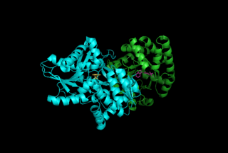
Tryptophan synthase or tryptophan synthetase is an enzyme that catalyses the final two steps in the biosynthesis of tryptophan. It is commonly found in Eubacteria, Archaebacteria, Protista, Fungi, and Plantae. However, it is absent from Animalia. It is typically found as an α2β2 tetramer. The α subunits catalyze the reversible formation of indole and glyceraldehyde-3-phosphate (G3P) from indole-3-glycerol phosphate (IGP). The β subunits catalyze the irreversible condensation of indole and serine to form tryptophan in a pyridoxal phosphate (PLP) dependent reaction. Each α active site is connected to a β active site by a 25 angstrom long hydrophobic channel contained within the enzyme. This facilitates the diffusion of indole formed at α active sites directly to β active sites in a process known as substrate channeling. The active sites of tryptophan synthase are allosterically coupled.

<span class="mw-page-title-main">Aminolevulinic acid synthase</span> Class of enzymes

Aminolevulinic acid synthase (ALA synthase, ALAS, or delta-aminolevulinic acid synthase) is an enzyme (EC 2.3.1.37) that catalyzes the synthesis of δ-aminolevulinic acid (ALA) the first common precursor in the biosynthesis of all tetrapyrroles such as hemes, cobalamins and chlorophylls. The reaction is as follows:

<span class="mw-page-title-main">Pyridoxal phosphate</span> Active form of vitamin B6

Pyridoxal phosphate (PLP, pyridoxal 5'-phosphate, P5P), the active form of vitamin B6, is a coenzyme in a variety of enzymatic reactions. The International Union of Biochemistry and Molecular Biology has catalogued more than 140 PLP-dependent activities, corresponding to ~4% of all classified activities. The versatility of PLP arises from its ability to covalently bind the substrate, and then to act as an electrophilic catalyst, thereby stabilizing different types of carbanionic reaction intermediates.

<span class="mw-page-title-main">Aspartate transaminase</span> Enzyme involved in amino acid metabolism

Aspartate transaminase (AST) or aspartate aminotransferase, also known as AspAT/ASAT/AAT or (serum) glutamic oxaloacetic transaminase, is a pyridoxal phosphate (PLP)-dependent transaminase enzyme that was first described by Arthur Karmen and colleagues in 1954. AST catalyzes the reversible transfer of an α-amino group between aspartate and glutamate and, as such, is an important enzyme in amino acid metabolism. AST is found in the liver, heart, skeletal muscle, kidneys, brain, red blood cells and gall bladder. Serum AST level, serum ALT level, and their ratio are commonly measured clinically as biomarkers for liver health. The tests are part of blood panels.

<span class="mw-page-title-main">Triosephosphate isomerase</span> Enzyme involved in glycolysis

Triose-phosphate isomerase is an enzyme that catalyzes the reversible interconversion of the triose phosphate isomers dihydroxyacetone phosphate and D-glyceraldehyde 3-phosphate.

<span class="mw-page-title-main">Catalytic triad</span> Set of three coordinated amino acids

A catalytic triad is a set of three coordinated amino acids that can be found in the active site of some enzymes. Catalytic triads are most commonly found in hydrolase and transferase enzymes. An acid-base-nucleophile triad is a common motif for generating a nucleophilic residue for covalent catalysis. The residues form a charge-relay network to polarise and activate the nucleophile, which attacks the substrate, forming a covalent intermediate which is then hydrolysed to release the product and regenerate free enzyme. The nucleophile is most commonly a serine or cysteine amino acid, but occasionally threonine or even selenocysteine. The 3D structure of the enzyme brings together the triad residues in a precise orientation, even though they may be far apart in the sequence.

<span class="mw-page-title-main">Methylmalonyl-CoA mutase</span> Mammalian protein found in Homo sapiens

Methylmalonyl-CoA mutase (EC 5.4.99.2, MCM), mitochondrial, also known as methylmalonyl-CoA isomerase, is a protein that in humans is encoded by the MUT gene. This vitamin B12-dependent enzyme catalyzes the isomerization of methylmalonyl-CoA to succinyl-CoA in humans. Mutations in MUT gene may lead to various types of methylmalonic aciduria.

<span class="mw-page-title-main">Enzyme catalysis</span> Catalysis of chemical reactions by specialized proteins known as enzymes

Enzyme catalysis is the increase in the rate of a process by a biological molecule, an "enzyme". Most enzymes are proteins, and most such processes are chemical reactions. Within the enzyme, generally catalysis occurs at a localized site, called the active site.

<span class="mw-page-title-main">Serine hydroxymethyltransferase</span>

Serine hydroxymethyltransferase (SHMT) is a pyridoxal phosphate (PLP) (Vitamin B6) dependent enzyme (EC 2.1.2.1) which plays an important role in cellular one-carbon pathways by catalyzing the reversible, simultaneous conversions of L-serine to glycine and tetrahydrofolate (THF) to 5,10-Methylenetetrahydrofolate (5,10-CH2-THF). This reaction provides the largest part of the one-carbon units available to the cell.

Lysine 2,3-aminomutase is a radical SAM enzyme that facilitates the conversion of the amino acid lysine to beta-lysine. It accomplishes this interconversion using three cofactors and a 5'-deoxyadenosyl radical formed in a S-Adenosyl methionine (SAM) activated radical reaction pathway.[1] The generalized reaction is shown below:

<span class="mw-page-title-main">Cystathionine beta synthase</span> Mammalian protein found in humans

Cystathionine-β-synthase, also known as CBS, is an enzyme (EC 4.2.1.22) that in humans is encoded by the CBS gene. It catalyzes the first step of the transsulfuration pathway, from homocysteine to cystathionine:

<span class="mw-page-title-main">Cystathionine gamma-lyase</span> Protein-coding gene in the species Homo sapiens

The enzyme cystathionine γ-lyase (EC 4.4.1.1, CTH or CSE; also cystathionase; systematic name L-cystathionine cysteine-lyase (deaminating; 2-oxobutanoate-forming)) breaks down cystathionine into cysteine, 2-oxobutanoate (α-ketobutyrate), and ammonia:

<span class="mw-page-title-main">Alanine racemase</span>

In enzymology, an alanine racemase is an enzyme that catalyzes the chemical reaction

In enzymology, a beta-lysine 5,6-aminomutase is an enzyme that catalyzes the chemical reaction

<span class="mw-page-title-main">Cystathionine beta-lyase</span> Enzyme

Cystathionine beta-lyase, also commonly referred to as CBL or β-cystathionase, is an enzyme that primarily catalyzes the following α,β-elimination reaction

<span class="mw-page-title-main">Methionine gamma-lyase</span>

The enzyme methionine γ-lyase (EC 4.4.1.11, MGL) is in the γ-family of PLP-dependent enzymes. It degrades sulfur-containing amino acids to α-keto acids, ammonia, and thiols:

<span class="mw-page-title-main">Arginine decarboxylase</span>

The enzyme Acid-Induced Arginine Decarboxylase (AdiA), also commonly referred to as arginine decarboxylase, catalyzes the conversion of L-arginine into agmatine and carbon dioxide. The process consumes a proton in the decarboxylation and employs a pyridoxal-5'-phosphate (PLP) cofactor, similar to other enzymes involved in amino acid metabolism, such as ornithine decarboxylase and glutamine decarboxylase. It is found in bacteria and virus, though most research has so far focused on forms of the enzyme in bacteria. During the AdiA catalyzed decarboxylation of arginine, the necessary proton is consumed from the cell cytoplasm which helps to prevent the over-accumulation of protons inside the cell and serves to increase the intracellular pH. Arginine decarboxylase is part of an enzymatic system in Escherichia coli, Salmonella Typhimurium, and methane-producing bacteria Methanococcus jannaschii that makes these organisms acid resistant and allows them to survive under highly acidic medium.

<span class="mw-page-title-main">Diaminopimelate decarboxylase</span>

The enzyme diaminopimelate decarboxylase (EC 4.1.1.20) catalyzes the cleavage of carbon-carbon bonds in meso 2,6 diaminoheptanedioate to produce CO2 and L-lysine, the essential amino acid. It employs the cofactor pyridoxal phosphate, also known as PLP, which participates in numerous enzymatic transamination, decarboxylation and deamination reactions.

<span class="mw-page-title-main">Serine C-palmitoyltransferase</span>

In enzymology, a serine C-palmitoyltransferase (EC 2.3.1.50) is an enzyme that catalyzes the chemical reaction:

Glutamate 2,3-aminomutase is an enzyme that belongs to the radical s-adenosyl methionine (SAM) superfamily. Radical SAM enzymes facilitate the reductive cleavage of S-adenosylmethionine (SAM) through the use of radical chemistry and an iron-sulfur cluster. This enzyme family is implicated in the biosynthesis of DNA precursors, vitamin, cofactor, antibiotic and herbicides and in biodegradation pathways. In particular, glutamate 2,3 aminomutase is involved in the conversion of L-alpha-glutamate to L-beta-glutamate in Clostridium difficile. The generalized reaction is shown below:

References

  1. 1 2 Sandala GM, Smith DM, Radom L (December 2006). "In search of radical intermediates in the reactions catalyzed by lysine 2,3-aminomutase and lysine 5,6-aminomutase". Journal of the American Chemical Society. 128 (50): 16004–5. doi:10.1021/ja0668421. PMID   17165731.
  2. Morley CG, Stadtman TC (December 1970). "Studies on the fermentation of D-alpha-lysine. Purification and properties of an adenosine triphosphate regulated B 12-coenzyme-dependent D-alpha-lysine mutase complex from Clostridium sticklandii". Biochemistry. 9 (25): 4890–900. doi:10.1021/bi00827a010. PMID   5480154.
  3. 1 2 Stadtman TC, White FH (June 1954). "Tracer studies on ornithine, lysine, and formate metabolism in an amino acid fermenting Clostridium". Journal of Bacteriology. 67 (6): 651–7. doi:10.1128/JB.67.6.651-657.1954. PMC   357300 . PMID   13174491.
  4. 1 2 Stadtman TC, Tsai L (September 1967). "A cobamide coenzyme dependent migration of the epsilon-amino group of D-lysine". Biochemical and Biophysical Research Communications. 28 (6): 920–6. doi:10.1016/0006-291x(67)90067-8. PMID   4229021.
  5. Berkovitch F, Behshad E, Tang KH, Enns EA, Frey PA, Drennan CL (November 2004). "A locking mechanism preventing radical damage in the absence of substrate, as revealed by the x-ray structure of lysine 5,6-aminomutase". Proceedings of the National Academy of Sciences of the United States of America. 101 (45): 15870–5. doi: 10.1073/pnas.0407074101 . PMC   528771 . PMID   15514022.
  6. Lo HH, Lin HH, Maity AN, Ke SC (May 2016). "The molecular mechanism of the open-closed protein conformational cycle transitions and coupled substrate binding, activation and product release events in lysine 5,6-aminomutase". Chemical Communications. 52 (38): 6399–402. doi:10.1039/c6cc01888b. PMID   27086547.
  7. Chen YH, Maity AN, Pan YC, Frey PA, Ke SC (November 2011). "Radical stabilization is crucial in the mechanism of action of lysine 5,6-aminomutase: role of tyrosine-263α as revealed by electron paramagnetic resonance spectroscopy". Journal of the American Chemical Society. 133 (43): 17152–5. doi:10.1021/ja207766c. PMID   21939264.
  8. 1 2 Morley CG, Stadtman TC (June 1971). "Studies on the fermentation of p-alpha-lysine. On the hydrogen shift catalyzed by the B 12 coenzyme dependent D-alpha-lysine mutase". Biochemistry. 10 (12): 2325–9. doi:10.1021/bi00788a023. PMID   5114991.
  9. 1 2 Maity AN, Ke S (October 2013). "5-Fluorolysine as alternative substrate of lysine 5,6-aminomutase: A computational study". Computational and Theoretical Chemistry. 1022: 1–5. doi:10.1016/j.comptc.2013.08.007.
  10. Chen Y, Maity AN, Frey PA, Ke S (January 2013). "Mechanism-based Inhibition Reveals Transitions between Two Conformational States in the Action of Lysine 5,6-Aminomutase: A Combination of Electron Paramagnetic Resonance Spectroscopy, Electron Nuclear Double Resonance Spectroscopy, and Density Functional Theory Study". Journal of the American Chemical Society. 135 (2): 788–794. doi:10.1021/ja309603a. PMID   23231091.
  11. Berkovitch F, Behshad E, Tang KH, Enns EA, Frey PA, Drennan CL (November 2004). "A locking mechanism preventing radical damage in the absence of substrate, as revealed by the x-ray structure of lysine 5,6-aminomutase". Proceedings of the National Academy of Sciences of the United States of America. 101 (45): 15870–5. doi: 10.1073/pnas.0407074101 . PMC   528771 . PMID   15514022.
  12. Wetmore SD, Smith DM, Radom L (September 2001). "Enzyme catalysis of 1,2-amino shifts: the cooperative action of B6, B12, and aminomutases". Journal of the American Chemical Society. 123 (36): 8678–89. doi:10.1021/ja010211j. PMID   11535072.
  13. Maity AN, Ke SC (February 2015). "4'-CyanoPLP presents better prospect for the experimental detection of elusive cyclic intermediate radical in the reaction of lysine 5,6-aminomutase". Biochemical and Biophysical Research Communications. 457 (2): 161–4. doi:10.1016/j.bbrc.2014.12.076. PMID   25542154.
  14. Maity AN, Lin H, Chiang H, Lo H, Ke S (May 2015). "Reaction of Pyridoxal-5′-phosphate-N-oxide with Lysine 5,6-Aminomutase: Enzyme Flexibility toward Cofactor Analog". ACS Catalysis. 5 (5): 3093–3099. doi:10.1021/acscatal.5b00671.
This article incorporates text from the public domain Pfam and InterPro: IPR015130