
X-ray fluorescence (XRF) is the emission of characteristic "secondary" X-rays from a material that has been excited by being bombarded with high-energy X-rays or gamma rays. The phenomenon is widely used for elemental analysis and chemical analysis, particularly in the investigation of metals, glass, ceramics and building materials, and for research in geochemistry, forensic science, archaeology and art objects such as paintings.

A synchrotron light source is a source of electromagnetic radiation (EM) usually produced by a storage ring, for scientific and technical purposes. First observed in synchrotrons, synchrotron light is now produced by storage rings and other specialized particle accelerators, typically accelerating electrons. Once the high-energy electron beam has been generated, it is directed into auxiliary components such as bending magnets and insertion devices in storage rings and free electron lasers. These supply the strong magnetic fields perpendicular to the beam that are needed to stimulate the high energy electrons to emit photons.
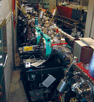
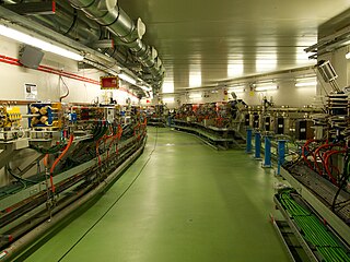
In accelerator physics, a beamline refers to the trajectory of the beam of particles, including the overall construction of the path segment along a specific path of an accelerator facility. This part is either
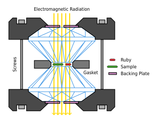
A diamond anvil cell (DAC) is a high-pressure device used in geology, engineering, and materials science experiments. It enables the compression of a small (sub-millimeter-sized) piece of material to extreme pressures, typically up to around 100–200 gigapascals, although it is possible to achieve pressures up to 770 gigapascals.
A diffractometer is a measuring instrument for analyzing the structure of a material from the scattering pattern produced when a beam of radiation or particles interacts with it.
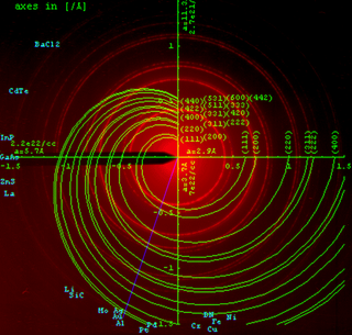
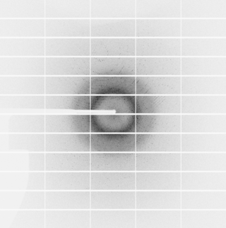
Powder diffraction is a scientific technique using X-ray, neutron, or electron diffraction on powder or microcrystalline samples for structural characterization of materials. An instrument dedicated to performing such powder measurements is called a powder diffractometer.
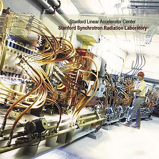
The Stanford Synchrotron Radiation Lightsource, a division of SLAC National Accelerator Laboratory, is operated by Stanford University for the Department of Energy. SSRL is a National User Facility which provides synchrotron radiation, a name given to electromagnetic radiation in the x-ray, ultraviolet, visible and infrared realms produced by electrons circulating in a storage ring at nearly the speed of light. The extremely bright light that is produced can be used to investigate various forms of matter ranging from objects of atomic and molecular size to man-made materials with unusual properties. The obtained information and knowledge is of great value to society, with impact in areas such as the environment, future technologies, health, biology, basic research, and education.
The National Synchrotron Light Source (NSLS) at Brookhaven National Laboratory (BNL) in Upton, New York was a national user research facility funded by the U.S. Department of Energy (DOE). Built from 1978 through 1984, and officially shut down on September 30, 2014, the NSLS was considered a second-generation synchrotron.
High-energy X-rays or HEX-rays are very hard X-rays, with typical energies of 80–1000 keV (1 MeV), about one order of magnitude higher than conventional X-rays used for X-ray crystallography. They are produced at modern synchrotron radiation sources such as the beamlines ID15 and BM18 at the European Synchrotron Radiation Facility (ESRF). The main benefit is the deep penetration into matter which makes them a probe for thick samples in physics and materials science and permits an in-air sample environment and operation. Scattering angles are small and diffraction directed forward allows for simple detector setups.
ALBA is a third-generation synchrotron light source facility located in the Barcelona Synchrotron Park in Cerdanyola del Vallès near Barcelona, in Catalonia (Spain). It was constructed and is operated by CELLS, and co-financed by the Spanish central administration and regional Catalan Government.
Australian Nuclear Science and Technology Organisation's Australian Synchrotron is a 3 GeV national synchrotron radiation facility located in Clayton, in the south-eastern suburbs of Melbourne, Victoria, which opened in 2007.
Multi-wavelength anomalous diffraction is a technique used in X-ray crystallography that facilitates the determination of the three-dimensional structure of biological macromolecules via solution of the phase problem.
Diffraction topography is a imaging technique based on Bragg diffraction. Diffraction topographic images ("topographies") record the intensity profile of a beam of X-rays diffracted by a crystal. A topography thus represents a two-dimensional spatial intensity mapping of reflected X-rays, i.e. the spatial fine structure of a Laue reflection. This intensity mapping reflects the distribution of scattering power inside the crystal; topographs therefore reveal the irregularities in a non-ideal crystal lattice. X-ray diffraction topography is one variant of X-ray imaging, making use of diffraction contrast rather than absorption contrast which is usually used in radiography and computed tomography (CT). Topography is exploited to a lesser extends with neutrons, and has similarities to dark field imaging in the electron microscope community.
PILATUS is the name of a series of x-ray detectors originally developed by the Paul Scherrer Institute at the Swiss Light Source and further developed and commercialized by DECTRIS. The PILATUS detectors are based on hybrid photon counting (HPC) technology, by which X-rays are converted to electrical signals by the photoelectric effect in a semiconductor sensor layer—either silicon or cadmium telluride—which is subject to a substantial bias voltage. The electric signals are counted directly by a series of cells in an ASIC bonded to the sensor. Each cell—or pixel—is a complete detector in itself, equipped with an amplifier, discriminator and counter circuit. This is possible thanks to contemporary CMOS integrated circuit technology.

Single-wavelength anomalous diffraction (SAD) is a technique used in X-ray crystallography that facilitates the determination of the structure of proteins or other biological macromolecules by allowing the solution of the phase problem. In contrast to multi-wavelength anomalous diffraction (MAD), SAD uses a single dataset at a single appropriate wavelength.

The National Synchrotron Light Source II (NSLS-II) at Brookhaven National Laboratory (BNL) in Upton, New York is a national user research facility funded primarily by the U.S. Department of Energy's (DOE) Office of Science. NSLS-II is one of the world's most advanced synchrotron light sources, designed to produce x-rays 10,000 times brighter than BNL's original light source, the National Synchrotron Light Source (NSLS). NSLS-II supports basic and applied research in energy security, advanced materials synthesis and manufacturing, environment, and human health.
Three-dimensional X-ray diffraction (3DXRD) is a microscopy technique using hard X-rays to investigate the internal structure of polycrystalline materials in three dimensions. For a given sample, 3DXRD returns the shape, juxtaposition, and orientation of the crystallites ("grains") it is made of. 3DXRD allows investigating micrometer- to millimetre-sized samples with resolution ranging from hundreds of nanometers to micrometers. Other techniques employing X-rays to investigate the internal structure of polycrystalline materials include X-ray diffraction contrast tomography (DCT) and high energy X-ray diffraction (HEDM).

X-ray emission spectroscopy (XES) is a form of X-ray spectroscopy in which a core electron is excited by an incident x-ray photon and then this excited state decays by emitting an x-ray photon to fill the core hole. The energy of the emitted photon is the energy difference between the involved electronic levels. The analysis of the energy dependence of the emitted photons is the aim of the X-ray emission spectroscopy.
ANAXAM stands for "Analytics with Neutrons And X-rays for Advanced Manufacturing" and it is a knowledge and technology transfer centre in Switzerland. It is a non-profit organisation. ANAXAM provides industry access to modern, applied material analytics with neutron and synchrotron radiation (X-rays) in the field of non-destructive material testing. The analytical services offered by ANAXAM are based on imaging (CT), diffraction, small-angle scattering and spectroscopy methods.

