Forensic science, also known as criminalistics, is the application of science to criminal and civil laws, mainly—on the criminal side—during criminal investigation, as governed by the legal standards of admissible evidence and criminal procedure. Forensic science is a broad field that includes; DNA analysis, fingerprint analysis, blood stain pattern analysis, firearms examination and ballistics, tool mark analysis, serology, toxicology, hair and fiber analysis, entomology, questioned documents, anthropology, odontology, pathology, epidemiology, footwear and tire tread analysis, drug chemistry, paint and glass analysis, digital audio video and photo analysis.

A CT scan, also known as computed tomography scan is a medical imaging technique used in radiology (x-ray) to obtain detailed internal images of the body noninvasively for diagnostic purposes. The personnel that perform CT scans are called radiographers or radiology technologists.
Forensic pathology is pathology that focuses on determining the cause of death by examining a corpse. A post mortem examination is performed by a medical examiner or forensic pathologist, usually during the investigation of criminal law cases and civil law cases in some jurisdictions. Coroners and medical examiners are also frequently asked to confirm the identity of remains.and

Radiography is an imaging technique using X-rays, gamma rays, or similar ionizing radiation and non-ionizing radiation to view the internal form of an object. Applications of radiography include medical radiography and industrial radiography. Similar techniques are used in airport security. To create an image in conventional radiography, a beam of X-rays is produced by an X-ray generator and is projected toward the object. A certain amount of the X-rays or other radiation is absorbed by the object, dependent on the object's density and structural composition. The X-rays that pass through the object are captured behind the object by a detector. The generation of flat two dimensional images by this technique is called projectional radiography. In computed tomography an X-ray source and its associated detectors rotate around the subject which itself moves through the conical X-ray beam produced. Any given point within the subject is crossed from many directions by many different beams at different times. Information regarding attenuation of these beams is collated and subjected to computation to generate two dimensional images in three planes which can be further processed to produce a three dimensional image.

Radiology is the medical discipline that uses medical imaging to diagnose diseases and guide their treatment, within the bodies of humans and other animals. It began with radiography, but today it includes all imaging modalities, including those that use no electromagnetic radiation, as well as others that do, such as computed tomography (CT), fluoroscopy, and nuclear medicine including positron emission tomography (PET). Interventional radiology is the performance of usually minimally invasive medical procedures with the guidance of imaging technologies such as those mentioned above.

Medical imaging is the technique and process of imaging the interior of a body for clinical analysis and medical intervention, as well as visual representation of the function of some organs or tissues (physiology). Medical imaging seeks to reveal internal structures hidden by the skin and bones, as well as to diagnose and treat disease. Medical imaging also establishes a database of normal anatomy and physiology to make it possible to identify abnormalities. Although imaging of removed organs and tissues can be performed for medical reasons, such procedures are usually considered part of pathology instead of medical imaging.
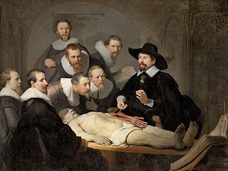
An autopsy is a surgical procedure that consists of a thorough examination of a corpse by dissection to determine the cause, mode, and manner of death or to evaluate any disease or injury that may be present for research or educational purposes.. Autopsies are usually performed by a specialized medical doctor called a pathologist. In most cases, a medical examiner or coroner can determine the cause of death, however, only a small portion of deaths require an autopsy to be performed, under certain circumstances.
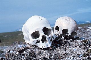
Forensic anthropology is the application of the anatomical science of anthropology and its various subfields, including forensic archaeology and forensic taphonomy, in a legal setting. A forensic anthropologist can assist in the identification of deceased individuals whose remains are decomposed, burned, mutilated or otherwise unrecognizable, as might happen in a plane crash. Forensic anthropologists are also instrumental in the investigation and documentation of genocide and mass graves. Along with forensic pathologists, forensic dentists, and homicide investigators, forensic anthropologists commonly testify in court as expert witnesses. Using physical markers present on a skeleton, a forensic anthropologist can potentially determine a person's age, sex, stature, and race. In addition to identifying physical characteristics of the individual, forensic anthropologists can use skeletal abnormalities to potentially determine cause of death, past trauma such as broken bones or medical procedures, as well as diseases such as bone cancer.

Tomography is imaging by sections or sectioning that uses any kind of penetrating wave. The method is used in radiology, archaeology, biology, atmospheric science, geophysics, oceanography, plasma physics, materials science, astrophysics, quantum information, and other areas of science. The word tomography is derived from Ancient Greek τόμος tomos, "slice, section" and γράφω graphō, "to write" or, in this context as well, "to describe." A device used in tomography is called a tomograph, while the image produced is a tomogram.

Forensic dentistry or forensic odontology involves handling, examination and evaluation of dental evidence in criminal justice cases. Forensic dentists are involved in assisting investigative agencies to identify recovered human remains in addition to the identification of whole or fragmented bodies. Forensic dentists have also been known to use their investigative techniques to identify burn victims by using the victims previous dental records. Forensic dentists may also be asked to assist in determining age, race, occupation, previous dental history and socioeconomic status of unidentified human beings.

Angiography or arteriography is a medical imaging technique used to visualize the inside, or lumen, of blood vessels and organs of the body, with particular interest in the arteries, veins, and the heart chambers. Modern angiography is performed by injecting a radio-opaque contrast agent into the blood vessel and imaging using X-ray based techniques such as fluoroscopy.
The Hounsfield scale, named after Sir Godfrey Hounsfield, is a quantitative scale for describing radiodensity. It is frequently used in CT scans, where its value is also termed CT number.

A diener is a morgue worker responsible for handling, moving, and cleaning the corpse. In the UK, the equivalent job title is 'Mortuary Assistant', whilst the preparation, evisceration and reconstruction of the deceased is performed by an Anatomical Pathology Technician. In the US, Dieners are also referred to as "mortuary assistants" or "autopsy technicians". The word is derived from the German word Leichendiener, which literally means corpse servant.

Digital subtraction angiography (DSA) is a fluoroscopy technique used in interventional radiology to clearly visualize blood vessels in a bony or dense soft tissue environment. Images are produced using contrast medium by subtracting a "pre-contrast image" or mask from subsequent images, once the contrast medium has been introduced into a structure. Hence the term "digital subtraction angiography. Subtraction angiography was first described in 1935 and in English sources in 1962 as a manual technique. Digital technology made DSA practical starting in the 1970s.

Quantitative computed tomography (QCT) is a medical technique that measures bone mineral density (BMD) using a standard X-ray Computed Tomography (CT) scanner with a calibration standard to convert Hounsfield Units (HU) of the CT image to bone mineral density values. Quantitative CT scans are primarily used to evaluate bone mineral density at the lumbar spine and hip.
The following outline is provided as an overview of and topical guide to forensic science:
Virtopsy is a virtual alternative to a traditional autopsy, conducted with scanning and imaging technology. The name is a portmanteau of 'virtual' and 'autopsy' and is a trademark registered to Prof. Richard Dirnhofer (de), the former head of the Institute of Forensic Medicine of the University of Bern, Switzerland.

Cone beam computed tomography is a medical imaging technique consisting of X-ray computed tomography where the X-rays are divergent, forming a cone.
A digital autopsy is a non-invasive autopsy in which digital imaging technology, such as with Computerized Tomography (CT) or Magnetic Resonance Imaging (MRI) scans, is used to develop three-dimensional images for a virtual exploration of a human body.
Human identification by forensic scientists can be done by three primary methods; friction ridge Analysis, DNA analysis and comparative dental analysis which is one of the roles of a forensic odontologist. It is the process of identification by a post-mortem dental examination of a deceased individual comparing these findings with the ante-mortem dental records, radiographs, study casts, etc. Believed to be those of the individual implicated assessing the concordance and/or discrepancy of the comparison of results. Teeth are resilient, and along with the highly specific and unique type, location and configuration of restorations, might be the only features used for the identification of bodies found in burnt, decomposed, skeletonised, or macerated condition.
