Gephyrin

Last updated
GPHN
Protein GPHN PDB 1ihc.png
Available structures
PDB Ortholog search: PDBe RCSB
Identifiers
Aliases GPHN , GEPH, GPH, GPHRYN, HKPX1, MOCODC, gephyrin
External IDs OMIM: 603930 MGI: 109602 HomoloGene: 10820 GeneCards: GPHN
Orthologs
SpeciesHumanMouse
Entrez
Ensembl
UniProt
RefSeq (mRNA)

NM_145965
NM_172952

RefSeq (protein)

NP_666077
NP_766540

Location (UCSC) Chr 14: 66.51 – 67.18 Mb Chr 12: 78.27 – 78.73 Mb
PubMed search [3] [4]
Wikidata
View/Edit Human View/Edit Mouse

Gephyrin is a protein that in humans is encoded by the GPHN gene. [5] [6] [7] [8] [9]

Contents

This gene encodes a neuronal assembly protein that anchors inhibitory neurotransmitter receptors to the postsynaptic cytoskeleton via high affinity binding to a receptor subunit domain and tubulin dimers. In nonneuronal tissues, the encoded protein is also required for molybdenum cofactor biosynthesis. Mutations in this gene may be associated with the neurological condition hyperekplexia and also lead to molybdenum cofactor deficiency.

Gene

Numerous alternatively spliced transcript variants encoding different isoforms have been described; however, the full-length nature of all transcript variants is not currently known. [8] The production of alternatively spliced variants is affected by noncoding regions within the gene. A ‘yin-yang’ noncoding sequence pair encompassing gephyrin has been identified. [10] These sequences are opposites of each other - consisting of hundreds of divergent nucleotide states. Both of these patterns are uniquely human and evolved rapidly after splitting from their ancestral DNA pattern. The gephyrin yin and yang sequences are prevalent today in populations representing every major human ancestry.

Function

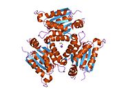
Gephyrin is a 93kDa multi-functional protein that is a component of the postsynaptic protein network of inhibitory synapses. It consists of 3 domains: N terminal G domain, C terminal E domain, and a large unstructured linker domain which connects the two. Although there are structures available for trimeric G and dimeric E domains, there is no structure available for the full length protein, which may be due to the large unstructured region which makes the protein hard to crystallize. But a recent study of the full length gephyrin by small-angle X-ray scattering shows that it predominantly forms trimers, and that because of its long linker region, it can exist in either a compact state or either of two extended states. [11]

Positive antibody staining for gephyrin at a synapse is most of the time consistent with the presence of glycine and/or GABAA receptors. Nevertheless, some exceptions can occur like in neurons of Dorsal Root Ganglions where gephyrin is absent despite the presence of GABAA receptors. [9] Gephyrin is considered a major scaffolding protein at inhibitory synapses, analogous in its function to that of PSD-95 at glutamatergic synapses. [12] [13] Gephyrin was identified by its interaction with the glycine receptor, the main receptor protein of inhibitory synapses in the spinal cord and brainstem. In addition to its interaction with the glycine receptor, recent publications have shown that gephyrin also interacts with the intracellular loop between the transmembrane helices TM3 and TM4 of alpha and beta subunits of the GABAA receptor. [14]

Gephyrin displaces GABA receptors from the GABARAP/P130 complex, then brings the receptors to the synapse. [15] Once at the synapse, the protein binds to collybistin [16] and neuroligin 2. [17] In cells, gephyrin appears to form oligomers of at least three subunits. Several splice variants have been described that prevent this oligomerization without influencing the affinity for receptors. They nevertheless affect the composition of inhibitory synapses and can even play a role in diseases like epilepsy. [18]

The gephyrin protein is also required for insertion of molybdenum into molybdopterin. [19]

As aforementioned, gephyrin also catalyzes terminal two steps of Moco biosynthesis. In the penultimate step, N-terminal G domain adenylate the apo form of the molybdopterin to form the intermediate adenylated molybdopterin. In the terminal step, the C-terminal E domain catalyzes the deadenylation and also the metal insertion mechanism.

Clinical significance

Humans with temporal lobe epilepsy have been found to have abnormally low levels of gephyrin in their temporal lobes. [20] In animal models, a total lack of gephyrin results in stiff muscles and death immediately after birth. Stiff muscles are also a symptom of startle disease, that can be caused by a mutation in the gephyrin gene. And if a person produces auto-antibodies against gephyrin, this can even result in stiff person syndrome. [18]

Yin-yang sequences

Yin-yang DNA sequences encompassing human gephyrin gene. Yin-yang haplotypes arise when a stretch of DNA evolves to present two divergent forms. This image shows the states for ~1000 markers in the genomic region centered on gephyrin for 934 individuals in eight global populations. Humans carry pairs of chromosomes, so each individual possesses two copies of the gephyrin gene. Dark blue and red horizontal lines in the yin-yang region represent individuals carrying two yin and two yang haplotypes, respectively, and light blue represents individuals carrying both a yin and a yang haplotype. Yin-yang DNA sequences encompassing human gephyrin gene.jpg
Yin-yang DNA sequences encompassing human gephyrin gene. Yin-yang haplotypes arise when a stretch of DNA evolves to present two divergent forms. This image shows the states for ~1000 markers in the genomic region centered on gephyrin for 934 individuals in eight global populations. Humans carry pairs of chromosomes, so each individual possesses two copies of the gephyrin gene. Dark blue and red horizontal lines in the yin-yang region represent individuals carrying two yin and two yang haplotypes, respectively, and light blue represents individuals carrying both a yin and a yang haplotype.

At some point in human history, there was a DNA sequence encompassing gephyrin that split and followed two divergent evolutionary paths. [10] These types of splits can occur when two populations become isolated from each other or when a chromosomal region does not experience recombination events. The two sequences that split from the ancestral sequence each acquired more than a hundred mutations that subsequently became common. This happened in a relatively short time on an evolutionary scale, as hundreds of mutations were fixed in distinct ‘yin’ and ‘yang’ sequences prior to human migration to Asia. It has been reported that currently Asians carry nearly equal numbers of yin and yang sequences and global populations representing every major human ancestry possess both yin and yang sequences. [10] The existence of this massive yin-yang pattern suggests that two completely divergent evolutionary paths rapidly progressed during human history, presumably achieving the common goal of enhancing regulation of gephyrin.

Interactions

GPHN has been shown to interact with Mammalian target of rapamycin [6] and ARHGEF9. [16]

Related Research Articles

<span class="mw-page-title-main">Chemical synapse</span> Biological junctions through which neurons signals can be sent

Chemical synapses are biological junctions through which neurons' signals can be sent to each other and to non-neuronal cells such as those in muscles or glands. Chemical synapses allow neurons to form circuits within the central nervous system. They are crucial to the biological computations that underlie perception and thought. They allow the nervous system to connect to and control other systems of the body.

In neurophysiology, long-term depression (LTD) is an activity-dependent reduction in the efficacy of neuronal synapses lasting hours or longer following a long patterned stimulus. LTD occurs in many areas of the CNS with varying mechanisms depending upon brain region and developmental progress.

<span class="mw-page-title-main">Glycine receptor</span> Widely distributed inhibitory receptor in the central nervous system

The glycine receptor is the receptor of the amino acid neurotransmitter glycine. GlyR is an ionotropic receptor that produces its effects through chloride current. It is one of the most widely distributed inhibitory receptors in the central nervous system and has important roles in a variety of physiological processes, especially in mediating inhibitory neurotransmission in the spinal cord and brainstem.

<span class="mw-page-title-main">Neurotransmission</span> Impulse transmission between neurons

Neurotransmission is the process by which signaling molecules called neurotransmitters are released by the axon terminal of a neuron, and bind to and react with the receptors on the dendrites of another neuron a short distance away. A similar process occurs in retrograde neurotransmission, where the dendrites of the postsynaptic neuron release retrograde neurotransmitters that signal through receptors that are located on the axon terminal of the presynaptic neuron, mainly at GABAergic and glutamatergic synapses.

<span class="mw-page-title-main">Hyperekplexia</span> Genetic disorder causing an exaggerated startle response

Hyperekplexia is a very rare neurologic disorder, classically characterised by a pronounced startle responses to tactile or acoustic stimuli and an ensuing period of hypertonia. The hypertonia may be predominantly truncal, attenuated during sleep, or less prominent after one year of age.

<span class="mw-page-title-main">Synapse</span> Structure connecting neurons in the nervous system

In the nervous system, a synapse is a structure that permits a neuron to pass an electrical or chemical signal to another neuron or to the target effector cell.

<span class="mw-page-title-main">Neurexin</span> Protein family

Neurexins (NRXN) are a family of presynaptic cell adhesion proteins that have roles in connecting neurons at the synapse. They are located mostly on the presynaptic membrane and contain a single transmembrane domain. The extracellular domain interacts with proteins in the synaptic cleft, most notably neuroligin, while the intracellular cytoplasmic portion interacts with proteins associated with exocytosis. Neurexin and neuroligin "shake hands," resulting in the connection between the two neurons and the production of a synapse. Neurexins mediate signaling across the synapse, and influence the properties of neural networks by synapse specificity. Neurexins were discovered as receptors for α-latrotoxin, a vertebrate-specific toxin in black widow spider venom that binds to presynaptic receptors and induces massive neurotransmitter release. In humans, alterations in genes encoding neurexins are implicated in autism and other cognitive diseases, such as Tourette syndrome and schizophrenia.

<span class="mw-page-title-main">DLG4</span> Mammalian protein found in Homo sapiens

PSD-95 also known as SAP-90 is a protein that in humans is encoded by the DLG4 gene.

<span class="mw-page-title-main">DLG2</span> Protein-coding gene in the species Homo sapiens

Disks large homolog 2 (DLG2) also known as channel-associated protein of synapse-110 (chapsyn-110) or postsynaptic density protein 93 (PSD-93) is a protein that in humans is encoded by the DLG2 gene.

<span class="mw-page-title-main">Glycine—tRNA ligase</span> Protein-coding gene in the species Homo sapiens

Glycine—tRNA ligase also known as glycyl–tRNA synthetase is an enzyme that in humans is encoded by the GARS1 gene.

<span class="mw-page-title-main">Glycine receptor, alpha 1</span> Protein-coding gene in the species Homo sapiens

Glycine receptor subunit alpha-1 is a protein that in humans is encoded by the GLRA1 gene.

<span class="mw-page-title-main">MOCS1</span> Protein-coding gene in the species Homo sapiens

Molybdenum cofactor biosynthesis protein 1 is a protein that in humans and other animals, fungi, and cellular slime molds, is encoded by the MOCS1 gene.

<span class="mw-page-title-main">Chloride potassium symporter 5</span> Protein-coding gene in the species Homo sapiens

Potassium-chloride transporter member 5 is a neuron-specific chloride potassium symporter responsible for establishing the chloride ion gradient in neurons through the maintenance of low intracellular chloride concentrations. It is a critical mediator of synaptic inhibition, cellular protection against excitotoxicity and may also act as a modulator of neuroplasticity. Potassium-chloride transporter member 5 is also known by the names: KCC2 for its ionic substrates, and SLC12A5 for its genetic origin from the SLC12A5 gene in humans.

<span class="mw-page-title-main">Sodium- and chloride-dependent glycine transporter 2</span> Protein-coding gene in the species Homo sapiens

Sodium- and chloride-dependent glycine transporter 2, also known as glycine transporter 2 (GlyT2), is a protein that in humans is encoded by the SLC6A5 gene.

<span class="mw-page-title-main">GRID2</span> Protein-coding gene in the species Homo sapiens

Glutamate receptor, ionotropic, delta 2, also known as GluD2, GluRδ2, or δ2, is a protein that in humans is encoded by the GRID2 gene. This protein together with GluD1 belongs to the delta receptor subtype of ionotropic glutamate receptors. They possess 14–24% sequence homology with AMPA, kainate, and NMDA subunits, but, despite their name, do not actually bind glutamate or various other glutamate agonists.

<span class="mw-page-title-main">GLRB</span> Protein-coding gene in the species Homo sapiens

Glycine receptor subunit beta is a protein that in humans is encoded by the GLRB gene.

<span class="mw-page-title-main">Neuroligin</span> Protein

Neuroligin (NLGN), a type I membrane protein, is a cell adhesion protein on the postsynaptic membrane that mediates the formation and maintenance of synapses between neurons. Neuroligins act as ligands for β-neurexins, which are cell adhesion proteins located presynaptically. Neuroligin and β-neurexin "shake hands", resulting in the connection between two neurons and the production of a synapse. Neuroligins also affect the properties of neural networks by specifying synaptic functions, and they mediate signalling by recruiting and stabilizing key synaptic components. Neuroligins interact with other postsynaptic proteins to localize neurotransmitter receptors and channels in the postsynaptic density as the cell matures. Additionally, neuroligins are expressed in human peripheral tissues and have been found to play a role in angiogenesis. In humans, alterations in genes encoding neuroligins are implicated in autism and other cognitive disorders. Antibodies in a mother from previous male pregnancies against neuroligin 4 from the Y chromosome increase the probability of homosexuality in male offspring.

<span class="mw-page-title-main">ARHGEF9</span> Protein-coding gene in humans

Rho guanine nucleotide exchange factor 9 is a protein that in humans is encoded by the ARHGEF9 gene.

Collybistin is a protein identified as a regulator of the localization of gephyrin, inducing the formation of submembrane gephyrin aggregates that accumulate glycine and GABA receptors. In 2000 it was identified as a gephyrin binding partner, and an important determinant of inhibitory postsynaptic membrane formation and plasticity. Gephyrin and collybistin are recruited to developing postsynaptic membranes of inhibitory synapses by the trans-synaptic adhesion molecule neuroligin-2, where they provide the scaffold for the clustering of inhibitory postsynaptic receptors to form a functioning inhibitory synapse.

Antoine Triller, born on 23 May 1952, is research director at the Institut national de la santé et de la recherche médicale (Inserm). He is a researcher in neurobiology.

References

  1. 1 2 3 GRCh38: Ensembl release 89: ENSG00000171723 - Ensembl, May 2017
  2. 1 2 3 GRCm38: Ensembl release 89: ENSMUSG00000047454 - Ensembl, May 2017
  3. "Human PubMed Reference:". National Center for Biotechnology Information, U.S. National Library of Medicine.
  4. "Mouse PubMed Reference:". National Center for Biotechnology Information, U.S. National Library of Medicine.
  5. Prior P, Schmitt B, Grenningloh G, Pribilla I, Multhaup G, Beyreuther K, Maulet Y, Werner P, Langosch D, Kirsch J (Jul 1992). "Primary structure and alternative splice variants of gephyrin, a putative glycine receptor-tubulin linker protein". Neuron. 8 (6): 1161–70. doi: 10.1016/0896-6273(92)90136-2 . PMID   1319186.
  6. 1 2 Sabatini DM, Barrow RK, Blackshaw S, Burnett PE, Lai MM, Field ME, Bahr BA, Kirsch J, Betz H, Snyder SH (Jun 1999). "Interaction of RAFT1 with gephyrin required for rapamycin-sensitive signaling". Science. 284 (5417): 1161–4. Bibcode:1999Sci...284.1161S. doi:10.1126/science.284.5417.1161. PMID   10325225.
  7. Fritschy JM, Harvey RJ, Schwarz G (May 2008). "Gephyrin: where do we stand, where do we go?". Trends Neurosci. 31 (5): 257–64. doi:10.1016/j.tins.2008.02.006. PMID   18403029. S2CID   6885626.
  8. 1 2 "Entrez Gene: GPHN gephyrin".
  9. 1 2 Lorenzo LE, Godin AG, Wang F, St-Louis M, Carbonetto S, Wiseman PW, Ribeiro-da-Silva A, De Koninck Y (June 2014). "Gephyrin Clusters Are Absent from Small Diameter Primary Afferent Terminals Despite the Presence of GABAA Receptors". J. Neurosci. 34 (24): 8300–17. doi:10.1523/JNEUROSCI.0159-14.2014. PMC   6608243 . PMID   24920633.
  10. 1 2 3 Climer S, Templeton AR, Zhang W (2015). "Human gephyrin is encompassed within giant functional noncoding yin-yang sequences". Nature Communications. 6: 6534. doi:10.1038/ncomms7534. PMC   4380243 . PMID   25813846.*Lay summary in: "Big data allows computer engineers to find genetic clues in humans". ScienceDaily. March 27, 2015.
  11. Sander B, Tria G, Shkumatov AV, Kim EY, Grossmann JG, Tessmer I, Svergun DI, Schindelin H (Oct 2013). "Structural characterization of gephyrin by AFM and SAXS reveals a mixture of compact and extended states". Acta Crystallographica Section D. 69 (Pt 10): 2050–60. doi:10.1107/S0907444913018714. PMID   24100323.
  12. Giesemann T, Schwarz G, Nawrotzki R, Berhörster K, Rothkegel M, Schlüter K, Schrader N, Schindelin H, Mendel RR, Kirsch J, Jockusch BM (September 2003). "Complex formation between the postsynaptic scaffolding protein gephyrin, profilin, and Mena: a possible link to the microfilament system". J. Neurosci. 23 (23): 8330–9. doi:10.1523/JNEUROSCI.23-23-08330.2003. PMC   6740687 . PMID   12967995.
  13. Ehrensperger MV, Hanus C, Vannier C, Triller A, Dahan M (May 2007). "Multiple association states between glycine receptors and gephyrin identified by SPT analysis". Biophys. J. 92 (10): 3706–18. doi:10.1529/biophysj.106.095596. PMC   1853151 . PMID   17293395.
  14. Maric HM, Mukherjee J, Tretter V, Moss SJ, Schindelin H (December 2011). "Gephyrin-mediated γ-aminobutyric acid type A and glycine receptor clustering relies on a common binding site". J. Biol. Chem. 286 (49): 42105–14. doi: 10.1074/jbc.M111.303412 . PMC   3234978 . PMID   22006921.
  15. Thiriet M (2013). Intracellular Signaling Mediators in the Circulatory and Ventilatory Systems. New York, NY: Springer New York. p. 605. ISBN   978-1-4614-4370-4.
  16. 1 2 Kins S, Betz H, Kirsch J (January 2000). "Collybistin, a newly identified brain-specific GEF, induces submembrane clustering of gephyrin". Nat. Neurosci. 3 (1): 22–9. doi:10.1038/71096. PMID   10607391. S2CID   24878249.
  17. Poulopoulos A, Aramuni G, Meyer G, Soykan T, Hoon M, Papadopoulos T, Zhang M, Paarmann I, Fuchs C, Harvey K, Jedlicka P, Schwarzacher SW, Betz H, Harvey RJ, Brose N, Zhang W, Varoqueaux F (September 2009). "Neuroligin 2 drives postsynaptic assembly at perisomatic inhibitory synapses through gephyrin and collybistin". Neuron. 63 (5): 628–42. doi: 10.1016/j.neuron.2009.08.023 . PMID   19755106. S2CID   15911502.
  18. 1 2 Tretter V, Mukherjee J, Maric HM, Schindelin H, Sieghart W, Moss SJ (2012). "Gephyrin, the enigmatic organizer at GABAergic synapses". Front Cell Neurosci. 6: 23. doi: 10.3389/fncel.2012.00023 . PMC   3351755 . PMID   22615685.
  19. Reiss J, Johnson JL (June 2003). "Mutations in the molybdenum cofactor biosynthetic genes MOCS1, MOCS2, and GEPH". Hum. Mutat. 21 (6): 569–76. doi: 10.1002/humu.10223 . PMID   12754701. S2CID   41013043.
  20. Fang M, Shen L, Yin H, Pan YM, Wang L, Chen D, Xi ZQ, Xiao Z, Wang XF, Zhou SN (October 2011). "Downregulation of gephyrin in temporal lobe epilepsy neurons in humans and a rat model". Synapse. 65 (10): 1006–14. doi:10.1002/syn.20928. PMID   21404332. S2CID   12025675.

Further reading