In primates, and specifically in humans, the labia majora, also known as the outer lips or outer labia, are two prominent longitudinal skin folds that extend downward and backward from the mons pubis to the perineum. Together with the labia minora, they form the labia of the vulva.

The levator labii superioris alaeque nasi muscle is, translated from Latin, the "lifter of both the upper lip and of the wing of the nose". The muscle is attached to the upper frontal process of the maxilla and inserts into the skin of the lateral part of the nostril and upper lip. At 44 characters, its name is longer than any other muscle's.

The facial artery is a branch of the external carotid artery that supplies structures of the superficial face.

Labiaplasty is a plastic surgery procedure for creating or altering the labia minora and the labia majora, the folds of skin of the human vulva. It is a type of vulvoplasty. There are two main categories of women seeking cosmetic genital surgery: those with conditions such as intersex, and those with no underlying condition who experience physical discomfort or wish to alter the appearance of their vulvas because they believe they do not fall within a normal range.

In neuroanatomy, the maxillary nerve (V2) is one of the three branches or divisions of the trigeminal nerve, the fifth (CN V) cranial nerve. It comprises the principal functions of sensation from the maxilla, nasal cavity, sinuses, the palate and subsequently that of the mid-face, and is intermediate, both in position and size, between the ophthalmic nerve and the mandibular nerve.

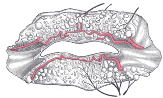
The inferior labial artery arises near the angle of the mouth as a branch of the facial artery; it passes upward and forward beneath the triangularis and, penetrating the orbicularis oris, runs in a tortuous course along the edge of the lower lip between this muscle and the mucous membrane.

The superior labial artery is larger and more egregious than the inferior labial artery.

The angular vein is a vein of the face. It is the upper part of the facial vein, above its junction with the superior labial vein. It is formed by the junction of the supratrochlear vein and supraorbital vein, and joins with the superior labial vein. It drains the medial canthus, and parts of the nose and the upper lip. It can be a route of spread of infection from the danger triangle of the face to the cavernous sinus.

The superior labial branches, the largest and most numerous, descend behind the quadratus labii superioris, and are distributed to the skin of the upper lip, the mucous membrane of the mouth, and labial glands.

The infraorbital nerve is a branch of the maxillary nerve. It arises in the pterygopalatine fossa. It passes through the inferior orbital fissure to enter the orbit. It travels through the orbit, then enters and traverses the infraorbital canal, exiting the canal at the infraorbital foramen to reach the face. It provides sensory innervation to the skin and mucous membranes around the middle of the face.

The posterior scrotal branches are two in number, medial and lateral. They are branches of the perineal nerve, which is itself a branch of the pudendal nerve. The pudendal nerve arises from spinal roots S2 through S4, travels through the pudendal canal on the fascia of the obturator internus muscle, and gives off the perineal nerve in the perineum. The major branch of the perineal nerve is the posterior scrotal/posterior labial.

The submental artery is the largest branch of the facial artery in the neck. It first runs forward under the mouth, then turns upward upon reaching the chin.

The deep external pudendal artery is one of the pudendal arteries that is more deeply seated than the superficial external pudendal artery, passes medially across the pectineus and the adductor longus muscles; it is covered by the fascia lata, which it pierces at the medial side of the thigh, and is distributed, in the male, to the integument of the scrotum and perineum, in the female to the labia majora; its branches anastomose with the scrotal or labial branches of the perineal artery.

The superficial perineal pouch is a compartment of the perineum.

The posterior labial nerves are branches of the pudendal nerve. They supply the female labia majora.

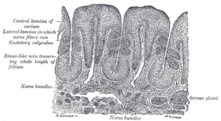
Mucous gland, also known as muciparous glands, are found in several different parts of the body, and they typically stain lighter than serous glands during standard histological preparation. Most are multicellular, but goblet cells are single-celled glands.

The labial glands are minor salivary glands situated between the mucous membrane and the orbicularis oris around the orifice of the mouth.

The superior labial vein is the vein receiving blood from the upper lip.

The inferior labial vein is the vein receiving blood from the lower lip.
Crossing the under surface of the sphenoid, the sphenopalatine artery ends on the nasal septum as the posterior septal branches; these anastomose with the ethmoidal arteries and the septal branch of the superior labial; one branch descends in a groove on the vomer to the incisive canal and anastomoses with the descending palatine artery.


