The superior labial branches (labial branches), the largest and most numerous, descend behind the Quadratus labii superioris, and are distributed to the skin of the upper lip, the mucous membrane of the mouth, and labial glands.

A mucous membrane or mucosa is a membrane that lines various cavities in the body and covers the surface of internal organs. It consists of one or more layers of epithelial cells overlying a layer of loose connective tissue. It is mostly of endodermal origin and is continuous with the skin at various body openings such as the eyes, ears, inside the nose, inside the mouth, lip, vagina, the urethral opening and the anus. Some mucous membranes secrete mucus, a thick protective fluid. The function of the membrane is to stop pathogens and dirt from entering the body and to prevent bodily tissues from becoming dehydrated.

In animal anatomy, the mouth, also known as the oral cavity, buccal cavity, or in Latin cavum oris, is the opening through which many animals take in food and issue vocal sounds. It is also the cavity lying at the upper end of the alimentary canal, bounded on the outside by the lips and inside by the pharynx and containing in higher vertebrates the tongue and teeth. This cavity is also known as the buccal cavity, from the Latin bucca ("cheek").
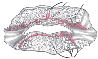
The labial glands are minor salivary glands situated between the mucous membrane and the orbicularis oris around the orifice of the mouth.
They are joined, immediately beneath the orbit, by filaments from the facial nerve, forming with them the infraorbital plexus.

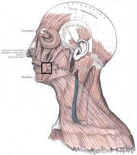
The facial nerve is the seventh cranial nerve, or simply CN VII. It emerges from the pons of the brainstem, controls the muscles of facial expression, and functions in the conveyance of taste sensations from the anterior two-thirds of the tongue. The nerves typically travels from the pons through the facial canal in the temporal bone and exits the skull at the stylomastoid foramen. It arises from the brainstem from an area posterior to the cranial nerve VI and anterior to cranial nerve VIII.

The superior labial branches descend behind the Quadratus labii superioris, and are distributed to the skin of the upper lip, the mucous membrane of the mouth, and labial glands. They are joined, immediately beneath the orbit, by filaments from the facial nerve, forming with them the infraorbital plexus.
This page is based on this
Wikipedia article Text is available under the
CC BY-SA 4.0 license; additional terms may apply.
Images, videos and audio are available under their respective licenses.

The facial artery is a branch of the external carotid artery that supplies structures of the superficial face.

The maxillary nerve (CN V2) is one of the three branches or divisions of the trigeminal nerve, the fifth (V) cranial nerve. It comprises the principal functions of sensation from the maxillary, nasal cavity, sinuses, the palate and subsequently that of the mid-face, and is intermediate, both in position and size, between the ophthalmic nerve and the mandibular nerve.
In facial anatomy, the modiolus is a chiasma of facial muscles held together by fibrous tissue, located lateral and slightly superior to each angle of the mouth. It is important in moving the mouth, facial expression and in dentistry. It is extremely important in relation to stability of lower denture, because of the strength and variability of movement of the area. It derives its motor nerve supply from the facial nerve, and its blood supply from labial branches of the facial artery.

Kiesselbach's plexus, which lies in Kiesselbach's area, Kiesselbach's triangle, or Little's area, is a region in the anteroinferior part of the nasal septum where four arteries anastomose to form a vascular plexus. The arteries are:

The inferior alveolar artery is an artery of the face. It is a branch of the first portion of the maxillary artery.
The inferior labial artery arises near the angle of the mouth as a branch of the facial artery; it passes upward and forward beneath the triangularis and, penetrating the orbicularis oris, runs in a tortuous course along the edge of the lower lip between this muscle and the mucous membrane.
The superior labial artery is larger and more egregious than the inferior labial artery.

Mental nerve is a sensory nerve which provides sensation to the front of the chin and lower lip as well as the labial gingivae of the mandibular anterior teeth and the premolars. It is a branch of the posterior trunk of the inferior alveolar nerve, which is itself a branch of the mandibular division of the trigeminal nerve.

The perineal nerve is a nerve arising from the pudendal nerve that supplies the perineum.

The angular vein is the upper most segment of the facial vein, above its junction with the superior labial vein. It is formed by the junction of the supratrochlear vein and supraorbital vein, runs obliquely downward by the side of the nose, passes under zygomaticus major and joins with the superior labial vein.

The posterior scrotal branches or posterior labial branches are two in number, medial and lateral. They are branches of the perineal nerve, which is itself is a branch of the pudendal nerve. The pudendal nerve arises from spinal roots S2 through S4, travels through the pudendal canal on the fascia of the obturator internus muscle, and gives off the perineal nerve in the perineum. The major branch of the perineal nerve is the posterior scrotal/posterior labial.
In anatomy, arterial tree is used to refer to all arteries and/or the branching pattern of the arteries. This article regards the human arterial tree. Starting from the aorta:

The submental artery is a branch of the facial artery that runs on the underside of the chin.

The deep external pudendal artery, is one of the pudendal arteries that is more deeply seated than the superficial external pudendal artery, passes medially across the pectineus and the adductor longus muscles; it is covered by the fascia lata, which it pierces at the medial side of the thigh, and is distributed, in the male, to the integument of the scrotum and perineum, in the female to the labia majora; its branches anastomose with the scrotal branches of the perineal artery.
The posterior labial nerves are branches of the pudendal nerve.
The superior labial vein is the vein receiving blood from the upper lip.
Crossing the under surface of the sphenoid the sphenopalatine artery ends on the nasal septum as the posterior septal branches; these anastomose with the ethmoidal arteries and the septal branch of the superior labial; one branch descends in a groove on the vomer to the incisive canal and anastomoses with the descending palatine artery.

In human anatomy, the mouth is the first portion of the alimentary canal that receives food and produces saliva. The oral mucosa is the mucous membrane epithelium lining the inside of the mouth.

