Klebsiella

Last updated

Klebsiella
Klebsiella-pneumoniae.jpg
Electromicrograph of Klebsiella pneumoniae
Scientific classification OOjs UI icon edit-ltr.svg
Domain: Bacteria
Phylum: Pseudomonadota
Class: Gammaproteobacteria
Order: Enterobacterales
Family: Enterobacteriaceae
Genus: Klebsiella
Trevisan 1885 [1]
Species [2]

Klebsiella is a genus of Gram-negative, oxidase-negative, rod-shaped bacteria with a prominent polysaccharide-based capsule. [3]

Contents

Klebsiella is named after German-Swiss microbiologist Edwin Klebs (1834–1913). Carl Friedlander described Klebsiella bacillus which is why it was termed Friedlander bacillus for many years. The species of Klebsiella are all gram-negative and usually non-motile. They tend to be shorter and thicker when compared to others in the family Enterobacteriaceae.

Klebsiella species are found everywhere in nature. This is thought to be due to distinct sublineages developing specific niche adaptations, with associated biochemical adaptations which make them better suited to a particular environment. They can be found in water, soil, plants, insects and other animals including humans, [4] [5] including as part of the human and animal's normal flora in the nose, mouth and intestines.

List of species

Features

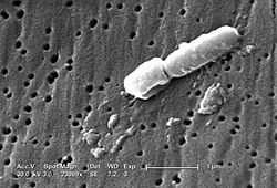
Klebsiella bacteria tend to be rounder and thicker than other members of the family Enterobacteriaceae. They typically occur as straight rods with rounded or slightly pointed ends. They can be found singly, in pairs, or in short chains. Diplobacillary forms are commonly found in vivo. [6]

They have no specific growth requirements and grow well on standard laboratory media, but grow best between 35 and 37 °C and at pH 7.2. [7] The species are facultative anaerobes, and most strains can survive with citrate and glucose as their sole carbon sources and ammonia as their sole nitrogen source. [6]

Members of the genus produce a prominent capsule, or slime layer, which can be used for serologic identification, but molecular serotyping may replace this method. [8]

Members of the genus Klebsiella typically express two types of antigens on their cell surfaces. The first, O antigen, is a component of the lipopolysaccharide (LPS), of which 9 varieties exist. The second is K antigen, a capsular polysaccharide with more than 80 varieties. [9] Both contribute to pathogenicity and form the basis for serogrouping. Based on those two major antigenic determinants several vaccines have been designed. [10]

In humans

Klebsiella species are routinely found in the human nose, mouth, and gastrointestinal tract as normal flora; however, they can also behave as opportunistic human pathogens. [6] Klebsiella species are known to also infect a variety of other animals, both as normal flora and opportunistic pathogens. [4]

Klebsiella organisms can lead to a wide range of disease states, notably pneumonia, urinary tract infections, sepsis, meningitis, diarrhea, peritonitis and soft tissue infections. [6] [11] Klebsiella species have also been implicated in the pathogenesis of ankylosing spondylitis and other spondyloarthropathies. [12] The majority of human Klebsiella infections are caused by K. pneumoniae , followed by K. oxytoca . Infections are more common in the very young, very old, and those with other underlying diseases, such as cancer, [4] and most infections involve contamination of an invasive medical device. [6]

During the last 40 years, many trials for constructing effective K. pneumoniae vaccines have been tried, [13] and new techniques were followed to construct vaccines against Klebsiella. [14] However, currently, no Klebsiella vaccine has been licensed for use in the US. K. pneumoniae is the most common cause of nosocomial respiratory tract and premature intensive care infections, and the second-most frequent cause of Gram-negative bacteraemia and urinary tract infections . Drug-resistant isolates remain an important hospital-acquired bacterial pathogen, add significantly to hospital stays, and are especially problematic in high-impact medical areas such as intensive care units. This antimicrobial resistance is thought to be attributable mainly to multidrug efflux pumps. [15] The ability of K. pneumoniae to colonize the hospital environment, including carpeting, sinks, flowers, and various surfaces, as well as the skin of patients and hospital staff, has been identified as a major factor in the spread of hospital-acquired infections. [4]

In animals

In addition to certain Klebsiella spp. being discovered as human pathogens, others such as K. variicola have been identified as emerging pathogens in humans and animals alike. For instance, K. variicola has been identified as one of the causes of bovine mastitis. [16] [17]

In plants

In plant systems, Klebsiella can be found in a variety of plant hosts. K. pneumoniae and K. oxytoca are able to fix atmospheric nitrogen into a form that can be used by plants, thus are called associative nitrogen fixers or diazotrophs. [5] [18] The bacteria attach strongly to root hairs and less strongly to the surface of the zone of elongation and the root cap mucilage. [19] They are bacteria of interest in an agricultural context, due to their ability to increase crop yields under agricultural conditions. [20] Their high numbers in plants are thought to be at least partly attributable to their lack of a flagellum, as flagella are known to induce plant defenses. [21] Additionally, K. variicola is known to associate with a number of different plants including banana trees, [22] sugarcane [23] and has been isolated from the fungal gardens of leaf-cutter ants. [24]

See also

Related Research Articles

<span class="mw-page-title-main">Enterobacteriaceae</span> Family of bacteria

Enterobacteriaceae is a large family of Gram-negative bacteria. It includes over 30 genera and more than 100 species. Its classification above the level of family is still a subject of debate, but one classification places it in the order Enterobacterales of the class Gammaproteobacteria in the phylum Pseudomonadota. In 2016, the description and members of this family were emended based on comparative genomic analyses by Adeolu et al.

<i>Klebsiella pneumoniae</i> Species of bacterium

Klebsiella pneumoniae is a Gram-negative, non-motile, encapsulated, lactose-fermenting, facultative anaerobic, rod-shaped bacterium. It appears as a mucoid lactose fermenter on MacConkey agar.

<i>Serratia</i> Genus of bacteria

Serratia is a genus of Gram-negative, facultatively anaerobic, rod-shaped bacteria of the family Enterobacteriaceae. According to the List of Prokaryotic names with Standing Nomenclature (LPSN), there are currently 19 species of Serratia that are credibly published with accurate names as of 2020: S. aquatilis, S. entomophila, S. ficaria, S. fonticola, S. grimesii, S. liquefaciens, S. marcescens, S. microhaemolytica, S. myotis, S. nematodiphila, S. odoriferae, S. oryzae, S. plymuthica, S. proteamaculans, S. quinivorans corrig, S. rubidaea, S. symbiotica, S. ureilytica, S. vespertilionis. They are typically 1–5 μm in length, do not produce spores, and can be found in water, soil, plants, and animals. Some members of this genus produce a characteristic red pigment, prodigiosin, and can be distinguished from other members of the order Enterobacterales by their unique production of three enzymes: DNase (nucA), lipase, and gelatinase (serralysin). Serratia was thought to be a harmless environmental bacteria until it was discovered that the most common species in the genus, S. marcescens, is an opportunistic pathogen of many animals, including humans. In humans, S. marcescens is mostly associated with nosocomial, or hospital-acquired, infections, but can also cause urinary tract infections, pneumonia, and endocarditis. S. marcescens is frequently found in showers, toilet bowls, and around wet tiles as a pinkish to red biofilm but only causes disease in immunocompromised individuals. Aside from S. marcescens, some rare strains of the Serratia species – S. plymuthica, S. liquefaciens, S. rubidaea, and S. odoriferae – have been shown to cause infection such as osteomyelitis and endocarditis.

<i>Proteus</i> (bacterium) Genus of bacteria

Proteus is a genus of Gram-negative bacteria. It is a rod shaped, aerobic and motile bacteria, which is able to migrate across surfaces due its “swarming” characteristic in temperatures between 20 and 37 °C. Their size generally ranges from 0.4–0.8 μm in diameter and 1.0–3.0 μm in length. They tend to have an ammonia smell. Proteus bacilli are widely distributed in nature as saprophytes, being found in decomposing animal matter, sewage, manure soil, the mammalian intestine, and human and animal feces. They are opportunistic pathogens, commonly responsible for urinary and septic infections, often nosocomial.

<span class="mw-page-title-main">Bacterial capsule</span> Polysaccharide layer that lies outside the cell envelope in many bacteria

The bacterial capsule is a large structure common to many bacteria. It is a polysaccharide layer that lies outside the cell envelope, and is thus deemed part of the outer envelope of a bacterial cell. It is a well-organized layer, not easily washed off, and it can be the cause of various diseases.

<span class="mw-page-title-main">Coliform bacteria</span> Group of bacterial species

Coliform bacteria are defined as either motile or non-motile Gram-negative non-spore forming bacilli that possess β-galactosidase to produce acids and gases under their optimal growth temperature of 35–37 °C. They can be aerobes or facultative aerobes, and are a commonly used indicator of low sanitary quality of foods, milk, and water. Coliforms can be found in the aquatic environment, in soil and on vegetation; they are universally present in large numbers in the feces of warm-blooded animals as they are known to inhabit the gastrointestinal system. While coliform bacteria are not normally causes of serious illness, they are easy to culture, and their presence is used to infer that other pathogenic organisms of fecal origin may be present in a sample, or that said sample is not safe to consume. Such pathogens include disease-causing bacteria, viruses, or protozoa and many multicellular parasites.

<span class="mw-page-title-main">Serotype</span> Distinct variation within a species of bacteria or virus or among immune cells

A serotype or serovar is a distinct variation within a species of bacteria or virus or among immune cells of different individuals. These microorganisms, viruses, or cells are classified together based on their surface antigens, allowing the epidemiologic classification of organisms to a level below the species. A group of serovars with common antigens is called a serogroup or sometimes serocomplex.

<span class="mw-page-title-main">Carbapenem</span> Class of highly effective antibiotic agents

Carbapenems are a class of very effective antibiotic agents most commonly used for treatment of severe bacterial infections. This class of antibiotics is usually reserved for known or suspected multidrug-resistant (MDR) bacterial infections. Similar to penicillins and cephalosporins, carbapenems are members of the beta-lactam antibiotics drug class, which kill bacteria by binding to penicillin-binding proteins, thus inhibiting bacterial cell wall synthesis. However, these agents individually exhibit a broader spectrum of activity compared to most cephalosporins and penicillins. Furthermore, carbapenems are typically unaffected by emerging antibiotic resistance, even to other beta-lactams.

<i>Klebsiella oxytoca</i> Species of bacterium

Klebsiella oxytoca is a Gram-negative, rod-shaped bacterium that is closely related to K. pneumoniae, from which it is distinguished by being indole-positive; it also has slightly different growth characteristics in that it is able to grow on melezitose, but not 3-hydroxybutyrate. It was first described in 1886 when it was isolated from sour milk and named Bacillus oxytocus perniciosus.

Klebsiella aerogenes, previously known as Enterobacter aerogenes, is a Gram-negative, oxidase-negative, catalase-positive, citrate-positive, indole-negative, rod-shaped bacterium. Capable of motility via peritrichous flagella, the bacterium is approximately 1–3 microns in length.

<i>Morganella morganii</i> Species of bacterium

Morganella morganii is a species of Gram-negative bacteria. It has a commensal relationship within the intestinal tracts of humans, mammals, and reptiles as normal flora. Although M. morganii has a wide distribution, it is considered an uncommon cause of community-acquired infection, and it is most often encountered in postoperative and other nosocomial infections, such as urinary tract infections.

<span class="mw-page-title-main">Mastitis in dairy cattle</span>

Bovine mastitis is the persistent, inflammatory reaction of the udder tissue due to physical trauma or microorganisms infections. Mastitis, a potentially fatal mammary gland infection, is the most common disease in dairy cattle in the United States and worldwide. It is also the most costly disease to the dairy industry. Milk from cows suffering from mastitis has an increased somatic cell count. Prevention and control of mastitis requires consistency in sanitizing the cow barn facilities, proper milking procedure and segregation of infected animals. Treatment of the disease is carried out by penicillin injection in combination with sulphar drug.

<i>Raoultella</i> Genus of bacteria

The genus Raoultella is composed of Gram-negative, oxidase-negative, aerobic, nonmotile, capsulated, facultatively anaerobic rods in the family Enterobacteriaceae. It is named after the French bacteriologist Didier Raoult. The existence of this genus renders the genus Klebsiella paraphyletic, and it has been repeatedly proposed to consider Raoultella a junior synonym of Klebsiella.

<span class="mw-page-title-main">New Delhi metallo-beta-lactamase 1</span> Enzyme

NDM-1 is an enzyme that makes bacteria resistant to a broad range of beta-lactam antibiotics. These include the antibiotics of the carbapenem family, which are a mainstay for the treatment of antibiotic-resistant bacterial infections. The gene for NDM-1 is one member of a large gene family that encodes beta-lactamase enzymes called carbapenemases. Bacteria that produce carbapenemases are often referred to in the news media as "superbugs" because infections caused by them are difficult to treat. Such bacteria are usually sensitive only to polymyxins and tigecycline.

Pathogenic <i>Escherichia coli</i> Strains of E. coli that can cause disease

Escherichia coli is a gram-negative, rod-shaped bacterium that is commonly found in the lower intestine of warm-blooded organisms (endotherms). Most E. coli strains are harmless, but pathogenic varieties cause serious food poisoning, septic shock, meningitis, or urinary tract infections in humans. Unlike normal flora E. coli, the pathogenic varieties produce toxins and other virulence factors that enable them to reside in parts of the body normally not inhabited by E. coli, and to damage host cells. These pathogenic traits are encoded by virulence genes carried only by the pathogens.

Carbapenem-resistant Enterobacteriaceae (CRE) or carbapenemase-producing Enterobacteriaceae (CPE) are Gram-negative bacteria that are resistant to the carbapenem class of antibiotics, considered the drugs of last resort for such infections. They are resistant because they produce an enzyme called a carbapenemase that disables the drug molecule. The resistance can vary from moderate to severe. Enterobacteriaceae are common commensals and infectious agents. Experts fear CRE as the new "superbug". The bacteria can kill up to half of patients who get bloodstream infections. Tom Frieden, former head of the Centers for Disease Control and Prevention has referred to CRE as "nightmare bacteria". Examples of enzymes found in certain types of CRE are KPC and NDM. KPC and NDM are enzymes that break down carbapenems and make them ineffective. Both of these enzymes, as well as the enzyme VIM have also been reported in Pseudomonas.

<span class="mw-page-title-main">Eravacycline</span> Chemical compound

Eravacycline is a synthetic halogenated tetracycline class antibiotic by Tetraphase Pharmaceuticals. It is closely related to tigecycline. It has a broad spectrum of activity including many multi-drug resistant strains of bacteria. Phase III studies in complicated intra-abdominal infections (cIAI) and complicated urinary tract infections (cUTI) were recently completed with mixed results. Eravacycline was granted fast track designation by the FDA and is currently available in USA.

Klebsiella variicola is a species of bacteria which was originally identified as a benign endosymbiont in plants, but has since been associated with disease in humans and cattle as well.

ESKAPE is an acronym comprising the scientific names of six highly virulent and antibiotic resistant bacterial pathogens including: Enterococcus faecium, Staphylococcus aureus, Klebsiella pneumoniae, Acinetobacter baumannii, Pseudomonas aeruginosa, and Enterobacter spp. The acronym is sometimes extended to ESKAPEE to include Escherichia coli. This group of Gram-positive and Gram-negative bacteria can evade or 'escape' commonly used antibiotics due to their increasing multi-drug resistance (MDR). As a result, throughout the world, they are the major cause of life-threatening nosocomial or hospital-acquired infections in immunocompromised and critically ill patients who are most at risk. P. aeruginosa and S. aureus are some of the most ubiquitous pathogens in biofilms found in healthcare. P. aeruginosa is a Gram-negative, rod-shaped bacterium, commonly found in the gut flora, soil, and water that can be spread directly or indirectly to patients in healthcare settings. The pathogen can also be spread in other locations through contamination, including surfaces, equipment, and hands. The opportunistic pathogen can cause hospitalized patients to have infections in the lungs, blood, urinary tract, and in other body regions after surgery. S. aureus is a Gram-positive, cocci-shaped bacterium, residing in the environment and on the skin and nose of many healthy individuals. The bacterium can cause skin and bone infections, pneumonia, and other types of potentially serious infections if it enters the body. S. aureus has also gained resistance to many antibiotic treatments, making healing difficult. Because of natural and unnatural selective pressures and factors, antibiotic resistance in bacteria usually emerges through genetic mutation or acquires antibiotic-resistant genes (ARGs) through horizontal gene transfer - a genetic exchange process by which antibiotic resistance can spread.

References

  1. Trevisan, V. "Caratteri di alcuni nuovi generi di Batteriaceae [Characteristics of some new genera of Bacteriaceae]." Atti. Accad. Fis.-Med.-Stat. Milano (Ser 4) (1885) 3:92-106.
  2. "Klebsiella". NCBI taxonomy. Bethesda, MD: National Center for Biotechnology Information. Retrieved 24 April 2019.
  3. Ryan KJ; Ray CG, eds. (2004). Sherris Medical Microbiology (4th ed.). McGraw Hill. p. 370. ISBN   978-0-8385-8529-0.
  4. 1 2 3 4 Bagley S (1985). "Habitat association of Klebsiella species". Infect Control. 6 (2): 52–8. doi:10.1017/S0195941700062603. PMID   3882590. S2CID   22799991.
  5. 1 2 Brisse S, Grimont F, Grimont PD (2006). Prokaryotes . New York, NY: Springer New York. pp.  159–196. ISBN   9783540325246.
  6. 1 2 3 4 5 Ristuccia, Patricia A; Cunha Burke A (1984). "Klebsiella". Topics in Clinical Microbiology. 5 (7): 343–348. JSTOR   30144997.
  7. Ristucci, Patricia; Cunha, Burke (July 1984). "Klebsiella". Infection Control. 5 (7): 343–348. doi:10.1017/S0195941700060549. JSTOR   30144997. PMID   6564087. S2CID   248999074.
  8. Brisse, Sylvain; S Issenhuth-Jeanjean; P AD Grimont (2004). "Molecular Serotyping of Klebsiella Species Isolates by Restriction of the Amplified Capsular Antigen Gene Cluster". Journal of Clinical Microbiology. 42 (8): 3388–3398. doi:10.1128/jcm.42.8.3388-3398.2004. PMC   497587 . PMID   15297473.
  9. Podschun, R; Ullmann, U (October 1998). "Klebsiella spp. as Nosocomial Pathogens: Epidemiology, Taxonomy, Typing Methods, and Pathogenicity Factors". Clinical Microbiology Reviews . 11 (4): 589–603. PMC   88898 .
  10. Ahmad, TA; El-Sayed, LH; Medhat, H; Hessen, A; El-Ashry, ESH (2012). "Development of immunization trials against Klebsiella pneumoniae". Vaccine. 30 (14): 2411–2420. doi:10.1016/j.vaccine.2011.11.027. PMID   22100884.
  11. Podschun R, Ullmann U (1998). "Klebsiella spp. as nosocomial pathogens: epidemiology, taxonomy, typing methods, and pathogenicity factors". Clin Microbiol Rev. 11 (4): 589–603. doi:10.1128/CMR.11.4.589. PMC   88898 . PMID   9767057.
  12. Sieper, Joachim; Braun, Jürgen (2011). Ankylosing Spondylitis in Clinical Practice. London: Springer-Verlag. p. 9. ISBN   978-0-85729-179-0.
  13. Ahmad, TA; El-Sayed, LH; Haroun,M; Hussin, A; El-Ashry, ESH (2012). "Development of immunization trials against Klebsiella pneumoniae". Vaccine. 30 (14): 2411–2420. doi:10.1016/j.vaccine.2011.11.027. PMID   22100884.
  14. Ahmad, TA; El-Sayed, LH; Haroun,M; Hussin, A; El-Ashry, ESH (2012). "Development of a new trend conjugate vaccine for the prevention of Klebsiella pneumoniae". Infectious Disease Reports. 4 (2): e33. doi: 10.4081/idr.2012.e33 . PMC   3892636 . PMID   24470947.
  15. Ogawa, Wakano; Li, Dai-Wei; Yu, Ping; Begum, Anowara; Mizushima, Tohru; Kuroda, Teruo; Tsuchiya, Tomofusa (2005). "Multidrug resistance in Klebsiella pneumoniae MGH78578 and cloning of genes responsible for the resistance". Biological & Pharmaceutical Bulletin. 28 (8): 1505–1508. doi: 10.1248/bpb.28.1505 . PMID   16079502.
  16. Davidson, Fraser W.; Whitney, Hugh G.; Tahlan, Kapil (2015-10-29). "Genome Sequences of Klebsiella variicola Isolates from Dairy Animals with Bovine Mastitis from Newfoundland, Canada". Genome Announcements. 3 (5): e00938–15. doi:10.1128/genomeA.00938-15. ISSN   2169-8287. PMC   4566169 . PMID   26358587.
  17. Podder, Milka P.; Rogers, Laura; Daley, Peter K.; Keefe, Greg P.; Whitney, Hugh G.; Tahlan, Kapil (2014). "Klebsiella Species Associated with Bovine Mastitis in Newfoundland". PLOS ONE. 9 (9): e106518. Bibcode:2014PLoSO...9j6518P. doi: 10.1371/journal.pone.0106518 . PMC   4152263 . PMID   25180510.
  18. Cakmaki ML, Evans HJ, Seidler RJ (1981). "Characteristics of a nitrogen-fixing Klebsiella oxytoca isolated from wheat roots". Plant and Soil. 61 (1–2): 53–64. doi:10.1007/BF02277362. S2CID   21625282.
  19. Haahtela, K; Laakso T; Korhonen TK (1986). "Associative nitrogen fixation by Klebsiella spp.: Adhesion sites and inoculation effects on grass roots". Applied and Environmental Microbiology. 52 (5): 1074–1079. Bibcode:1986ApEnM..52.1074H. doi:10.1128/aem.52.5.1074-1079.1986. PMC   239175 . PMID   16347205.
  20. Riggs, PJ; Chelius MK; Iniguez AL; Kaeppler SM; Triplett EW (2001). "Enhanced maize productivity by inoculation with diazotrophic bacteria". Australian Journal of Plant Physiology. 28 (9): 829–836. doi:10.1071/PP01045.
  21. Fouts, Derrick E.; Tyler, Heather L.; Deboy, Robert T.; Daugherty, Sean; Ren, Qinghu; Badger, Jonathan H.; Durkin, Anthony S.; Huot, Heather; Shrivastava, Susmita; Kothari, Sagar; Dodson, Robert J.; Mohamoud, Yasmin; Khouri, Hoda; Roesch, Luiz F. W.; Krogfelt, Karen A.; Struve, Carsten; Triplett, Eric W.; Methé, Barbara A. (2008). "Complete Genome Sequence of the N2-Fixing Broad Host Range Endophyte Klebsiella pneumoniae 342 and Virulence Predictions Verified in Mice". PLOS Genetics. 4 (7): e1000141. doi: 10.1371/journal.pgen.1000141 . PMC   2453333 . PMID   18654632. Open Access logo PLoS transparent.svg
  22. Rosenblueth, Mónica; Martínez, Lucía; Silva, Jesús; Martínez-Romero, Esperanza (2004-01-01). "Klebsiella variicola, A Novel Species with Clinical and Plant-Associated Isolates". Systematic and Applied Microbiology. 27 (1): 27–35. doi:10.1078/0723-2020-00261. PMID   15053318. S2CID   40606728.
  23. Wei, Chun-Yan; Lin, Li; Luo, Li-Jing; Xing, Yong-Xiu; Hu, Chun-Jin; Yang, Li-Tao; Li, Yang-Rui; An, Qianli (2013-11-19). "Endophytic nitrogen-fixing Klebsiella variicola strain DX120E promotes sugarcane growth". Biology and Fertility of Soils. 50 (4): 657–666. doi:10.1007/s00374-013-0878-3. ISSN   0178-2762. S2CID   15594459.
  24. Pinto-Tomás, Adrián A.; Anderson, Mark A.; Suen, Garret; Stevenson, David M.; Chu, Fiona S. T.; Cleland, W. Wallace; Weimer, Paul J.; Currie, Cameron R. (2009-11-20). "Symbiotic Nitrogen Fixation in the Fungus Gardens of Leaf-Cutter Ants". Science. 326 (5956): 1120–1123. Bibcode:2009Sci...326.1120P. doi:10.1126/science.1173036. ISSN   0036-8075. PMID   19965433. S2CID   3119587.