| Instrument | Uses |
|---|
| Toric Marker | to mark 0 to 180 degree reference mark for Toric IOL implant |
| Pre-chopper | to chop lens into pieces before implantation new lens and reduce phaco time |
| Spectacles (glasses) | to correct refractive errors of the eye; not invasive |
| Contact lenses | to correct refractive errors of the eye; a little invasive |
| Phoropter | used in refraction testing |
| Tonometers | used to determine the intraocular pressure (IOP) - useful in glaucoma; video link for various types of tonometers. |
| Speculum: | to keep the eyes open during any operation |
| Universal eye speculum | -do-; heavy instrument and can not keep eyelashes out of the operating field |
| •Guarded eye speculum (left and right) | -do-; heavy instrument but can keep eyelashes out of the operating field with its "guard" and hence left or right ones are required |
| •Wire Speculum | to keep the eyes open during any operation; light wire instrument |
| Needle holders: | holding the needle in position while applying sutures |
| •Silcock's needle holder | -do-; has a catch and is used for heavier gauge needles; used mainly for skin, muscle and corneal incisions |
| •Arruga's needle holder | -do-; has a catch (lock) and is used for heavier gauge needles (thicker than 6–0); used mainly for skin, muscle and corneal incisions |
| •Barraquer's needle holder | -do-; small instrument with a spring action with or without a catch used for finer gauge needles (5-0 or finer); used mainly for intraoccular incisions |
| Forceps: | to hold anything |
| •Artery forceps (haemostat) | medium-sized, with a serrated tip and a catch; used to hold bleeding vessels and compress them in order to make them stop bleeding and also to hold or crush structures. |
| •Fixation forceps | has a few teeth at the tip; for holding structures and restricting their movement or to hold small swabs |
| •Plain dissecting forceps | blunt untoothed with a serrated tip; for holding structures and restricting their movement or to hold small swabs |
| •Iris forceps | fine tipped (straight or otherwise) with small teeth; to hold the iris tissue during procedures |
| •Elschnig's intracapsular forceps | fine untoothed forceps for holding tissue, swabs, sutures, etc.; removing things like clots, capsule fragments, lens, etc.; used in cataract surgery |
| •Arruga's intracapsular forceps | fine untoothed forceps holding tissue, swabs, sutures, etc.; removing things like clots, capsule fragments, lens, etc.; used in cataract surgery |
| •Colibri forceps | fine toothed forceps for holding flaps of cornea or sclera and rarely the iris |
| •Saint Martin's forceps | holding flaps of cornea or sclera and rarely the iris |
| •Superior rectus holding forceps | specially curved (to fit into the orbit of the eye) forceps for catching hold of the muscle bellies of the intraorbital muscles and sutures |
| •Suture tier forceps | fine limbed untoothed forceps to hold fine sutures or hairs |
| •Capsulotomy forceps | to tear the anterior capsule of the lens during cataract surgery |
| •Disc holding forceps | used in glaucoma surgery (obsolete) |
| •Capsulorhexis forceps | fine sharp-tipped untoothed forceps for doing a continuous curvilinear incision and removal of the anterior capsule of the lens ("continuous curvilinear capsulorhexis - ccc") |
| •MacPherson's forceps | fine sharp-tipped untoothed forceps with an angulation for holding parts of the lens, the intraocular lens, 10-0 (very fine) sutures, etc. |
| •Chalazion forceps (clamp) | self-retaining with discoid ends; used to hold and prevent a chalazion from bleeding during its surgery |
| Diamond knife | used to perform microincisions on the cornea in the Radial keratotomy and Mini Asymmetric Radial Keratotomy (M.A.R.K.) |
| •Epilation forceps (Cilia forceps) | stout flat-ended blunt forceps with a thickened end to remove eyelashes |
| •Entropion forceps | self-retaining with big discoid ends used to hold and prevent an entropion from bleeding during its surgery |
| Chalazion scoop | to remove the granulation tissue from a chalazion during surgery |
| Entropion clamp | right and left varieties exist; large clamp with two limbs; self-retaining with big discoid ends used to hold and prevent an entropion from bleeding during its surgery |
| Nettleship's punctum dilator | to dilate the lacrimal punctum of the lacrimal apparatus of the eye for syringing or operations |
| Cystotome | a 26 gauge needle bent twice used for incising the anterior capsule of the lens in lens extraction |
| Wire vectis | a loop of wire attached to a stack used to extract cataract affected lenses |
| Irrigating vectis | a small hollow instrument with a used to introduce fluid into the anterior chamber to raise its pressure to aid cataract extraction [12] |
| Canula | used to carry fluid |
| •Irrigation-aspiration two-way canula | effectively two small canulae fitted together, one to introduce fluid and the other to extract the cortical materials, blood, etc. in eye operations |
| •Lacrimal canula | small curved canula the size of a syringe needle used to introduce fluids or drugs into the nasolacrimal passage to test its patency or during surgery (dacrocystography, dacrocystectomy, dacryocystorhinostomy(DCR), etc. |
| Lang's lacrimal dissector with scoop | for blunt dissections and cleaning during operations like dacryocystorhinostomy |
| Rougine | dissection of lacrimal sac |
| Retractor | to pull and hold overlying tissue out of the operating field |
| •Muller's self retaining adjustable haemostatic retractor | -do-; self retaining haemostatic |
| •Cat's paw retractor | -do- |
| •Desmarre's lid retractor | -do-; specially for noncooperative patients and to see the fornices (see human eye) |
| Bone punch | to fracture pieces from a thin bone in facial surgery and during operations like dacryocystorhinostomy |
| Evisceration spoon or scoop | removing all the contents of the eyeball during evisceration (complete removal of all structures within the eye in diseases like endophthalmitis |
| Lid plate | flat large instrument that has a groove and is placed between the lid and globe of the eye to provide a solid support for eyelid surgery |
| Hammer, chisel and bone gouge | bone cutting and shaping |
| Bowmen's discussion needle | microsurgery of the lens capsule [13] |
| Knives | to cut structures |
| •Surgical scalpel with small blades | general purpose instrument |
| •von Graefe's cataract knife | cutting out of the anterior chamber from the inside through the limbus |
| •Tookes' knife (Sclero-corneal splitter) | making sclerocorneal tunnels in "small incision cataract surgery (SICS)" and keratoplasty |
| •Crescent knife (Sclero-corneal splitter) | making sclerocorneal tunnels in "small incision cataract surgery" |
| •Angular keratome | making sclerocorneal tunnels in "small incision cataract surgery"; larger one used to increase the size of the incision |
| •Side-port blade | making sclerocorneal "side port" (a secondary tunnel) tunnels in "small incision cataract surgery" |
| •Beer's knife | incise the conjunctiva or the eyelid skin |
| •Keratotome | small triangular blade with two sharp edges used to incise the limbus (sclerocorneal junction) |
| •Zeigler's knife | very tiny knife for intaoccular maneuvers specially when space is less |
| Scissors | - |
| •Conjunctival sac scissors | flat small curved scissors to cut the conjunctive |
| •Corneal spring scissors | medium spring-open used to cut the external side of the cornea, fine sutures; iris, etc. |
| •de' Wecker's iris scissors | small slender spring-open scissors for intraocular maneuvers (iris and deeper and more delicate structures); has two wings to operate it and one sharp and one blunt blade. |
| •Vannas' scissors | small slender spring-open scissors for intraocular maneuvers (iris and deeper and more delicate structures); has two wings to operate it and one sharp and one blunt blade. |
| •Enucleation scissors | thick scissors used to cut the optic nerve in enucleation operation |
| Bowman's lacrimal probe | probing the nasolacrimal duct |
| Lens expressor | used to force out the lens in extracapsular or intracapsular cataract extraction |
| McNamar's spoon | used to force out the lens in intracapsular cataract extraction |
| Iris repositor | two limbed instrument used to remove the iris during posterior chamber maneuvers |
| Sinsky's hook intraocular lens dialler | angulated round hook with a handle used in insertion of an intraocular lens |
| Strabismus hook | muscle hook or squint hook; sharp tip or knobbed tip; used in squint surgery |
| Foreign body spud and needle | Spud to remove superficial and needle for the deep foreign bodies in the eye |
| Elliot's trephine with handle | used in corneal donation (eye donation) to cut out the cornea in a circular fashion |
| Castroveijo's calipers | various measurements are taken |
| Castroveijo's corneal trephine | used in corneal donation (eye donation) to cut out the cornea in a circular fashion |
| Pin-hole | testing visual acuity |
| Red green goggles | (red - right side & green - left side) used in Worth 4 dot test, diplopia testing |
| Prisms | to measure the degree of squints; in other instruments; refractive correction; etc. |
| Placido's disc | to assess the condition of the corneal surface |
| Retinoscope | objective determination of refractive error and for looking inside the eye |
| Loupe | used to search for magnified examination of the anterior segment of the eye (uniocular or binocular) |
| Jackson's cross cylinder | used to check the power and axis of a cylindrical lens |
| Maddox rod | used to test for latent squint and retinal function |
| Refraction box | has lenses of different powers for refraction testing |
| Slit lamp bio microscope | used for examining the anteriorly placed structures the eye; video link |
| Charts for vision | - |
| •Distant vision | to determine visual acuity of distant vision |
| ••Snellen's distant vision chart | -do-; for those who can read in English |
| ••Regional language charts | -do-; for those who can read in their local language |
| ••E Chart | -do-; for those who can not read |
| ••Landolt's broken ring chart | -do-; for those who can not read |
| ••Toys pr picture chart | -do-; for children |
| •Near vision | -do-; to determine visual acuity of near vision |
| ••Jager's chart | -do- |
| ••Printer's types of N series | -do- |
| ••Snellen's near chart (1/17th reduction of distant chart) | -do-; standard chart of alphabets; video link |
| •Colour vision: | to test colour vision |
| ••Ishihara's chart | to determine the type of colour blindness |
| Stenopaeic slit | detection of axis of the cylindrical (astigmatism) power of the eye; glaucoma testing |
| Implants | - |
| •Intraocular lens | prosthetic lenses implanted after lens (anatomy) removal |
| •Artificial eyes | as non-functional cosmetic implants into the eye socket |
| Blade breaker | to break disposable blade after use to prevent reuse |
| Thermo-cautery | to coagulate blood vessels and prevent haemorrhage |
| Cryoprobe | to freeze and extract the lens |
| Yttrium aluminium garnet laser (YAG laser) | to correct posterior capsular opacification (specially after removal of a cataract, if required), peripheral iridotomy, retinal surgery, laser-assisted sub-epithelial keratectomy (LASEK) [14] etc. |
| Electrolysis | used for permanent hair removal |
| Electrocautery | for electrosurgery |
| Phacoemulsification | used for extraction of a cataract affected lens after emulsifying it using a high frequency (energy) ultrasound probe [15] |
 Akahoshi Combo II Prechopper
 Glasses
 Contact lenses
 Plain dissecting forceps
 Artery forceps or Haemostat
 Mosquito forceps
 Linen holding forceps
 Bowman's lacrimal probe
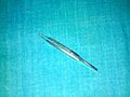 Saint Martin's forceps
 Eye Lens expressor
 Nettleship's punctum dilator
 Small scissors
 Scalpel with blade attached
 Conjunctival sac scissors
 Barraquer's needle holder
 Lacrimal sac dilator with scoop
 Muller's retractor, top view
 Muller's retractor, bottom view
 Angular keratotome
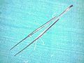 Long dissecting forceps
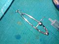 Universal eye speculum
 Rougine
 Iris repositor
 Irrigating vectis
 Lacrimal dissector with scoop
 Special blades
 von Graefe's cataract knife
 Foreign body spud and needle
 Cystitome
 Angular keratotomes
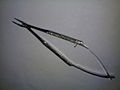 Barraquer's needle holder
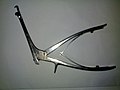 A bone punch
 Callipers
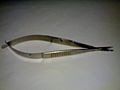 Corneal spring scissors
 Intraoccular lenses in their cases
 Intraoccular lens in place
 Intraoccular lens "dialer" or Sinsky hook
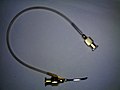 Irrigating aspirating bi-way cannula
 Lenses used for refraction testing
 Suture tying forceps for fine sutures like 8-0
 Upper one: Suture tying forceps; Lower one: Iris forceps; For comparison
 Upper right: Corneal spring scissors; Lower left: Vanna's scissors; for comparison
 Vanna's scissors
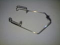 Wire speculum
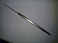 Wire vectis
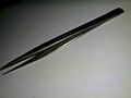 Plain dissecting forceps
 Thermocautery
 A standard illuminated E chart
 A standard illuminated Snellen's chart for distant vision
 A set of lenses used in refraction testing
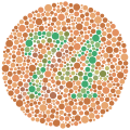 Ishihara Plate 9
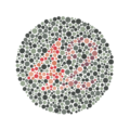 Ishihara Plate 23
 A phoropter
 NdYAG Laser
 Lacrimal canula


