Mammomonogamus is a genus of parasitic nematodes of the family Syngamidae that parasitise the respiratory tracts of cattle, sheep, goats, deer, cats, orangutans, and elephants. The nematodes can also infect humans and cause the disease called mammomonogamiasis.[1] Several known species fall under the genus Mammomonogamus, but the most common species found to infest humans is M. laryngeus. Infection in humans is very rare, with only about 100 reported cases worldwide, and is assumed to be largely accidental.[2] Cases have been reported from the Caribbean,[3] China,[4] Korea,[5] Thailand,[6] and Philippines.[7]
The worm usually inhabits the upper-respiratory region in the trachea, bronchus, or larynx, and can elicit chronic coughing and asthma-like symptoms.[8] One interesting case from Thailand reported finding worms in the patient's duodenum, suggesting M. laryngeus can also be a gastrointestinal parasite.[6] More research is needed because the life cycle is not completely known. Diagnosis is made by recovering the worms on bronchoscopy or oesophagogastroduodenoscopy. Due to the scant amount of information available on this parasite in literature, increased awareness is necessary, especially in endemic areas near M. laryngeus’ reservoir hosts, for clinicians, the local population in the endemic area, and traveling tourists, to effectively recognize and prevent mammomonogamiasis.[citation needed]
Taxonomic classification
Taxonomic family tree of Mammomonogamus
Mammomonogamus is classified in the family Syngamidae. The Syngamidae are in the superfamily Strongyloidae and order Strongylata, making them close relatives to hookworms and other nematodes.[6]
The generic name Mammomonogamus is derived from the Latin term mamma ('breast'), and the Greek terms monos (μόνος 'single') and gamos (γάμος 'marriage'),[9] which most likely is referring to the distinct characteristic of the male and female worm acting as a single unit through the male being joined in permanent copulation to the middle portion of the female's body.
Species within this genus are M. laryngeus, M. nasicola, M. gangguiensis,[2] and M. auris.[10] Only M. laryngeus is known to infect and cause disease in humans.[6] Because of the close resemblance of M. laryngeus to the gapeworm from the genus Syngamus that commonly infect birds, M. laryngeus was originally called Syngamus laryngeus and Syngamus kingi.[6] The classification was revised in 1948 when Ryzhikov reconstructed the phylogenetic relationship of the family Syngamidae and recategorized the parasite as M. laryngeus.[11]
Infestation with M. laryngeus has been called mammomonogamiasis, mammomonogamosis, syngamosis', or 'syngamiasis.[12]
History
The parasite in this genus of greatest significance to humans is M. laryngeus, due to the occasional, incidental cases reported in humans. While M. laryngeus commonly infects domestic ungulates and ruminants, the first reported case of a human infection was by Dr. King, who diagnosed the parasite in a woman from St. Lucia, Antilles.[3] The next case originated in Brazil in the 1920s, where the connection with bovine species was confirmed.[13]
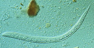
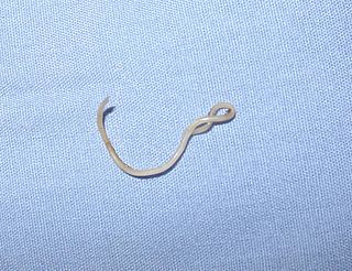
Morphology
The most distinct feature of M. laryngeus is the “Y” shape formed when the male is joined to the female in copula.[14] The smaller male uses its posterior bursa to attach to the female vulva located on the side near the middle of the female worm.[15] The adult worms usually remain permanently joined in this “Y” formation as they settle on the mucosal epithelium of the larynx, trachea, or bronchi.[14]
Adult M. laryngeus worms are red to reddish-brown in color due to their hemophagous nature.[5][9][14] They possess spicules ranging from 23-30 μm in length and cup-shaped buccal capsules (mouths) that open at the anterior end.[2][14][15] Located deep in the buccal cavity are eight to 10 teeth that are not thought to be used for attachment.[1]
The adult male is about half the length of the female. Case reports have found male worms ranging from 3–6.3mm in length and 360-380 μm in width. The larger females were reported to be 8.7-23.5mm long and 550-570 μm wide.[2][14] The female also has a pointed posterior end with a long or short tail. The female while in copula lays many ellipsoid-shaped eggs that are about 40 x 80 μm in size, not operculated, and usually possess thicker shells than hookworm eggs.[5][15]
Life cycle
Hypothesized lifecycle of Mammomonogamus laryngeus
Although the complete lifecycle of M. laryngeus is not fully known due to the rareness of the parasite in humans, the parasite is thought to adopt a lifecycle similar to Syngamus trachea,[16] the common gapeworm infection in birds that was initially thought to be mammomonogamiasis. Currently, two existing hypotheses may help aid medical diagnostics, especially in endemic areas such as the tropics, the Caribbean, and Brazil.[citation needed]
Hypothesis #1: Infection initially begins by the ingestion of foods, water, or intermediate hosts contaminated by adult worms. The infective adults migrate to the larynx or trachea and attach to the mucosal walls. Sexual reproduction occurs here, and the females begin to lay eggs in the upper respiratory region. Eggs do not develop at body temperature, and are expelled in sputum or reswallowed and excreted in feces.[5]
Hypothesis #2: The infective agent may be embryonated eggs or infective larvae, and infection is due to ingestion of contaminated food, water, or intermediate hosts. As larvae are released into the intestinal area, they can burrow through intestinal walls, travel into the mesenteric veins, and migrate to the alveoli. Here, they undergo a pulmonary cycle, where the larvae develop into adult worms in a process that may take seven days. After reaching adulthood, M. laryngeus migrates upwards to the trachea, larynx, or bronchi, where sexual reproduction occurs. Egg production begins about three weeks later, and eggs are coughed up and expelled in sputum, or excreted in feces. Larvae may hatch from embryonated eggs outside of the mammalian host.[5][17]
More research is needed to fully elucidate the lifecycle, but both larvae and adults appear to be infective. One recent case reported finding adult worms in the duodenum,[6] which is the first presentation of adult worms not in the upper respiratory region. The adult worms might have been coughed up and reswallowed before settling in the duodenum. The development from larvae to adults is about three weeks,[8] but the existence of a larval pulmonary cycle is uncertain. Intermediate hosts, although not fully known, may be earthworms (an intermediate host for the genus Syngamus),[15]snails, or arthropods.[14] Other than intermediate hosts, no mention of other biological or mechanical vectors has been made.
Symptoms of infection in humans
Symptoms usually began to appear six to 11 days after initial infection, beginning with a fever and cough. Most cases reported a progression to a persistent cough, leading to expectoration and sometimes hemoptysis.[2][14] Worms in the bronchial region can trigger a chronic, nonproductive cough and asthma-like symptoms due to the obstruction of airways by the worms.[2][9][16] These symptoms, along with a low-grade fever, can last several months if not initially diagnosed correctly. A scratching or crawling sensation can be felt in the throat if the worms are attached in the larynx.[8][14][16] Weight loss[18] and pneumonitis[16] have been reported as possible long-term consequences, but not anemia.
Recently, M. laryngeus worms were found in the duodenum of a Thai patient, which was the first gastrointestinal case of ammomonogamiasis. The patient complained of chest pain, haematemesia, melaena, and abdominal bloating, but no respiratory symptoms. Although nothing conclusive was determined, the adult worms perhaps were dislodged from the larynx, reswallowed, and later found in the duodenum.[6]
Pathogenesis
Little is known about how M. laryngeus causes disease. Symptoms do not arise until the worms have reached the adult stage and obstruct the bronchial airways leading to asthma-like symptoms and coughing.[8] Similar symptoms are seen in humans, as well as the domestic ungulates and ruminant hosts. Bronchial inflammation or hemotypsis may occur due to the worms attaching to the mucosal walls and ingesting red blood cells.[14]
The incubation period is usually six to 11 days after infection.[8] This supports the second hypothesis of a possible pulmonary cycle that explains the one- to two-week delay in the presentation of symptoms.[citation needed]
Eosinophilia is not a reliable measure of extent of infection because it varies from individual to individual. Some cases with multiple pairs of worms have reported low eosinophil levels, while other cases with a single pair had very high eosinophil counts. Such variation may be due to the lack of host tissue invasion by the parasite, since M. laryngeus attaches to the mucosal epithelium in the tracheolaryngeal region.[2] A singular case even found worms within a cyst.[19]
Past cases have demonstrated that simple removal of the worms from upper respiratory area led to a cessation of symptoms and was a sufficient cure with or without antihelmintics.[2][14] No lasting pathological tissue damage was reported.
Thus far, no reinfection mechanism for M. laryngeus has been posited, so all adult worms found inside a human must be the result of ingested embryonated eggs, larvae, or adult worms. Most of the time, only one pair of worms is found, but occasionally, the patient may have multiple pairs that must all be removed.[2][20]
Diagnosis
The definitive diagnosis is the recovery of adult worms either by coughing them up[9] or removing them with forceps, a bronchoscope, or endoscopic instruments. However, worms might be difficult to remove if firmly attached to the bronchial walls.[5] Finding M. laryngeus eggs in the sputum or feces[2][14] is another sure sign of infection.
The eggs closely resemble hookworm eggs, but Mammomonogamus eggs have a much thicker shell.[15]
Treatment
Mammomonogamiasis is relatively easy to treat. Manual or bronchoscopic removal of worms has been successful.[14] One case followed up with aspiration.[5] Although no controlled study on the efficacy of antihelmintics in treating mammomonogamiasis has been conducted, most patients were given albendazole, mebendazole or thiabendazole with no adverse effects. Patients given albendazole were instructed to take 400mg for three days[5] or if given a combination of drugs, albendazole is given 200mg, three times a day for three days, with mebendazole at 100mg, three times a day for three days.[14] The drug regimens ranged from 200–3000mg/day for three to 20 days.[2]
Epidemiology
Mammomonogamiasis is a very rare human infection yet a common veterinary parasite. Only 100 human cases of M. laryngeus have been reported, thus far.[2] Its reservoir hosts are largely in tropical regions, most commonly the domestic cattle,[21] cats,[1] orangutans,[22] and other ruminants and ungulates. Therefore, humans are accidental hosts,[14] where infections are most likely to due close exposure to bovine or feline species.[citation needed]
While the complete life cycle is still not fully known, transmission is thought to be oral-fecal, where infection comes from ingesting contaminated food or water containing embryonated eggs, hatched larvae, or intermediate hosts. Possible intermediate host candidates include earthworms, snails, and arthropods.[14] Eggs are expelled in sputum or feces. Most often, travelers to tropical climate places become exposed to contaminated sources and are diagnosed upon return to their country. The best preventative measure is to ensure proper food preparation and water sanitation.
From the numerous case reports, endemic areas include Martinique, Brazil, Puerto Rico, Dominica, Santa Lucia, Trinidad, Guyana, Guadeloupe,[15] India, tropic regions in Africa, Malaysia,[20] the Philippines,[7][23] Vietnam,[17] China,[24][25] Korea,[5] and Thailand.[6][20][26]
Due to the frequency of travelers contracting this disease, countries such as Australia,[18] Canada,[27] United States,[2][8][19] UK,[28][29] France[30][31] have reported cases, although these countries are not considered to be endemic for the parasite.
Public health
Mammomonogamiasis is not considered an emerging disease[14] because of the rarity of cases. Consequently, no control measures are currently being implemented. Very little information about this disease can be found in literature, so physicians especially in endemic areas should be aware of the parasitic disease, the clinical presentations, and treatment for humans.[6]
Related Research Articles
Ascaris lumbricoides is a large parasitic roundworm of the genus Ascaris. It is the most common parasitic worm in humans. An estimated 807 million–1.2 billion people are infected with A. lumbricoides worldwide. People living in tropical and subtropical countries are at greater risk of infection. Infection by Ascaris lumbricoides is known as ascariasis.
Strongyloides stercoralis is a human pathogenic parasitic roundworm causing the disease strongyloidiasis. Its common name in the US is threadworm. In the UK and Australia, however, the term threadworm can also refer to nematodes of the genus Enterobius, otherwise known as pinworms.
Trichuriasis, also known as whipworm infection, is an infection by the parasitic worm Trichuris trichiura (whipworm). If the infection is only with a few worms, there are often no symptoms. In those who are infected with many worms, there may be abdominal pain, fatigue and diarrhea. The diarrhea sometimes contains blood. Infections in children may cause poor intellectual and physical development. Low red blood cell levels may occur due to loss of blood.
Gnathostomiasis, also known as larva migrans profundus, is the human infection caused by the nematode Gnathostoma spinigerum and/or Gnathostoma hispidum, which infects vertebrates.
Hookworm infection is an infection by a type of intestinal parasite known as a hookworm. Initially, itching and a rash may occur at the site of infection. Those only affected by a few worms may show no symptoms. Those infected by many worms may experience abdominal pain, diarrhea, weight loss, and tiredness. The mental and physical development of children may be affected. Anemia may result.
Taenia saginata, commonly known as the beef tapeworm, is a zoonotic tapeworm belonging to the order Cyclophyllidea and genus Taenia. It is an intestinal parasite in humans causing taeniasis and cysticercosis in cattle. Cattle are the intermediate hosts, where larval development occurs, while humans are definitive hosts harbouring the adult worms. It is found globally and most prevalently where cattle are raised and beef is consumed. It is relatively common in Africa, Europe, Southeast Asia, South Asia, and Latin America. Humans are generally infected as a result of eating raw or undercooked beef which contains the infective larvae, called cysticerci. As hermaphrodites, each body segment called proglottid has complete sets of both male and female reproductive systems. Thus, reproduction is by self-fertilisation. From humans, embryonated eggs, called oncospheres, are released with faeces and are transmitted to cattle through contaminated fodder. Oncospheres develop inside muscle, liver, and lungs of cattle into infective cysticerci.
Paragonimus westermani is the most common species of lung fluke that infects humans, causing paragonimiasis. Human infections are most common in eastern Asia and in South America. Paragonimiasis may present as a sub-acute to chronic inflammatory disease of the lung. It was discovered by Dutch zoologist Coenraad Kerbert in 1878.
Paragonimiasis is a food-borne parasitic disease caused by several species of lung flukes belonging to genus Paragonimus. Infection is acquired by eating crustaceans such as crabs and crayfishes which host the infective forms called metacercariae, or by eating raw or undercooked meat of mammals harboring the metacercariae from crustaceans.
Parasitic bronchitis, also known as hoose, husk, or verminous bronchitis, is a disease of sheep, cattle, goats, and swine caused by the presence of various species of parasite, commonly known as lungworms, in the bronchial tubes or in the lungs. It is marked by cough, dyspnea, anorexia and constipation. Lungworms which cause parasitic bronchitis include nematodes of the genera Dictyocaulus, Metastrongylus, and Protostrongylus. Hoose is essentially an infantile disease, almost always afflicting animals under one year of age.
A gapeworm, also known as a red worm and forked worm, is a parasitic nematode worm that infects the tracheas of certain birds. The resulting disease, known as "gape", occurs when the worms clog and obstruct the airway. The worms are also known as "red worms" or "forked worms" due to their red color and the permanent procreative conjunction of males and females. Gapeworms are common in young, domesticated chickens and turkeys.
Toxocara canis is a worldwide-distributed helminth parasite that primarily infects dogs and other canids, but can also infect other animals including humans. The name is derived from the Greek word toxon 'bow, quiver' and the Latin word caro 'flesh'. T. canis live in the small intestine of the definitive host. This parasite is very common in puppies and somewhat less common in adult dogs. In adult dogs, infection is usually asymptomatic but may be characterized by diarrhea. By contrast, untreated infection with Toxocara canis can be fatal in puppies, causing diarrhea, vomiting, pneumonia, enlarged abdomen, flatulence, poor growth rate, and other complications.
Angiostrongyliasis is an infection by a roundworm of the Angiostrongylus type. Symptoms may vary from none to mild, to meningitis.
Gongylonema pulchrum is the only parasite of the genus Gongylonema capable of infecting humans.
Lungworms are parasitic nematode worms of the order Strongylida that infest the lungs of vertebrates. The name is used for a variety of different groups of nematodes, some of which also have other common names; what they have in common is that they migrate to their hosts' lungs or respiratory tracts, and cause bronchitis or pneumonia. The lungworm will gradually damage the airways or lung tissue by inciting an inflammatory reaction inside the tissue. Ultimately, the parasites survive and reproduce in the respiratory tissues. The category is thus more a descriptive than a precisely taxonomic one.
Toxocara cati, also known as the feline roundworm, is a parasite of cats and other felids. It is one of the most common nematodes of cats, infecting both wild and domestic felids worldwide. Adult worms are localised in the gut of the host. In adult cats, the infection – which is called toxocariasis – is usually asymptomatic. However, massive infection in juvenile cats can be fatal.
The Syngamidae are a family of nematodes which commonly parasitize mammals, birds, and rarely humans. They are classified in the Strongyloidae superfamily and Strongylata order.
Gnathostoma hispidum is a nematode (roundworm) that infects many vertebrate animals including humans. Infection of Gnathostoma hispidum, like many species of Gnathostoma causes the disease gnathostomiasis due to the migration of immature worms in the tissues.
Hookworms are intestinal, blood-feeding, parasitic roundworms that cause types of infection known as helminthiases. Hookworm infection is found in many parts of the world, and is common in areas with poor access to adequate water, sanitation, and hygiene. In humans, infections are caused by two main species of roundworm, belonging to the genera Ancylostoma and Necator. In other animals the main parasites are species of Ancylostoma. Hookworm is closely associated with poverty because it is most often found in impoverished areas, and its symptoms promote poverty through the educational and health effects it has on children. It is the leading cause of anemia and undernutrition in developing countries, while being one of the most commonly occurring diseases among poor people. Hookworm thrives in areas where rainfall is sufficient and keeps the soil from drying out, and where temperatures are higher, making rural, coastal areas prime conditions for the parasite to breed.
Cat worm infections, the infection of cats (Felidae) with parasitic worms, occur frequently. Most worm species occur worldwide in both domestic and other cats, but there are regional, species and lifestyle differences in the frequency of infestation. According to the classification of the corresponding parasites in the zoological system, infections can be divided into those caused by nematode and flatworms - in the case of the latter, mainly cestoda and trematoda - while other strains are of no veterinary significance. While threadworms usually do not require an intermediate host for their reproduction, the development cycle of flatworms always proceeds via alternate hosts.
Nematode infection in dogs - the infection of dogs with parasitic nemamotodes - are, along with tapeworm infections and infections with protozoa, frequent parasitoses in veterinary practice. Nematodes, as so-called endoparasites, colonize various internal organs - most of them the digestive tract - and the skin. To date, about 30 different species of nematode have been identified in domestic dogs; they are essentially also found in wild dog species. However, the majority of them often cause no or only minor symptoms of disease in adult animals. The infection therefore does not necessarily have to manifest itself in a worm disease (helminthosis). For most nematodes, an infection can be detected by examining the feces for eggs or larvae. Roundworm infection in dogs and the hookworm in dogs is of particular health significance in Central Europe, as they can also be transmitted to humans (zoonosis). Regular deworming can significantly reduce the frequency of infection and thus the risk of infection for humans and dogs.
References
1 2 3 Anderson RC, Chabaud AG, Willmott S. CIH keys to the nematode parasites of vertebrates, no 7. Keys to genera of superfamily Strongyloidea. Commonwealth Agricultural Bureaux, England, 1980.
1 2 3 4 5 6 7 8 9 10 11 12 13 Nosanchuk, J.S., Wade, S.E., and Landolf, M (1995). Case Report of and Description of Parasite in Mammomonogamus laryngeus (Human Syngamosis) Infection. J of Clinical Microbiology. 33: 998–1000.
1 2 Leiper RJ (1913). "Gapes in man, an occasional helminthic infection: a notice of its discovery by Dr A King in St Lucia". Lancet. i (4664): 170. doi:10.1016/S0140-6736(00)76203-9.
↑ Li D, Li G, Zhan X (1997). "Three cases of human Mammomonogamus laryngeus infection". Chin J Parasitol Parasit Dis. 15: 281–4.
1 2 3 4 5 6 7 8 9 Eamsobhana P, Mongkolporn T, Punthuprapasa P, Yoolek A (2006). "Mammomonogamus roundworm (Nematoda: Syngamidae) recovered from the duodenum of a Thai patient: a first and unusual case originating in Thailand". Trans R Soc Trop Med Hyg. 100 (4): 387–91. doi:10.1016/j.trstmh.2005.05.018. PMID16257022.
1 2 SaintJohn JH, Simmons JS, Gardner LL (1929). "Infestation of the lung by a nematode of the genus Cyathostoma". J Am Med Assoc. 92 (22): 1816–18. doi:10.1001/jama.1929.02700480006003.
1 2 3 4 5 6 Weinstein, L., and A. Molovi. 1971. Syngamus laryngeus infection (Syngamosis) with chronic cough. Ann. Intern. Med. 74:577–580.
1 2 3 4 de Lara, T. de A., Barbosa, M.A., de Oliveira, M.R., de Godoy, I., and Queluz, T.T. (1993). Human syngamosis: Two cases of chronic cough caused by Mammomonogamus laryngeus. Chest. 103(1): 264-5.
↑ Bowman, Dwight D.; Hendrix, Charles M.; Lindsay, David S.; Barr, Stephen C. (2001). Feline clinical parasitology. Hoboken: John Wiley & Sons. pp.258–259. doi:10.1002/9780470376805. ISBN9780470376591.
↑ Ryzhikov, K. M. (1948). "[Phylogenetic interrelationships of nematodes of the family Syngamidae and an attempt to reconstruct their systematics]". Doklady Akademii Nauk SSSR (in Russian). 62: 733.
↑ Freitas, A.L., De Carli, G., and Blankenhein, M.H. “Mammonomonogamus (Syngamus) laryngeus infection: a new Brazilian human case. Rev. Inst. Med. Trop. S. Paulo, 37 (2): 177-179, 1995.
1 2 3 4 5 6 Gutierrez, Yezid. Diagnostic Pathology of Parasitic Infections with Clinical Correlations. 2nd ed. New York: Oxford University Press, 2000.
1 2 3 4 Severo LC, Conci LMA, Camargo JJP, Andre-Alves MR, Palombini BC. Syngamosis: two new Brazilian cases and evidence of possible pulmonary cycle. Trans R Soc Trop Med Hyg. 1988;82: 467–8.
1 2 Acha PN, Szyfres B. Mammomonogamiasis. Zoonosis and communicable diseases common to man and animals. Washington (DC): Pan American Health Organization; 2003. Scientific and Technical Publication No. 580.
1 2 Birrell, D.J., Moorhouse, D.E., Gardnert, M.A.H., and May, C.S (1978). Chronic Cough and Haemoptysis due to a Nematode, "Syngamus Laryngeus." Aust. N.Z. J. Med.8: 168-170.
1 2 Gardiner, C.H. and Schantz, P.M (1983). Mammomonogamus Infection in A Human: Report of A Case. Am. J. Trop. Med. Hyg.32(5) 995-997.
1 2 3 Pipitogool V, Chaisiri K, Visetsuspakarn P, Srigan V, Maleewong W. Mammomonogamus (syngamus) laryngeus. First case report in Thailand. Southeast Asian J Trop Med Public Health. 1992;23:336–7.
↑ Van Aken, D., Lagapa, J.T., Dargantes, A.P., Vercruysse, J., 1996. Mammomonogamus laryngeus (Railliet, 1899) infections in cattle in Mindanao. Philippines. Vet. Parasitol.64, 329—332.
↑ Collet, J.Y., Galdikas, B.M., Sugarjito, J., Jojosudharmo, S., 1986. A coprological study of parasitism in orangutans (Pongo pygmaeus) in Indonesia. J. Med. Primatol. 15, 121—129.
↑ Beaver PC, Jung RC, Wayne E. Clinical parasitology. Philadelphia: Lea and Febiger; 1984.
↑ Li D, Li G, Zhan X (1997). Three cases of human Mammomonogamus laryngeus infection. Chin J Parasitol Parasit Dis15: 281–4.
↑ Qu, F.Y., 1997. First Mammomonogamus laryngeus infection case occurred in Shanghai. Chin. J. Parasitol. Parasit. Dis.15, 198—200
↑ Limawongpranee, S., Samanthai, S., Yoolek, A., 2004. Human Mammomonogamus laryngeus infection (syngamosis): the second case report in Thailand. Siriraj Hosp. Gaz.56, 82—86.
↑ Leers, W.-D., M. K. Sarin, and K. Arthurs. 1985. Syngamosis, an unusual cause of asthma: the first reported case in Canada. Can. Med. Assoc. J.132:269–270.
↑ Basden, R. D. E., J. W. Jackson, and E. I. Jones. 1974. Gapeworm infestation in man. Br. J. Dis. Chest68:207–209.
↑ Turner P, Turner CG, Bowers KM, Gibson DI, Chiodini PL (2003). A Case of Human Syngamosis. Travel Med Infect Dis.1(4): 231-3.
↑ Junod, C., M. Philbert, and H. T. Sang. 1970. Une observation de syngamose humaine a localization bronchique. Premier cas tracte et queri par le thiabendazole. Bull. Soc. Pathol. Exot.63:483–488.
↑ Sang, H. T., C. Junod, and M. Philbert. 1970. Notes parasitologiques sur Syngamus laryngeus Railliet, 1899 et la syngamose humaine. A propos d’un cas de syngamose bronchique chez l’homme. Bull. Soc. Pathol. Exot.63:488–497.
This page is based on this Wikipedia article Text is available under the CC BY-SA 4.0 license; additional terms may apply. Images, videos and audio are available under their respective licenses.