Moniliformis moniliformis

Last updated

Moniliformis moniliformis
Moniliformis moniliformis.jpg
Adult specimen
Scientific classification Red Pencil Icon.png
Kingdom: Animalia
Phylum: Acanthocephala
Class: Archiacanthocephala
Order: Moniliformida
Family: Moniliformidae
Genus: Moniliformis
Species:
M. moniliformis
Binomial name
Moniliformis moniliformis
(Bremser, 1811)

Moniliformis moniliformis is a parasite of the Acanthocephala phylum in the family Moniliformidae. The adult worms are usually found in intestines of rodents or carnivores such as cats and dogs. The species can also infest humans, though this is rare.

Contents

Distribution

Infested rats have been found world-wide. Cases of human infestation by Moniliformis moniliformis have been reported in the United States, Iran, Iraq, and Nigeria. [1]

Morphology

Acanthocephalans do not have digestive tracts and absorb nutrients through the tegument, the external layer. The scolex of this worm has a cylindrical proboscis and a multitude of curved hooks. The main parts of the worm body are the proboscis, neck, and trunk. Because of horizontal markings on the worm, there is the appearance of segmentation. Acanthocephalans are sexually dimorphic (dioecious) – adult males are generally 4 to 5 cm long while females are longer, ranging from lengths of 10 to 30 cm. Males also have copulatory bursas, used to hold on to the female during copulation and cement glands. Females have floating ovaries within a ligament sac where fertilization of the eggs occurs. [2] The eggs of this parasite are 90–125 μm long and 65 μm wide. They are oval in shape with a thick, clear outer coat. [3]

Reservoirs

Usually, the definitive hosts for M. moniliformis are rodents, cats, dogs and red foxes (in Poland); human infestations are rare. The intermediate hosts are usually beetles and cockroaches. [1]

Life cycle

In the life cycle of M. moniliformis, the intermediate hosts ingest the eggs of the parasite. In the intermediate host, the acanthor, or the parasite in its first larval stage, morphs into the acanthella, the second larval stage. After 6–12 weeks in this stage, the acanthella becomes a cystacanth. The cystacanth, or infective acanthella, of M. moniliformis are cyst-shaped and encyst in the tissues of the intermediate hosts. However, most other acanthocephalans have infective larvae that more closely resemble underdeveloped adult worms. The definitive hosts consume the cystacanths upon feeding on infested intermediate hosts. These cystacanths mature and mate in the small intestine in 8–12 weeks. After this time, the eggs are excreted with the feces, to be ingested yet again by another intermediate host and renew this cycle. [3]

The reproduction of the parasite only occurs in the definitive host. In acanthocephalans, adult males have cement glands in their posterior ends. The widely held theory is that the mucilaginous and proteinaceous substance that these glands secrete is used by males to seal up the females after copulation in order to prevent leakage of the inseminated sperm and further insemination by other males. It has also been found that these males may create this seal on other males in order to prevent them from copulating. [4] These seals, or copulatory caps, last for a week.

Behavioral changes in the intermediate host

In what is commonly known as "brain-jacking", the parasite induces a behavioral change in its intermediate host that increases the risk of predation for the host. It is thought that this behavioral change holds an evolutionary advantage for the parasite by increasing its chances of getting to its definitive host. When M. moniliformis infests its intermediate host, the cockroach species Periplaneta americana , it changes the cockroach's escape response. In one study, it was concluded that cockroaches infected by M. moniliformis took longer to respond to wind stimuli simulating the approach of a potential predator and displayed fewer escape responses implying that the parasite infection renders its intermediate host more vulnerable to predation by hindering its ability to detect and escape from its predator. It is thought that serotonin plays a role in upending the communication between giant interneurons and the thoracic interneurons and in turn hampers the escape response of the cockroach. [5] In a similar study, the effects of parasitism on three Periplaneta species are studied. The results show that Periplaneta australasiae uses substrates differently and moves around less when infected with Moniliformis moniliformis. [6] Another study concludes an increased vulnerability of infected Periplaneta americana due to increased phototaxis, more time spent moving (due to slower movement) and movement in response to light (uninfected cockroaches hesitated before moving). [7]

Human infection

As it requires consumption of raw infested beetles or cockroaches, human acanthocephaliasis is rare.

Clinical manifestations

In 1888 in Italy, Salvatore Calandruccio  [ it ] infected himself by ingesting larvae, reported gastrointestinal disturbances, and then shed the eggs in two weeks. This was the first report of the clinical manifestations of an M. moniliformis infestation in humans.

Calandruccio provided the first description of the clinical manifestations of acanthocephaliasis and similar accounts are found in the few case studies since; many of the patients described were asymptomatic. When they showed symptoms, they normally experienced abdominal pain, diarrhea, dizziness, edema, and anorexia. [8] In some patients, giddiness has also been reported. [1] In rodents, acanthocephaliasis is fatal and manifests itself through hemorrhaging and gastrointestinal disturbance.

Diagnosis

The proper diagnosis of acanthocephaliasis in humans is made through fecal analysis, which, if the host is infested, should contain adult worms or eggs. To obtain the worms from the host, piperazine citrate, levamisole and bithionol can be administered to the patient. [8]

Treatment

Acanthocephaliasis is treated with anti-helminthics. There is considerable debate over the efficacy of anti-helminth drugs on this parasite but, so far, the drugs seem to be working. Pyrantel pamoate and ivermectin have been shown to be particularly effective in treating patients. [8] Mebendazole and thiabendazole have also both been cited to work. [5] [9]

Prevention strategies

Because the only way of developing acanthocephaliasis is through ingesting the intermediate hosts, the most effective means of prevention is avoiding the consumption of uncooked beetles and cockroaches. This is especially difficult in children exhibiting pica and in areas with poor hygiene. Awareness campaigns on the risks of consuming infested beetles and cockroaches would be effective. Moreover, preventing entry of the intermediate hosts into the home, and especially the kitchen where it is at risk of getting into the food, would help curb the risk of becoming infested. [10]

Related Research Articles

Acanthocephala Phylum of parasitic thorny-headed worms

Acanthocephala is a phylum of parasitic worms known as acanthocephalans, thorny-headed worms, or spiny-headed worms, characterized by the presence of an eversible proboscis, armed with spines, which it uses to pierce and hold the gut wall of its host. Acanthocephalans have complex life cycles, involving at least two hosts, which may include invertebrates, fish, amphibians, birds, and mammals. About 1420 species have been described.

Hymenolepiasis is infestation by one of two species of tapeworm: Hymenolepis nana or H. diminuta. Alternative names are dwarf tapeworm infection and rat tapeworm infection. The disease is a type of helminthiasis which is classified as a neglected tropical disease.

Gnathostomiasis is the human infection caused by the nematode (roundworm) Gnathostoma spinigerum and/or Gnathostoma hispidum, which infects vertebrates.

<i>Echinococcus multilocularis</i> Species of flatworm

Echinococcus multilocularis is a small cyclophyllid tapeworm found extensively in the northern hemisphere. E. multilocularis, along with other members of the Echinococcus genus, produce diseases known as echinococcosis. Unlike E. granulosus,E. multilocularis produces many small cysts that spread throughout the internal organs of the infected animal. The resultant disease is called Alveolar echinococcosis, and is caused by ingesting the eggs of E. multilocularis.

<i>Hymenolepis diminuta</i> Species of flatworm

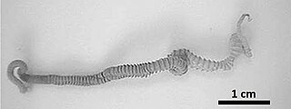
Hymenolepis diminuta, also known as rat tapeworm, is a species of Hymenolepis tapeworm that causes hymenolepiasis. It has slightly bigger eggs and proglottids than H. nana and infects mammals using insects as intermediate hosts. The adult structure is 20 to 60 cm long and the mature proglottid is similar to that of H. nana, except it is larger.

Mammomonogamus is a genus of parasitic nematodes of the family Syngamidae that parasitise the respiratory tracts of cattle, sheep, goats, deer, cats, orangutans, and elephants. The nematodes can also infect humans and cause the disease called mammomonogamiasis. Several known species fall under the genus Mammomonogamus, but the most common species found to infest humans is M. laryngeus. Infection in humans is very rare, with only about 100 reported cases worldwide, and is assumed to be largely accidental. Cases have been reported from the Caribbean, China, Korea, Thailand, and Philippines.

Pomphorhynchus laevis is an endo-parasitic acanthocephalan worm, with a complex life cycle, that can modify the behaviour of its intermediate host, the freshwater amphipod Gammarus pulex. P. laevis does not contain a digestive tract and relies on the nutrients provided by its host species. In the fish host this can lead to the accumulation of lead in P. laevis by feeding on the bile of the host species.

<i>Hymenolepis</i> (tapeworm) Genus of worms

Hymenolepis is a genus of cyclophyllid tapeworms responsible for hymenolepiasis. They are parasites of humans and other mammals. The focus in this article is in Hymenolepis commonly parasitizing humans.

<i>Gongylonema pulchrum</i> Species of human parasite

Gongylonema pulchrum is the only parasite of the genus Gongylonema capable of infecting humans.

<i>Nanophyetus</i> Genus of flukes

Nanophyetus salmincola is a food-borne intestinal trematode parasite prevalent on the Pacific Northwest coast. The species may be the most common trematode endemic to the United States.

<i>Profilicollis</i> Genus of thorny-headed worms

Profilicollis is a genus of acanthocephalan parasites of crustaceans. The status of the genus Profilicollis has been debated, and species placed in this genus were formerly included in the genus Polymorphus. However, research on the morphology of the group and their use of hosts has concluded that Profilicollis and Polymorphus should be regarded as distinct genera, and species previously described as Polymorphus altmani are now referred to as Profilicollis altmani in taxonomic and biological literature. Profilicollis parasites infect decapod crustaceans, usually shore crabs, as intermediate hosts, and use many species of shorebirds as definitive (final) hosts.

Angiostrongylus costaricensis is a species of parasitic nematode and is the causative agent of abdominal angiostrongyliasis in humans. It occurs in Latin America and the Caribbean.

<i>Gigantorhynchus</i> Genus of parasitic worms

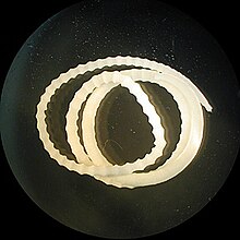
Gigantorhynchus is a genus of Acanthocephala that parasitize marsupials, anteaters, and possibly baboons by attaching themselves to the intestines using their hook-covered proboscis. Their life cycle includes an egg stage found in host feces, a cystacanth (larval) stage in an intermediate host such as termites, and an adult stage where cystacanths mature in the intestines of the host. This genus is characterized by a cylindrical proboscis with a crown of robust hooks at the apex followed by numerous small hooks on the rest of the proboscis, a long body with pseudosegmentation, filiform lemnisci, and ellipsoid testes. The largest known specimen is the female G. ortizi with a length of around 240 millimetres (9.4 in) and a width of 2 millimetres (0.08 in). Genetic analysis on one species of Gigantorhynchus places it with the related genus Mediorhynchus in the family Gigantorhynchidae. Six species in this genus are distributed across Central and South America and possibly Zimbabwe. Infestation by a Gigantorhynchus species may cause partial obstructions of the intestines, severe lesions of the intestinal wall, and may lead to death.

Mediorhynchus is a genus of small parasitic spiny-headed worms. Phylogenetic analysis has been conducted on two known species of Mediorhynchus and confirmed the placement along with the related genus Gigantorhynchus in the family Gigantorhynchida. The distinguishing features of this order among archiacanthocephalans is a divided proboscis. This genus contains fifty-eight species that are distributed globally. These worms exclusively parasitize birds by attaching themselves around the cloaca using their hook-covered proboscis. The bird hosts are of different orders.

Moniliformidae Family of worms

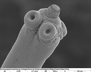
Moniliformidae is a family of parasitic spiny-headed worms. It is the only family in the Moniliformida order and contains three genera: Australiformis containing a single species, Moniliformis containing eighteen species and Promoniliformis containing a single species. Genetic analysis have determined that the clade is monophyletic despite being distributed globally. These worms primarily parasitize mammals, including humans in the case of Moniliformis moniliformis, and occasionally birds by attaching themselves into the intestinal wall using their hook-covered proboscis. The intermediate hosts are mostly cockroaches. The distinguishing features of this order among archiacanthocephalans is the presence of a cylindrical proboscis with long rows of hooks with posteriorly directed roots and proboscis retractor muscles that pierce both the posterior and ventral end or just posterior end of the receptacle. Infestation with Monoliformida species can cause moniliformiasis, an intestinal condition characterized as causing lesions, intestinal distension, perforated ulcers, enteritis, gastritis, crypt hypertrophy, goblet cell hyperplasia, and blockages.

Oligacanthorhynchidae Order of thorny-headed worms

Oligacanthorhynchida is an order containing a single parasitic worm family, Oligacanthorhynchidae, that attach themselves to the intestinal wall of terrestrial vertebrates.

<i>Hymenolepis microstoma</i> Species of flatworm

Hymenolepis microstoma, also known as the rodent tapeworm, is an intestinal dwelling parasite. Adult worms live in the bile duct and small intestines of mice and rats, and larvae metamorphose in the haemocoel of beetles. It belongs to the genus Hymenolepis; tapeworms that cause hymenolepiasis. H. microstoma is prevalent in rodents worldwide, but rarely infects humans.

Alaria americana is a species of a trematode in a family Diplostomidae. All of these species infect carnivorous mammals by living in their small intestines as mature worms. A. americana are most frequently found in temperate regions, predominately in northern North America. This organisms habit is extremely diverse, as it occupies four different hosts throughout its lifetime. This trematode thrives in areas close to water as it is needed for several developmental stages to occur. A. americana has been isolated to the different North American mammals with a wide range of definitive hosts, including cattle, lynx, martens, skunks, bobcats, foxes, coyotes, and wolves.

<i>Corynosoma australe</i> Species of thorny-headed worm

Corynosoma autrale is a species of acanthocephalan. This species usually infects pinnipeds; the semi-aquatic fin-footed marine mammals most commonly known as seals and sea lions. Pinniped infections are not exclusive, recently C. australe has been discovered in Magellanic penguins.

<i>Pachysentis</i> Genus of worms

Pachysentis is a genus of Acanthocephala that parasitize carnivourous mammals by attaching themselves to the intestines using their hook-covered proboscis. Their life cycle includes an egg stage found in host feces, a cystacanth (larval) stage in an intermediate host such as the Egyptian cobra, and an adult stage where cystacanths mature in the intestines of the host. This genus appears identical to the closely related Oncicola apart from a greater number of hooks on the proboscis. The eleven species in this genus are distributed across Africa and the Americas.

References

  1. 1 2 3 , "Acanthocephalan Worms." Gideon. Gideon Informatics. Web.
  2. , "Phylum: Acanthocephala." Lecture. Animal Parasitology. Kansas State, 14 March 2005. Web. 23 February 2010.
  3. 1 2 , Acanthocephaliasis. Parasites and Health. CDC, 20 July 2009. Web.
  4. Crompton, D.W.T. Reproduction. In Biology of the Acanthocephala (ed. Crompton, D. W. T. & Nickol, B. B.), pp. 213–271. Cambridge University Press, Cambridge. 1985
  5. 1 2 Berenji, F; Fata, A; Hosseininejad, Z (2007). "A case of Moniliformis moniliformis (Acanthocephala) infection in Iran". Korean J Parasitol. 45 (2): 145–8. doi:10.3347/kjp.2007.45.2.145. PMC   2526305 . PMID   17570979.
  6. Moore, J.; Freehling, M.; Gotelli, N. J. (1994). "Altered behavior in two species of blattid cockroaches infected with Moniliformis moniliformis (Acanthocephala)". Journal of Parasitology. 80 (2): 220–223. doi:10.2307/3283750. JSTOR   3283750. PMID   8158464.
  7. Moore, J (1983). "Altered behaviour in cockroaches (Periplaneta americana) infected with an archiacantho- cephalan Moniliformis moniliformis" (PDF). J. Parasitol. 69 (6): 1174–1177. doi:10.2307/3280893. JSTOR   3280893. Archived from the original (PDF) on 19 July 2011.
  8. 1 2 3 Richardson, Dennis J.; Krause, Peter J. (2003). North American Parasitic Zoonoses. Vol. 6. Boston: Springer Science & Business Media. ISBN   978-1-4020-7212-3.
  9. Salehabadi, Alireza (2008). "Human Infection with Moniliformis moniliformis (Bremser 1811) (Travassos 1915) in Iran: Another Case Report After Three Decades". Vector-Borne and Zoonotic Diseases. 8 (1): 101–104. doi:10.1089/vbz.2007.0150. PMID   18237263.
  10. Neafie, RC; Marty, AM (1993). "Unusual infections in humans". Clin Microbiol Rev. 66 (1): 34–56. doi:10.1128/CMR.6.1.34. PMC   358265 . PMID   8457979.