
Thyroid neoplasm is a neoplasm or tumor of the thyroid. It can be a benign tumor such as thyroid adenoma, or it can be a malignant neoplasm, such as papillary, follicular, medullary or anaplastic thyroid cancer. Most patients are 25 to 65 years of age when first diagnosed; women are more affected than men. The estimated number of new cases of thyroid cancer in the United States in 2023 is 43,720 compared to only 2,120 deaths. Of all thyroid nodules discovered, only about 5 percent are cancerous, and under 3 percent of those result in fatalities.
Renal cell carcinoma (RCC) is a kidney cancer that originates in the lining of the proximal convoluted tubule, a part of the very small tubes in the kidney that transport primary urine. RCC is the most common type of kidney cancer in adults, responsible for approximately 90–95% of cases. It is more common in men. It is most commonly diagnosed in the elderly.

Kidney cancer, also known as renal cancer, is a group of cancers that starts in the kidney. Symptoms may include blood in the urine, a lump in the abdomen, or back pain. Fever, weight loss, and tiredness may also occur. Complications can include spread to the lungs or brain.

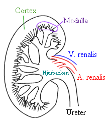
The collecting duct system of the kidney consists of a series of tubules and ducts that physically connect nephrons to a minor calyx or directly to the renal pelvis. The collecting duct participates in electrolyte and fluid balance through reabsorption and excretion, processes regulated by the hormones aldosterone and vasopressin.

Neprilysin is an enzyme that in humans is encoded by the MME gene. Neprilysin is a zinc-dependent metalloprotease that cleaves peptides at the amino side of hydrophobic residues and inactivates several peptide hormones including glucagon, enkephalins, substance P, neurotensin, oxytocin, and bradykinin. It also degrades the amyloid beta peptide whose abnormal folding and aggregation in neural tissue has been implicated as a cause of Alzheimer's disease. Synthesized as a membrane-bound protein, the neprilysin ectodomain is released into the extracellular domain after it has been transported from the Golgi apparatus to the cell surface.

Cadherin-16 is a protein that in humans is encoded by the CDH16 gene.

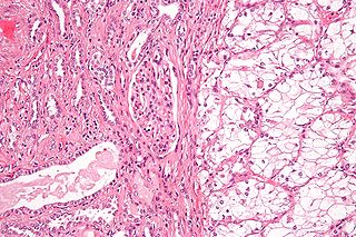
Collecting duct carcinoma (CDC) is a type of kidney cancer that originates in the papillary duct of the kidney. It is rare, accounting for 1-3% of all kidney cancers. It is also recently described; a 2002 review found just 40 case reports worldwide. Previously, due to its location, CDC was commonly diagnosed as renal cell carcinoma or a subtype of renal cell carcinoma. However, CDC does not respond well to chemotherapy drugs used for renal cell carcinoma, and progresses and spreads more quickly.
Juxtaglomerular cell tumor is an extremely rare kidney tumour of the juxtaglomerular cells, with fewer than 100 cases reported in literature. This tumor typically secretes renin, hence the former name of reninoma. It often causes severe hypertension that is difficult to control, in adults and children, although among causes of secondary hypertension it is rare. It develops most commonly in young adults, but can be diagnosed much later in life. It is generally considered benign, but its malignant potential is uncertain.
Renal medullary carcinoma is a rare type of cancer that affects the kidney. It tends to be aggressive, difficult to treat, and is often metastatic at the time of diagnosis. Most individuals with this type of cancer have sickle cell trait or rarely sickle cell disease, suggesting that the sickle cell trait may be a risk factor for this type of cancer.

Kidney tumours are tumours, or growths, on or in the kidney. These growths can be benign or malignant.

Metanephric adenoma (MA) is a rare, benign tumour of the kidney, that can have a microscopic appearance similar to a nephroblastoma, or a papillary renal cell carcinoma.

Sarcomatoid carcinoma, sometimes referred to as pleomorphic carcinoma, is a relatively uncommon form of cancer whose malignant cells have histological, cytological, or molecular properties of both epithelial tumors ("carcinoma") and mesenchymal tumors ("sarcoma"). It is believed that sarcomatoid carcinomas develop from more common forms of epithelial tumors.

Epithelial-myoepithelial carcinoma (EMCa) is a rare malignant tumour that typically arises in a salivary gland and consists of both an epithelial and myoepithelial component. They are predominantly found in the parotid gland and represent approximately 1% of salivary gland tumours.

Hyalinizing clear cell carcinoma (HCCC) is a rare malignant salivary gland tumour, with a good prognosis, that is usually found on the tongue or palate.

Hereditary leiomyomatosis and renal cell carcinoma (HLRCC) or Reed's syndrome is rare autosomal dominant disorder associated with benign smooth muscle tumors and an increased risk of renal cell carcinoma. It is characterised by multiple cutaneous leiomyomas and, in women, uterine leiomyomas. It predisposes individuals to renal cell cancer, an association denominated hereditary leiomyomatosis and renal cell cancer. It is also associated with increased risk of uterine leiomyosarcoma. The syndrome is caused by a mutation in the fumarate hydratase gene, which leads to an accumulation of fumarate. The inheritance pattern is autosomal dominant and screening can typically begin in childhood.
Acquired cystic kidney disease-associated renal cell carcinoma is rare subtype of renal cell carcinoma. It is most commonly seen in people with end-stage kidney disease who have a much higher risk of developing acquired cystic kidney disease (ACKD). Affected individuals have small kidneys with several cysts and their risk of renal cell carcinoma is 30 times higher than people without ACKD.
Mammary analogue secretory carcinoma (MASC), also termed MASCSG, is a salivary gland neoplasm. It is a secretory carcinoma which shares the microscopic pathologic features with other types of secretory carcinomas including mammary secretory carcinoma, secretory carcinoma of the skin, and salivary gland–type carcinoma of the thyroid. MASCSG was first described by Skálová et al. in 2010. The authors of this report found a chromosome translocation in certain salivary gland tumors, i.e. a (12;15)(p13;q25) fusion gene mutation. The other secretory carcinoma types carry this fusion gene.

Papillary renal cell carcinoma (PRCC) is a malignant, heterogeneous tumor originating from renal tubular epithelial cells of the kidney, which comprises approximately 10-15% of all kidney neoplasms. Based on its morphological features, PRCC can be classified into two main subtypes, which are type 1 (basophilic) and type 2 (eosinophilic).
Tubular carcinoma is a subtype of invasive ductal carcinoma of the breast. More rarely, tubular carcinomas may arise in the pancreas or kidney. Most tubular carcinomas begin in the milk duct of the breast and spread to healthy tissue around it.
Papillary carcinomas of the breast (PCB), also termed malignant papillary carcinomas of the breast, are rare forms of the breast cancers. The World Health Organization (2019) classified papillary neoplasms of the breast into 5 types: intraductal papilloma, papillary ductal carcinoma in situ (PDCIS), encapsulated papillary carcinoma (EPC), solid-papillary carcinoma (SPC), and invasive papillary carcinoma (IPC). The latter four carcinomas are considered here; intraductal papilloma is a benign neoplasm. The World Health Organization regarded solid papillary carcinoma as having two subtypes: in situ and invasive SPC.
