Neural encoding of sound

Last updated

The neural encoding of sound is the representation of auditory sensation and perception in the nervous system. [ citation needed ]The complexities of contemporary neuroscience are continually redefined. Thus what is known of the auditory system has been continually changing. The encoding of sounds includes the transduction of sound waves into electrical impulses (action potentials) along auditory nerve fibers, and further processing in the brain.

Contents

Basic physics of sound

Sound waves are what physicists call longitudinal waves, which consist of propagating regions of high pressure (compression) and corresponding regions of low pressure (rarefaction).

Waveform

Waveform is a description of the general shape of the sound wave. Waveforms are sometimes described by the sum of sinusoids, via Fourier analysis.

Amplitude

Graph of a simple sine wave Sine wave.jpg
Graph of a simple sine wave

Amplitude is the size (magnitude) of the pressure variations in a sound wave, and primarily determines the loudness with which the sound is perceived. In a sinusoidal function such as , C represents the amplitude of the sound wave.

Frequency and wavelength

The frequency of a sound is defined as the number of repetitions of its waveform per second, and is measured in hertz; frequency is inversely proportional to wavelength (in a medium of uniform propagation velocity, such as sound in air). The wavelength of a sound is the distance between any two consecutive matching points on the waveform. The audible frequency range for young humans is about 20 Hz to 20 kHz. Hearing of higher frequencies decreases with age, limiting to about 16 kHz for adults, and even down to 3 kHz for elders.[ citation needed ]

Anatomy of the ear

How sounds make their way from the source to the brain
Flowchart of sound passage - outer ear Outer Ear.jpg
Flowchart of sound passage - outer ear

Given the simple physics of sound, the anatomy and physiology of hearing can be studied in greater detail.

Outer ear

The Outer ear consists of the pinna or auricle (visible parts including ear lobes and concha), and the auditory meatus (the passageway for sound). The fundamental function of this part of the ear is to gather sound energy and deliver it to the eardrum. Resonances of the external ear selectively boost sound pressure with frequency in the range 2–5 kHz. [1]

The pinna as a result of its asymmetrical structure is able to provide further cues about the elevation from which the sound originated. The vertical asymmetry of the pinna selectively amplifies sounds of higher frequency from high elevation thereby providing spatial information by virtue of its mechanical design. [1] [2]

Middle ear

Flowchart of sound passage - middle ear Middle Ear.jpg
Flowchart of sound passage - middle ear

The middle ear plays a crucial role in the auditory process, as it essentially converts pressure variations in air to perturbations in the fluids of the inner ear. In other words, it is the mechanical transfer function that allows for efficient transfer of collected sound energy between two different media. [1] The three small bones that are responsible for this complex process are the malleus, the incus, and the stapes, collectively known as the ear ossicles. [3] [4] The impedance matching is done through via lever ratios and the ratio of areas of the tympanic membrane and the footplate of the stapes, creating a transformer-like mechanism. [3] Furthermore, the ossicles are arranged in such a manner as to resonate at 700–800 Hz while at the same time protecting the inner ear from excessive energy. [4] A certain degree of top-down control is present at the middle ear level primarily through two muscles present in this anatomical region: the tensor tympani and the stapedius. These two muscles can restrain the ossicles so as to reduce the amount of energy that is transmitted into the inner ear in loud surroundings. [2] [3]

Inner ear

Flowchart of sound passage - inner ear Inner Ear.jpg
Flowchart of sound passage - inner ear

The cochlea of the inner ear, a marvel of physiological engineering, acts as both a frequency analyzer and nonlinear acoustic amplifier. [1] The cochlea has over 32,000 hair cells. Outer hair cells primarily provide amplification of traveling waves that are induced by sound energy, while inner hair cells detect the motion of those waves and excite the (Type I) neurons of the auditory nerve.

The basal end of the cochlea, where sounds enter from the middle ear, encodes the higher end of the audible frequency range while the apical end of the cochlea encodes the lower end of the frequency range. This tonotopy plays a crucial role in hearing, as it allows for spectral separation of sounds. A cross section of the cochlea will reveal an anatomical structure with three main chambers (scala vestibuli, scala media, and scala tympani). [4] At the apical end of the cochlea, at an opening known as the helicotrema, the scala vestibuli merges with the scala tympani. The fluid found in these two cochlear chambers is perilymph, while scala media, or the cochlear duct, is filled with endolymph. [2]

Transduction

Auditory hair cells

The auditory hair cells in the cochlea are at the core of the auditory system's special functionality (similar hair cells are located in the semicircular canals). Their primary function is mechanotransduction, or conversion between mechanical and neural signals. The relatively small number of the auditory hair cells is surprising when compared to other sensory cells such as the rods and cones of the visual system. Thus the loss of a lower number (in the order of thousands) of auditory hair cells can be devastating while the loss of a larger number of retinal cells (in the order to hundreds of thousands) will not be as bad from a sensory standpoint. [5]

Cochlear hair cells are organized as inner hair cells and outer hair cells; inner and outer refer to relative position from the axis of the cochlear spiral. The inner hair cells are the primary sensory receptors and a significant amount of the sensory input to the auditory cortex occurs from these hair cells. Outer hair cells on the other hand boost the mechanical signal by using electromechanical feedback. [5]

Mechanotransduction

The apical surface of each cochlear hair cell contains a hair bundle. Each hair bundle contains approximately 300 fine projections known as stereocilia, formed by actin cytoskeletal elements. [6] The stereocilia in a hair bundle are arranged in multiple rows of different heights. In addition to the stereocilia, a true ciliary structure known as the kinocilium exists and is believed to play a role in hair cell degeneration that is caused by exposure to high frequencies. [1] [6]

A stereocilium is able to bend at its point of attachment to the apical surface of the hair cell. The actin filaments that form the core of a stereocilium are highly interlinked and cross linked with fibrin, and are therefore stiff and inflexible at positions other than the base. When stereocilia in the tallest row are deflected in the positive-stimulus direction, the shorter rows of stereocilia are also deflected. [6] These simultaneous deflections occur due to filaments called tip links that attach the side of each taller stereocilium to the top of the shorter stereocilium in the adjacent row. When the tallest stereocilia are deflected, tension is produced in the tip links and causes the stereocilia in the other rows to deflect as well. At the lower end of each tip link is one or more mechano-electrical transduction (MET) channels, which are opened by tension in the tip links. [7] These MET channels are cation-selective transduction channels that allow potassium and calcium ions to enter the hair cell from the endolymph that bathes its apical end.

The influx of cations, particularly potassium, through the open MET channels causes the membrane potential of the hair cell to depolarize. This depolarization opens voltage-gated calcium channels to allow the further influx of calcium. This results in an increase in the calcium concentration, which triggers the exocytosis of neurotransmitter vesicles at ribbon synapses at the basolateral surface of the hair cell. The release of neurotransmitter at a ribbon synapse, in turn, generates an action potential in the connected auditory-nerve fiber. [6] Hyperpolarization of the hair cell, which occurs when potassium leaves the cell, is also important, as it stops the influx of calcium and therefore stops the fusion of vesicles at the ribbon synapses. Thus, as elsewhere in the body, the transduction is dependent on the concentration and distribution of ions. [6] The perilymph that is found in the scala tympani has a low potassium concentration, whereas the endolymph found in the scala media has a high potassium concentration and an electrical potential of about 80 millivolts compared to the perilymph. [1] Mechanotransduction by stereocilia is highly sensitive and able to detect perturbations as small as fluid fluctuations of 0.3 nanometers, and can convert this mechanical stimulation into an electrical nerve impulse in about 10 microseconds.[ citation needed ]

Nerve fibers from the cochlea

There are two types of afferent neurons found in the cochlear nerve: Type I and Type II. Each type of neuron has specific cell selectivity within the cochlea. [8] The mechanism that determines the selectivity of each type of neuron for a specific hair cell has been proposed by two diametrically opposed theories in neuroscience known as the peripheral instruction hypothesis and the cell autonomous instruction hypothesis. The peripheral instruction hypothesis states that phenotypic differentiation between the two neurons are not made until after these undifferentiated neurons attach to hair cells which in turn will dictate the differentiation pathway. The cell autonomous instruction hypothesis states that differentiation into Type I and Type II neurons occur following the last phase of mitotic division but preceding innervations. [8] Both types of neuron participate in the encoding of sound for transmission to the brain.

Type I neurons

Type I neurons innervate inner hair cells. There is significantly greater convergence of this type of neuron towards the basal end in comparison with the apical end. [8] A radial fiber bundle acts as an intermediary between Type I neurons and inner hair cells. The ratio of innervation that is seen between Type I neurons and inner hair cells is 1:1 which results in high signal transmission fidelity and resolution. [8]

Type II neurons

Type II neurons on the other hand innervate outer hair cells. However, there is significantly greater convergence of this type of neuron towards the apex end in comparison with the basal end. A 1:30-60 ratio of innervation is seen between Type II neurons and outer hair cells which in turn make these neurons ideal for electromechanical feedback. [8] Type II neurons can be physiologically manipulated to innervate inner hair cells provided outer hair cells have been destroyed either through mechanical damage or by chemical damage induced by drugs such as gentamicin. [8]

Brainstem and midbrain

Levels of transmission of neural auditory signals Transmission of Nerve Impulse.jpg
Levels of transmission of neural auditory signals

The auditory nervous system includes many stages of information processing between the ear and cortex.

Auditory cortex

Primary auditory neurons carry action potentials from the cochlea into the transmission pathway shown in the adjacent image. Multiple relay stations act as integration and processing centers. The signals reach the first level of cortical processing at the primary auditory cortex (A1), in the superior temporal gyrus of the temporal lobe. [5] Most areas up to and including A1 are tonotopically mapped (that is, frequencies are kept in an ordered arrangement). However, A1 participates in coding more complex and abstract aspects of auditory stimuli without coding well the frequency content, including the presence of a distinct sound or its echoes. [9] Like lower regions, this region of the brain has combination-sensitive neurons that have nonlinear [ disambiguation needed ] responses to stimuli. [5]

Recent studies conducted in bats and other mammals have revealed that the ability to process and interpret modulation in frequencies primarily occurs in the superior and middle temporal gyri of the temporal lobe. [5] Lateralization of brain function exists in the cortex, with the processing of speech in the left cerebral hemisphere and environmental sounds in the right hemisphere of the auditory cortex. Music, with its influence on emotions, is also processed in the right hemisphere of the auditory cortex. While the reason for such localization is not quite understood, lateralization in this instance does not imply exclusivity as both hemispheres do participate in the processing, but one hemisphere tends to play a more significant role than the other. [5]

Recent ideas

Related Research Articles

<span class="mw-page-title-main">Inner ear</span> Innermost part of the vertebrate ear

The inner ear is the innermost part of the vertebrate ear. In vertebrates, the inner ear is mainly responsible for sound detection and balance. In mammals, it consists of the bony labyrinth, a hollow cavity in the temporal bone of the skull with a system of passages comprising two main functional parts:

<span class="mw-page-title-main">Cochlea</span> Snail-shaped part of inner ear involved in hearing

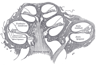
The cochlea is the part of the inner ear involved in hearing. It is a spiral-shaped cavity in the bony labyrinth, in humans making 2.75 turns around its axis, the modiolus. A core component of the cochlea is the organ of Corti, the sensory organ of hearing, which is distributed along the partition separating the fluid chambers in the coiled tapered tube of the cochlea.

<span class="mw-page-title-main">Vestibulocochlear nerve</span> Cranial nerve VIII, for hearing and balance

The vestibulocochlear nerve or auditory vestibular nerve, also known as the eighth cranial nerve, cranial nerve VIII, or simply CN VIII, is a cranial nerve that transmits sound and equilibrium (balance) information from the inner ear to the brain. Through olivocochlear fibers, it also transmits motor and modulatory information from the superior olivary complex in the brainstem to the cochlea.

<span class="mw-page-title-main">Basilar membrane</span> Stiff structural element within the cochlea of the inner ear which separates two liquid-filled tubes

The basilar membrane is a stiff structural element within the cochlea of the inner ear which separates two liquid-filled tubes that run along the coil of the cochlea, the scala media and the scala tympani. The basilar membrane moves up and down in response to incoming sound waves, which are converted to traveling waves on the basilar membrane.

<span class="mw-page-title-main">Saccule</span> Bed of sensory cells in the inner ear

The saccule is a bed of sensory cells in the inner ear. It translates head movements into neural impulses for the brain to interpret. The saccule detects linear accelerations and head tilts in the vertical plane. When the head moves vertically, the sensory cells of the saccule are disturbed and the neurons connected to them begin transmitting impulses to the brain. These impulses travel along the vestibular portion of the eighth cranial nerve to the vestibular nuclei in the brainstem.

<span class="mw-page-title-main">Organ of Corti</span> Receptor organ for hearing

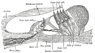
The organ of Corti, or spiral organ, is the receptor organ for hearing and is located in the mammalian cochlea. This highly varied strip of epithelial cells allows for transduction of auditory signals into nerve impulses' action potential. Transduction occurs through vibrations of structures in the inner ear causing displacement of cochlear fluid and movement of hair cells at the organ of Corti to produce electrochemical signals.

<span class="mw-page-title-main">Auditory system</span> Sensory system used for hearing

The auditory system is the sensory system for the sense of hearing. It includes both the sensory organs and the auditory parts of the sensory system.

<span class="mw-page-title-main">Hair cell</span> Auditory sensory receptor nerve cells

Hair cells are the sensory receptors of both the auditory system and the vestibular system in the ears of all vertebrates, and in the lateral line organ of fishes. Through mechanotransduction, hair cells detect movement in their environment.

In physiology, tonotopy is the spatial arrangement of where sounds of different frequency are processed in the brain. Tones close to each other in terms of frequency are represented in topologically neighbouring regions in the brain. Tonotopic maps are a particular case of topographic organization, similar to retinotopy in the visual system.

Presbycusis, or age-related hearing loss, is the cumulative effect of aging on hearing. It is a progressive and irreversible bilateral symmetrical age-related sensorineural hearing loss resulting from degeneration of the cochlea or associated structures of the inner ear or auditory nerves. The hearing loss is most marked at higher frequencies. Hearing loss that accumulates with age but is caused by factors other than normal aging is not presbycusis, although differentiating the individual effects of distinct causes of hearing loss can be difficult.

<span class="mw-page-title-main">Stereocilia (inner ear)</span> Mechanosensing organelles of hair cells

In the inner ear, stereocilia are the mechanosensing organelles of hair cells, which respond to fluid motion in numerous types of animals for various functions, including hearing and balance. They are about 10–50 micrometers in length and share some similar features of microvilli. The hair cells turn the fluid pressure and other mechanical stimuli into electric stimuli via the many microvilli that make up stereocilia rods. Stereocilia exist in the auditory and vestibular systems.

<span class="mw-page-title-main">Cochlear nerve</span> Nerve carrying auditory information from the inner ear to the brain

The cochlear nerve is one of two parts of the vestibulocochlear nerve, a cranial nerve present in amniotes, the other part being the vestibular nerve. The cochlear nerve carries auditory sensory information from the cochlea of the inner ear directly to the brain. The other portion of the vestibulocochlear nerve is the vestibular nerve, which carries spatial orientation information to the brain from the semicircular canals, also known as semicircular ducts.

<span class="mw-page-title-main">Cochlear nucleus</span> Two cranial nerve nuclei of the human brainstem

The cochlear nuclear (CN) complex comprises two cranial nerve nuclei in the human brainstem, the ventral cochlear nucleus (VCN) and the dorsal cochlear nucleus (DCN). The ventral cochlear nucleus is unlayered whereas the dorsal cochlear nucleus is layered. Auditory nerve fibers, fibers that travel through the auditory nerve carry information from the inner ear, the cochlea, on the same side of the head, to the nerve root in the ventral cochlear nucleus. At the nerve root the fibers branch to innervate the ventral cochlear nucleus and the deep layer of the dorsal cochlear nucleus. All acoustic information thus enters the brain through the cochlear nuclei, where the processing of acoustic information begins. The outputs from the cochlear nuclei are received in higher regions of the auditory brainstem.

<span class="mw-page-title-main">Tectorial membrane</span>

The tectoria membrane (TM) is one of two acellular membranes in the cochlea of the inner ear, the other being the basilar membrane (BM). "Tectorial" in anatomy means forming a cover. The TM is located above the spiral limbus and the spiral organ of Corti and extends along the longitudinal length of the cochlea parallel to the BM. Radially the TM is divided into three zones, the limbal, middle and marginal zones. Of these the limbal zone is the thinnest (transversally) and overlies the auditory teeth of Huschke with its inside edge attached to the spiral limbus. The marginal zone is the thickest (transversally) and is divided from the middle zone by Hensen's Stripe. It overlies the sensory inner hair cells and electrically-motile outer hair cells of the organ of Corti and during acoustic stimulation stimulates the inner hair cells through fluid coupling, and the outer hair cells via direct connection to their tallest stereocilia.

<span class="mw-page-title-main">Kinocilium</span> Sensory hair in the inner ear

A kinocilium is a special type of cilium on the apex of hair cells located in the sensory epithelium of the vertebrate inner ear. Contrasting with stereocilia, which are numerous, there is only one kinocilium on each hair cell. The kinocilium can be identified by its apical position as well as its enlarged tip.

<span class="mw-page-title-main">Hearing</span> Sensory perception of sound by living organisms

Hearing, or auditory perception, is the ability to perceive sounds through an organ, such as an ear, by detecting vibrations as periodic changes in the pressure of a surrounding medium. The academic field concerned with hearing is auditory science.

The cochlear amplifier is a positive feedback mechanism within the cochlea that provides acute sensitivity in the mammalian auditory system. The main component of the cochlear amplifier is the outer hair cell (OHC) which increases the amplitude and frequency selectivity of sound vibrations using electromechanical feedback.

Electrocochleography is a technique of recording electrical potentials generated in the inner ear and auditory nerve in response to sound stimulation, using an electrode placed in the ear canal or tympanic membrane. The test is performed by an otologist or audiologist with specialized training, and is used for detection of elevated inner ear pressure or for the testing and monitoring of inner ear and auditory nerve function during surgery.

Cochlea is Latin for “snail, shell or screw” and originates from the Greek word κοχλίας kokhlias. The modern definition, the auditory portion of the inner ear, originated in the late 17th century. Within the mammalian cochlea exists the organ of Corti, which contains hair cells that are responsible for translating the vibrations it receives from surrounding fluid-filled ducts into electrical impulses that are sent to the brain to process sound.

<span class="mw-page-title-main">A. James Hudspeth</span> American neutroscience professor

A. James Hudspeth is the F.M. Kirby Professor at Rockefeller University in New York City, where he is director of the F.M. Kirby Center for Sensory Neuroscience. His laboratory studies the physiological basis of hearing.

References

  1. 1 2 3 4 5 6 Hudspeth, AJ. (Oct 1989). "How the ear's works work". Nature. 341 (6241): 397–404. Bibcode:1989Natur.341..397H. doi:10.1038/341397a0. PMID   2677742. S2CID   33117543.
  2. 1 2 3 Hudspeth, AJ. (2001). "How the ear's works work: mechanoelectrical transduction and amplification by hair cells of the internal ear". Harvey Lect. 97: 41–54. PMID   14562516.
  3. 1 2 3 Hudde, H.; Weistenhofer, C. (2006). "Key features of the human middle ear". ORL J Otorhinolaryngol Relat Spec. 68 (6): 324–328. doi:10.1159/000095274. PMID   17065824. S2CID   42550955.
  4. 1 2 3 Hudspeth, AJ.; Konishi, M. (Oct 2000). "Auditory neuroscience: development, transduction, and integration". Proceedings of the National Academy of Sciences of the United States of America. 97 (22): 11690–1. doi: 10.1073/pnas.97.22.11690 . PMC   34336 . PMID   11050196.
  5. 1 2 3 4 5 6 Kaas, JH.; Hackett, TA.; Tramo, MJ. (Apr 1999). "Auditory processing in primate cerebral cortex" (PDF). Current Opinion in Neurobiology. 9 (2): 164–170. doi:10.1016/S0959-4388(99)80022-1. PMID   10322185. S2CID   22984374.
  6. 1 2 3 4 5 Fettiplace, R.; Hackney, CM. (Jan 2006). "The sensory and motor roles of auditory hair cells". Nat Rev Neurosci. 7 (1): 19–29. doi:10.1038/nrn1828. PMID   16371947. S2CID   10155096.
  7. Beurg, M.; Fettiplace, R.; Nam, JH.; Ricci, AJ. (May 2009). "Localization of inner hair cell mechanotransducer channels using high-speed calcium imaging". Nature Neuroscience. 12 (5): 553–558. doi:10.1038/nn.2295. PMC   2712647 . PMID   19330002.
  8. 1 2 3 4 5 6 Rubel, EW.; Fritzsch, B. (2002). "Auditory system development: primary auditory neurons and their targets". Annual Review of Neuroscience. 25: 51–101. doi:10.1146/annurev.neuro.25.112701.142849. PMID   12052904.
  9. 1 2 Chechik, Gal; Nelken (2012). "Auditory abstraction from spectro-temporal features to coding auditory entities". Proceedings of the National Academy of Sciences of the United States of America. 109 (44): 18968–73. Bibcode:2012PNAS..10918968C. doi: 10.1073/pnas.1111242109 . PMC   3503225 . PMID   23112145.
  10. Frisina, RD. (Aug 2001). "Subcortical neural coding mechanisms for auditory temporal processing". Hearing Research. 158 (1–2): 1–27. doi:10.1016/S0378-5955(01)00296-9. PMID   11506933. S2CID   36727875.
  11. Brigande, JV.; Heller, S. (Jun 2009). "Quo vadis, hair cell regeneration?". Nature Neuroscience. 12 (6): 679–685. doi:10.1038/nn.2311. PMC   2875075 . PMID   19471265.
  12. Lemus, L.; Hernández, A.; Romo, R. (Jun 2009). "Neural codes for perceptual discrimination of acoustic flutter in the primate auditory cortex". Proceedings of the National Academy of Sciences of the United States of America. 106 (23): 9471–9476. Bibcode:2009PNAS..106.9471L. doi: 10.1073/pnas.0904066106 . PMC   2684844 . PMID   19458263.
  13. Kandler, K.; Clause, A.; Noh, J. (Jun 2009). "Tonotopic reorganization of developing auditory brainstem circuits". Nature Neuroscience. 12 (6): 711–7. doi:10.1038/nn.2332. PMC   2780022 . PMID   19471270.