Oil Red O

Last updated
Oil Red O
Oil Red O.svg
Differentiated 3T3-L1 Cell line stained with Oil O Red.jpg
Differentiated adipocytes in a 3T3-L1 cell line stained with Oil Red O
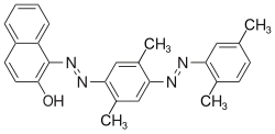
Names
IUPAC name
1-(2,5-dimethyl-4-(2,5-dimethylphenyl) phenyldiazenyl) azonapthalen-2-ol
Identifiers
3D model (JSmol)
ChEBI
ChemSpider
ECHA InfoCard 100.013.906 OOjs UI icon edit-ltr-progressive.svg
MeSH oil+red+O
PubChem CID
UNII
  • InChI=1S/C26H24N4O/c1-16-9-10-17(2)22(13-16)27-28-23-14-19(4)24(15-18(23)3)29-30-26-21-8-6-5-7-20(21)11-12-25(26)31/h5-15,31H,1-4H3/b28-27+,30-29+ Yes check.svgY
    Key: NPGIHFRTRXVWOY-XOXGWFOHSA-N Yes check.svgY
  • InChI=1/C26H24N4O/c1-16-9-10-17(2)22(13-16)27-28-23-14-19(4)24(15-18(23)3)29-30-26-21-8-6-5-7-20(21)11-12-25(26)31/h5-15,31H,1-4H3/b28-27+,30-29+
    Key: NPGIHFRTRXVWOY-XOXGWFOHBH
  • Cc4cc(/N=N/c1cc(C)c(cc1C)/N=N/c2c3ccccc3ccc2O)c(C)cc4
Properties
C26H24N4O
Molar mass 408.49496
Except where otherwise noted, data are given for materials in their standard state (at 25 °C [77 °F], 100 kPa).
X mark.svgN  verify  (what is  Yes check.svgYX mark.svgN ?)

Oil Red O (Solvent Red 27, Sudan Red 5B, C.I. 26125, C26H24N4O) is a lysochrome (fat-soluble dye) diazo dye used for staining of neutral triglycerides and lipids on frozen sections and some lipoproteins on paraffin sections. It has the appearance of a red powder with an absorbance maximum at 518 nanometers. [1]

Contents

Uses

Oil Red O is one of the dyes used for Sudan staining. Similar dyes include Sudan III, Sudan IV, and Sudan Black B. The staining has to be performed on fresh samples, as alcohol fixation removes most lipids. Oil Red O largely replaced Sudan III and Sudan IV, as it provides much deeper red color and the stains are therefore much easier to see.

Oil Red O can be used to mark lipid-containing vacuoles, particularly in cases of acute lymphoblastic leukemia or Burkitt's lymphoma. It can also be used to stain liver sections for histological analysis, quantify cell lipid content, and to stain the aorta to examine lesions from atherosclerosis. [2]

In pyrotechnics, Oil Red O is used in some compositions of red colored smokes.

Forensic

When staining, Oil Red O can make fat more visible in various cuts in pathology. [3]

It is also used in a technique (the method is called as the dye: Oil Red O), discovered in 2004 by Alexandre Beaudoin, for staining latent fingerprints. [4] This technique allows the development of latent fingerprints on porous exhibits (such as paper, cardboard, etc.) that are dry or wet.

It mainly targets fat deposits on the surface of porous exhibits. [5] It is a non-destructive technique (which does not destroy the exhibit nor prevents the use of other techniques).

It is a safe alternative to the Physical Developer method, [6] and is also used in sequence with other methods of fingerprints development. [7]

Related Research Articles

<span class="mw-page-title-main">Forensic science</span> Application of science to criminal and civil laws

Forensic science, also known as criminalistics, is the application of science to criminal and civil laws. During criminal investigation in particular, it is governed by the legal standards of admissible evidence and criminal procedure. It is a broad field utilizing numerous practices such as the analysis of DNA, fingerprints, bloodstain patterns, firearms, ballistics, and toxicology.

<span class="mw-page-title-main">Fingerprint</span> Biometric identifier

A fingerprint is an impression left by the friction ridges of a human finger. The recovery of partial fingerprints from a crime scene is an important method of forensic science. Moisture and grease on a finger result in fingerprints on surfaces such as glass or metal. Deliberate impressions of entire fingerprints can be obtained by ink or other substances transferred from the peaks of friction ridges on the skin to a smooth surface such as paper. Fingerprint records normally contain impressions from the pad on the last joint of fingers and thumbs, though fingerprint cards also typically record portions of lower joint areas of the fingers.

<span class="mw-page-title-main">Staining</span> Technique used to enhance visual contrast of specimens observed under a microscope

Staining is a technique used to enhance contrast in samples, generally at the microscopic level. Stains and dyes are frequently used in histology, in cytology, and in the medical fields of histopathology, hematology, and cytopathology that focus on the study and diagnoses of diseases at the microscopic level. Stains may be used to define biological tissues, cell populations, or organelles within individual cells.

<span class="mw-page-title-main">Steatosis</span> Medical condition

Steatosis, also called fatty change, is abnormal retention of fat (lipids) within a cell or organ. Steatosis most often affects the liver – the primary organ of lipid metabolism – where the condition is commonly referred to as fatty liver disease. Steatosis can also occur in other organs, including the kidneys, heart, and muscle. When the term is not further specified, it is assumed to refer to the liver.

Dye penetrant inspection (DP), also called liquid penetrate inspection (LPI) or penetrant testing (PT), is a widely applied and low-cost inspection method used to check surface-breaking defects in all non-porous materials. The penetrant may be applied to all non-ferrous materials and ferrous materials, although for ferrous components magnetic-particle inspection is often used instead for its subsurface detection capability. LPI is used to detect casting, forging and welding surface defects such as hairline cracks, surface porosity, leaks in new products, and fatigue cracks on in-service components.

<span class="mw-page-title-main">Histopathology</span> Microscopic examination of tissue in order to study and diagnose disease

Histopathology refers to the microscopic examination of tissue in order to study the manifestations of disease. Specifically, in clinical medicine, histopathology refers to the examination of a biopsy or surgical specimen by a pathologist, after the specimen has been processed and histological sections have been placed onto glass slides. In contrast, cytopathology examines free cells or tissue micro-fragments.

<span class="mw-page-title-main">Ziehl–Neelsen stain</span> A type of acid-fast stain

The Ziehl-Neelsen stain, also known as the acid-fast stain, is a bacteriological staining technique used in cytopathology and microbiology to identify acid-fast bacteria under microscopy, particularly members of the Mycobacterium genus. This staining method was initially introduced by Paul Ehrlich (1854–1915) and subsequently modified by the German bacteriologists Franz Ziehl (1859–1926) and Friedrich Neelsen (1854–1898) during the late 19th century.

<span class="mw-page-title-main">Sudan IV</span> Chemical compound

Sudan IV (C24H20N4O) is a lysochrome (fat-soluble dye) diazo dye used for the staining of lipids, triglycerides and lipoproteins on frozen paraffin sections. It has the appearance of reddish brown crystals with melting point 199 °C and maximum absorption at 520(357) nm.

<span class="mw-page-title-main">Sudan III</span> Chemical compound

Sudan III is a lysochrome diazo dye. It is structurally related to azobenzene.

Solvent Black 3 is an azo dye. It is a non-fluorescent, relatively thermostable lysochrome diazo dye used for staining of neutral triglycerides and lipids on frozen sections and some lipoproteins on paraffin sections. It has the appearance of a dark brown to black powder with maximum absorption at 596–605 nm and melting point 120–124 °C. It stains blue-black.

<span class="mw-page-title-main">Sudan II</span> Chemical compound

Sudan II (Solvent Orange 7, C.I. 12140, C18H16N2O) is a lysochrome (fat-soluble dye) azo dye used for staining of triglycerides in frozen sections, and some protein bound lipids and lipoproteins on paraffin sections. It has the appearance of red powder with melting point 156–158 °C and maximum absorption at 493(420) nm.

<span class="mw-page-title-main">H&E stain</span> Histological stain method

Hematoxylin and eosin stain is one of the principal tissue stains used in histology. It is the most widely used stain in medical diagnosis and is often the gold standard. For example, when a pathologist looks at a biopsy of a suspected cancer, the histological section is likely to be stained with H&E.

Sudan stains and Sudan dyes are synthetic organic compounds that are used as dyes for various plastics and are also used to stain sudanophilic biological samples, usually lipids. Sudan II, Sudan III, Sudan IV, Oil Red O, and Sudan Black B are important members of this class of compounds.

<span class="mw-page-title-main">Fingerprint powder</span> Chemical compound used to lift fingerprints from surfaces

Fingerprint powders are fine powders used, in conjunction with fingerprint brushes, by crime scene investigators and other law enforcement personnel to search for and enhance latent/invisible fingerprints that can be used to determine identification. This method of fingerprint development commonly referred to as dusting for fingerprints, involves the adherence of the powder particles to the moisture and sweat secretions deposited on to surfaces by the raised ridges on fingers, palms, or soles of feet designed for grip, called friction ridges.

<span class="mw-page-title-main">Lawsone</span> Chemical compound

Lawsone (2-hydroxy-1,4-naphthoquinone), also known as hennotannic acid, is a red-orange dye present in the leaves of the henna plant, for which it is named, as well as in the common walnut and water hyacinth. Humans have used henna extracts containing lawsone as hair and skin dyes for more than 5,000 years. Lawsone reacts chemically with the protein keratin in skin and hair via a Michael addition reaction, resulting in a strong permanent stain that lasts until the skin or hair is shed. Darker colored staining is due to more lawsone–keratin interactions occurring, which evidently break down as the concentration of lawsone decreases and the tattoo fades. Lawsone strongly absorbs UV light, and aqueous extracts can be effective sunless tanning agents and sunscreens. Lawsone is a 1,4-naphthoquinone derivative, an analog of hydroxyquinone containing one additional ring.

<span class="mw-page-title-main">Toluidine blue</span> Chemical compound

Toluidine blue, also known as TBO or tolonium chloride (INN) is a blue cationic (basic) dye used in histology and sometimes clinically.

<span class="mw-page-title-main">Alexandre Beaudoin</span> Canadian forensic scientist (born 1978)

Alexandre Beaudoin is a Quebec fingerprint scientist known for inventing a technique for developing latent fingerprints on dry and/or wet porous surfaces.

Brian E. Dalrymple is an Ontarian fingerprint scientist known for introducing for the first time the use of lasers as a forensic light sources for fingerprints and other evidence detection, using the Argon Ion Lasers to detect the inherent fluorescence of the latent fingerprints and finding fluorescing evidence. That was the beginning of a real revolution in the forensic identification field. Brian Dalrymple also become the first to use this forensic technique on a real case.

<span class="mw-page-title-main">Luxol fast blue stain</span>

Luxol fast blue stain, abbreviated LFB stain or simply LFB, is a commonly used stain to observe myelin under light microscopy, created by Heinrich Klüver and Elizabeth Barrera in 1953. LFB is commonly used to detect demyelination in the central nervous system (CNS), but cannot discern myelination in the peripheral nervous system.

<span class="mw-page-title-main">Glove prints</span>

Glove prints, also sometimes described as gloveprints or glove marks, are latent, fingerprint-like impressions that are transferred to a surface or object by an individual who is wearing gloves.

References

  1. Kraus, Nils A.; Ehebauer, Franziska; Zapp, Benedikt; et al. (2016). "Quantitative assessment of adipocyte differentiation in cell culture". Adipocyte. 5 (4): 351–358. doi: 10.1080/21623945.2016.1240137 . ISSN   2162-3945. PMC   5160397 . PMID   27994948.
  2. "Oil Red O". MilliporeSigma. Retrieved 15 February 2023.
  3. "Forensic Pathology".
  4. Triplett M, Fingerprint Dictionary, Two Rings Publishing, Bellevue, Washington.
  5. Beaudoin, A. New technique for revealing latent fingerprints on wet, porous surfaces: Oil Red O. Journal of Forensic Identification, 2004, 54 (4), 413-421.
  6. Rawji, A. ; Beaudoin, A. Oil Red O versus Physical Developer on wet papers: a comparative study. Journal of Forensic Identification, 2006, 56 (1), 33-54.
  7. Guigui, K.; Beaudoin, A. The use of Oil Red O in sequence with other methods of fingerprint development. Journal of Forensic Identification, 2007, 57 (4), 550-581.