Paecilomyces variotii

Last updated

Paecilomyces variotii
Paecilomyces variotii culture.jpg
Scientific classification OOjs UI icon edit-ltr.svg
Domain: Eukaryota
Kingdom: Fungi
Division: Ascomycota
Class: Eurotiomycetes
Order: Eurotiales
Family: Thermoascaceae
Genus: Paecilomyces
Species:
P. variotii
Binomial name
Paecilomyces variotii
Bainier (1907)
Synonyms
  • Byssochlamys spectabilis(Udagawa & Shoji Suzuki) Houbraken & Samson (2008) [1]
  • Paecilomyces aureocinnamomeum(Biourge) Thom (1930) [1]
  • Paecilomyces varioti [1]
  • Penicillium aureocinnamomeumBiourge (1923) [1]
  • Penicillium variotii(Bainier) Sacc. (1913) [1]
  • Talaromyces spectabilisUdagawa & Shoji Suzuki (1994) [1]

Paecilomyces variotii, also known by the name Byssochlamys spectabilis for the sexual state, is a common environmental mold from the Phylum Ascomycota (Family Thermoascaceae). [1] It is widespread in the environment and can be found in composts, soils and wood, [2] [3] [4] as well es a common environmental contaminant in indoor air and carpet dust. [5] Ascospores of the sexual state of P. variotii (B. spectabilis) are strongly heat-resistant. As such the fungus is a common contaminant of heat-treated foods and juices. Paecilomyces variotii has been associated with a number of infective diseases of humans and animals. [6]

Contents

Taxonomy

Paecilomyces variotii was first described by Georges Bainier in 1907, while its teleomorph was described in 1994 and ranked in Byssochlamys . P. variotii is closely related to P. brunneolus , P. formosus , P. divaricatus and P. dactylethromorphus . [7]

Morphology

Paecilomyces variotii is fast growing, producing powdery to suede-like colonies that are yellow-brown or sand-colored. [8] It is distinguishable from microscopically similar microfungi, such as the biverticillate members of the genus Penicillium (affiliated with the genus Talaromyces) by its broadly ellipsoidal to lemon-shaped conidia, loosely branched conidiophores and phialides with pointed tips.

The colonies are usually flat, powdery to suede-like and funiculose or tufted. [9] The color is initially white, and becomes yellow, yellow-brown, or sand-colored as they mature. A sweet aromatic odor may be associated with older cultures. [10] Colonies of P. variotii are fast growing and mature within 3 days. Colonies grown on Sabouraud's dextrose agar reach about 7–8 mm after one week. Colonies on CYA are flat, floccose in texture, produce brown or olive brown from conidia, and range in diameter from 30–79 mmn in one week. [11] Colonies on malt extract agar reach 70 mm diameter or more, otherwise very similar in appearance to those on CYA. Colonies on G25N media reach 8–16 mm diameter, similar to on CYA but with predominantly white mycelium. Microscopically, the spore-bearing structures of P. variotii consist of a loosely branched, [12] irregularly brush-like conidiophores with phialides at the tips. [13] [9] The phialides are swollen at the base, and gradually taper to a sharp point at the tip. [12] Conidia are single-celled, hyaline, and are borne in chains with the youngest at the base. [13] Chlamydospores (thick-walled vegetative resting structures) are occasionally produced singly or in short chains. [14]

The teleomorph of Paecilomyces variotii has also been described by the name Byssochlamys spectabilis. [7] However, the Byssochlamys state is rarely observed in culture due to the heterothallic nature of this species (i.e., it requires culturing of positive and negative strains in co-culture to produce the teleomorph).

Genetics

This fungus is heterothallic, and mating experiments have shown that P. variotii can form ascomata and ascospores in culture when compatible mating types are present. [2] [13]

The teleomorph of P. variotii, Byssochlamys spectabilis, is rarely observed in cultures from environmental or clinical specimens, which tend to be colonized by a single mating type. [13] The genome sequences of two isolates of P. variotii of opposite mating type have been generated. [15]

Ecology

This species is thermophilic, able to grow at high temperatures as high as 50–60 °C. [13] [10] It can withstand brief exposures of up to 15 min at 80–100 °C. [16] Accordingly, it typically causes spoilage of food products following pasteurization or other heat-treatments (e.g., curry sauces, fruit juices). [16] [17] It also has been reported as a contaminant in salami and margarine. [5] The fungus is known from a number of non-food items including compost, rubber, glue, urea-formaldehyde foam insulation and creosote-treated wooden poles. [5] [17] The combination of its ability to survive significant heat stress and its ability to break-down aromatic hydrocarbons has led to interest in P. variotii as a potential candidate organism to assist in bioremediation.

It is also known from decaying wood and creosote-treated wood utility poles. [13] [8]

Health significance

Although frequently encountered as a contaminant in clinical specimens, P. variotii is an uncommon causative agent of human and animal infections, but is considered to be an emerging agent of opportunistic disease, particularly in immunocompromised individuals. It has been suggested that the extremotolerant nature of the fungus contributes to its pathogenic potential. P. variotii is one of the most commonly encountered species in cases of cutaneous hyalohyphomycosis. [7] [18] [19] [20] Pneumonia due to P. variotii has been reported, albeit rarely, in the medical literature. [21] [22] Most cases are known from diabetics or individuals subject to long-term corticosteroid treatment for other diseases. [23] [24] P. variotii has also been reported as a causative agent of sinusitis, [25] [26] [27] endophthalmitis, [28] [29] [30] wound infection following tissue transplant, [31] onychomycosis, [32] osteomyelitis, [33] otitis media [34] and dialysis-related peritonitis. [6] [35] It has also been reported from mastitis in a goat, and as an agent of mycotic infections of dogs and horses. [ citation needed ] Besides clinical samples, the fungus is a common contaminant of moisture-damaged materials in the indoor environment including carpet, plaster and wood. [5] It is commonly found in indoor air samples and may contribute to indoor allergy. [5] This species produces the mycotoxin viriditoxin, [5] via the action of six enzymes encoded within a cluster of genes within the genome. [36]

Related Research Articles

<i>Penicillium</i> Genus of fungi

Penicillium is a genus of ascomycetous fungi that is part of the mycobiome of many species and is of major importance in the natural environment, in food spoilage, and in food and drug production.

<i>Aspergillus niger</i> Species of fungus

Aspergillus niger is a mold classified within the Nigri section of the Aspergillus genus. The Aspergillus genus consists of common molds found throughout the environment within soil and water, on vegetation, in fecal matter, on decomposing matter, and suspended in the air. Species within this genus often grow quickly and can sporulate within a few days of germination. A combination of characteristics unique to A. niger makes the microbe invaluable to the production of many acids, proteins and bioactive compounds. Characteristics including extensive metabolic diversity, high production yield, secretion capability, and the ability to conduct post-translational modifications are responsible for A. niger's robust production of secondary metabolites. A. niger's capability to withstand extremely acidic conditions makes it especially important to the industrial production of citric acid.

<i>Paecilomyces</i> Genus of fungi

Paecilomyces is a genus of fungi. A number of species in this genus are plant pathogens.
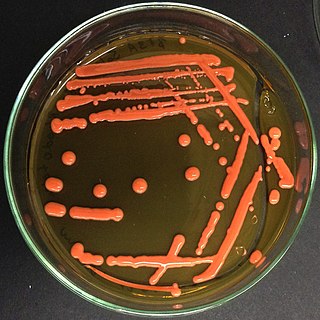
Hyphomycetes are a form classification of fungi, part of what has often been referred to as fungi imperfecti, Deuteromycota, or anamorphic fungi. Hyphomycetes lack closed fruit bodies, and are often referred to as moulds. Most hyphomycetes are now assigned to the Ascomycota, on the basis of genetic connections made by life-cycle studies or by phylogenetic analysis of DNA sequences; many remain unassigned phylogenetically.

<i>Acrophialophora fusispora</i> Species of ascomycete fungus found in soil, air and various plants

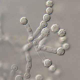
Acrophialophora fusispora is a poorly studied ascomycete fungus found in soil, air and various plants. A. fusispora is morphologically similar to the genera Paecilomyces and Masonia, but differ in the presence of pigmented conidiophores, verticillate phialides, and frequent sympodial proliferation. Moreover, A. fusispora is distinguished by its pigmented spindle-shaped conidia, covered with spiral bands. The fungus is naturally found in soils of tropical to temperate regions. The fungus has been identified as a plant and animal pathogen, and has recently been recognized as an emerging opportunistic human pathogen. A. fusispora infection in human is rare and has few documented clinical cases, but due to the rarity of the fungus and potential misidentification, the infections may be underdiagnosed. Clinical cases of A. fusispora include cases of keratitis, pulmonary colonization and infection, and cerebral infections. The fungus also has two documented cases of infection in dogs.

<i>Penicillium chrysogenum</i> Species of fungus

Penicillium chrysogenum is a species of fungus in the genus Penicillium. It is common in temperate and subtropical regions and can be found on salted food products, but it is mostly found in indoor environments, especially in damp or water-damaged buildings. It has been recognised as a species complex that includes P. notatum, P. meleagrinum, and P. cyaneofulvum. Molecular phylogeny has established that Alexander Fleming's first discovered penicillin producing strain is of a distinct species, P. rubens, and not of P. notatum. It has rarely been reported as a cause of human disease. It is the source of several β-lactam antibiotics, most significantly penicillin. Other secondary metabolites of P. chrysogenum include roquefortine C, meleagrin, chrysogine, 6-MSA YWA1/melanin, andrastatin A, fungisporin, secalonic acids, sorbicillin, and PR-toxin.

<i>Purpureocillium lilacinum</i> Species of fungus

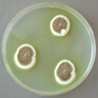
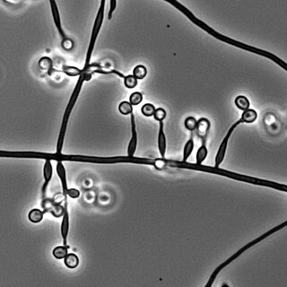
Purpureocillium lilacinum is a species of filamentous fungus in the family Ophiocordycipitaceae. It has been isolated from a wide range of habitats, including cultivated and uncultivated soils, forests, grassland, deserts, estuarine sediments and sewage sludge, and insects. It has also been found in nematode eggs, and occasionally from females of root-knot and cyst nematodes. In addition, it has frequently been detected in the rhizosphere of many crops. The species can grow at a wide range of temperatures – from 8 to 38 °C for a few isolates, with optimal growth in the range 26 to 30 °C. It also has a wide pH tolerance and can grow on a variety of substrates. P. lilacinum has shown promising results for use as a biocontrol agent to control the growth of destructive root-knot nematodes.

Coniochaeta hoffmannii, also known as Lecythophora hoffmannii, is an ascomycete fungus that grows commonly in soil. It has also been categorized as a soft-rot fungus capable of bringing the surface layer of timber into a state of decay, even when safeguarded with preservatives. Additionally, it has pathogenic properties, although it causes serious infection only in rare cases. A plant pathogen lacking a known sexual state, C. hoffmannii has been classified as a "dematiaceous fungus" despite its contradictory lack of pigmentation; both in vivo and in vitro, there is no correlation between its appearance and its classification.

<i>Microascus brevicaulis</i> Species of fungus

Microascus brevicaulis is a microfungus in the Ascomycota. It is the teleomorph form of Scopulariopsis brevicaulis.Microascus brevicaulis occurs world-wide as a saprotroph in soil, a common agent of biodeterioration, an irregular plant pathogen, and an occasional agent of human nail infection.

<i>Aspergillus ustus</i> Species of fungus

Aspergillus ustus is a microfungus and member of the division Ascomycota. It is commonly found in indoor environments and soil. Isolated cases of human infection resulting from A. ustus have been described; however the majority of these are nail infections.

Rasamsonia is a genus of fungi in the family Trichocomaceae, circumscribed in 2011 by mycologists Jos Houbraken and Jens Frisvad. It is characterized from other genera of the Trichocomaceae by the following combination of features: species are thermotolerant or thermophilic; their conidiophores have distinctly rough-walled stipes; conidia are olive brown; and ascomata, if present, have minimal covering. Rasamsonia phenotypically resembles Paecilomyces, in that both have thermotolerant species, produce olive-brown conidia, and form ascomata with no or scarce ascomatal covering; Rasamsonia, however, differs from Paecilomyces in having more regularly branched conidiophores with distinct rough-walled structures. The type species is Rasamsonia emersonii, a fungus formerly classified in the genus Talaromyces.

<i>Aspergillus tubingensis</i> Species of fungus

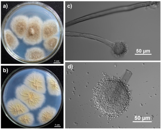
Aspergillus tubingensis is a darkly pigmented species of fungus in the genus Aspergillus section Nigri. It is often confused with Aspergillus niger due to their similar morphology and habitat. A. tubingensis is often involved in food spoilage of fruits and wheat, and industrial fermentation. This species is a rare agent of opportunistic infection.

Penicillium commune is an indoor fungus belonging to the genus Penicillium. It is known as one of the most common fungi spoilage moulds on cheese. It also grows on and spoils other foods such as meat products and fat-containing products like nuts and margarine. Cyclopiazonic acid and regulovasine A and B are the most important mycotoxins produced by P. commune. The fungus is the only known species to be able to produce both penitrem A and roquefortine. Although this species does not produce penicillin, it has shown to have anti-pathogenic activity. There are no known plant, animal or human diseases caused by P. commune.

<i>Penicillium spinulosum</i> Species of fungus

Penicillium spinulosum is a non-branched, fast-growing fungus with a swelling at the terminal of the stipe (vesiculate) in the genus Penicillium. P. spinulosum is able to grow and reproduce in environment with low temperature and low water availability, and is known to be acidotolerant. P. spinulosum is ubiquitously distributed, and can often be isolated from soil. Each individual strain of P. spinulosum differs from others in their colony morphology, including colony texture, amount of sporulation and roughness of conidia and conidiophores.

<i>Mariannaea elegans</i> Species of fungus

Mariannaea elegans an anamorphic fungus. It is mainly found on rotting wood and soil. M. elegans is not pathogenic to humans, animals, or plants.

<i>Metarhizium granulomatis</i> Species of fungus

Metarhizium granulomatis is a fungus in the family Clavicipitaceae associated with systemic mycosis in veiled chameleons. The genus Metarhizium is known to infect arthropods, and collectively are referred to green-spored asexual pathogenic fungi. This species grows near the roots of plants and has been reported as an agent of disease in captive veiled chameleons. The etymology of the species epithet, "granulomatis" refers to the ability of the fungus to cause granulomatous disease in susceptible reptiles.

Sarocladium kiliense is a saprobic fungus that is occasionally encountered as a opportunistic pathogen of humans, particularly immunocompromised and individuals. The fungus is frequently found in soil and has been linked with skin and systemic infections. This species is also known to cause disease in the green alga, Cladophora glomerata as well as various fruit and vegetable crops grown in warmer climates.

Microascus manginii is a filamentous fungal species in the genus Microascus. It produces both sexual (teleomorph) and asexual (anamorph) reproductive stages known as M. manginii and Scopulariopsis candida, respectively. Several synonyms appear in the literature because of taxonomic revisions and re-isolation of the species by different researchers. M. manginii is saprotrophic and commonly inhabits soil, indoor environments and decaying plant material. It is distinguishable from closely related species by its light colored and heart-shaped ascospores used for sexual reproduction. Scopulariopsis candida has been identified as the cause of some invasive infections, often in immunocompromised hosts, but is not considered a common human pathogen. There is concern about amphotericin B resistance in S. candida.

Aspergillus viridinutans is a species of fungus in the genus Aspergillus. The species was first isolated in Frankston, Victoria, Australia and described in 1954. It is from the Fumigati section of Aspergillus. Several fungi from this section produce heat-resistant ascospores, and the isolates from this section are frequently obtained from locations where natural fires have previously occurred. A. viridinutans has been identified as the cause of chronic aspergillosis. The mycotoxin viriditoxin was first identified in A. viridinutans. A draft genome sequence of the strain derived from the original species description has been generated.

Myriodontium keratinophilum is a fungus widespread in nature, most abundantly found in keratin-rich environments such as feathers, nails and hair. Despite its ability to colonize keratinous surfaces of human body, the species has been known to be non-pathogenic in man and is phylogentically distant to other human pathogenic species, such as anthropophilic dermatophytes. However, its occasional isolation from clinical specimens along with its keratinolytic properties suggest the possibility it may contribute to disease.

References

  1. 1 2 3 4 5 6 7 Page Paecilomyces variotii on "Mycobank Database". Westerdijk Fungal Biodiversity Institute . Retrieved 2023-09-06.
  2. 1 2 Houbraken J, Varga J, Rico-Munoz E, Johnson S, Samson RA (March 2008). "Sexual reproduction as the cause of heat resistance in the food spoilage fungus Byssochlamys spectabilis (anamorph Paecilomyces variotii)". Applied and Environmental Microbiology. 74 (5): 1613–9. Bibcode:2008ApEnM..74.1613H. doi:10.1128/aem.01761-07. PMC   2258620 . PMID   18192427.
  3. Pitt JL, Hocking AD (2009). Fungi and food spoilage (3rd ed.). Bibcode:2009ffs..book.....P.
  4. Guarro J, Chander J, Alvarez E, Stchigel AM, Robin K, Dalal U, Rani H, Punia RS, Cano JF (January 2011). "Apophysomyces variabilis infections in humans". Emerging Infectious Diseases. 17 (1): 134–5. doi:10.3201/eid1701.101139. PMC   3204648 . PMID   21192877.
  5. 1 2 3 4 5 6 Samson RA, Houbraken J, Summerbell RC, Flannigan B, Miller JD (2001). "Common and important species of fungi and actinomycetes in indoor environment". In Flannigan B, Samson RA, Miller JD (eds.). Microorganisms in Home and Indoor Work Environments. CRC Press. pp. 285–473. ISBN   978-0-203-30293-4.
  6. 1 2 Rinaldi S, Fiscarelli E, Rizzoni G (May 2000). "Paecilomyces variotii peritonitis in an infant on automated peritoneal dialysis". Pediatric Nephrology. 14 (5): 365–6. doi:10.1007/s004670050775. PMID   10805461. S2CID   21567918.
  7. 1 2 3 Houbraken J, Verweij PE, Rijs AJ, Borman AM, Samson RA (August 2010). "Identification of Paecilomyces variotii in clinical samples and settings". Journal of Clinical Microbiology. 48 (8): 2754–61. doi:10.1128/jcm.00764-10. PMC   2916617 . PMID   20519470.
  8. 1 2 Ellis D (May 2001). "Paecilomyces variotii." Mycology Online". The University of Adelaide.
  9. 1 2 Bainier D (Nov 2013). "Paecilomyces Species". Paecilomyces Species.
  10. 1 2 de Hoog GS, Guarro J, Gene J, Figueras MJ (2000). Atlas of Clinical Fungi. Vol. 1 (2nd ed.). Reus, Spain: Universitat Rovira i Virgili. ISBN   978-90-70351-43-4.
  11. Pitt J, Hocking AD (1985). "Fungi and Food Spoilage". Sydney: Academic: 186–96.
  12. 1 2 Samson RA (1974). Paecilomyces and some allied hyphomycetes. Vol. 6. pp. 1–119. ISBN   978-94-91751-00-4.{{cite book}}: |journal= ignored (help)
  13. 1 2 3 4 5 6 Houbraken J, Verweij PE, Rijs AJ, Borman AM, Samson RA (August 2010). "Identification of Paecilomyces variotii in clinical samples and settings". Journal of Clinical Microbiology. 48 (8): 2754–61. doi:10.1128/jcm.00764-10. PMC   2916617 . PMID   20519470.
  14. Sampson RA (1975). "Paecilomyces and Some Allied Hyphomycetes". Transactions of the British Mycological Society. 64: 174. doi:10.1016/s0007-1536(75)80098-2.
  15. Urquhart AS, Mondo SJ, Mäkelä MR, Hane JK, Wiebenga A, He G, Mihaltcheva S, Pangilinan J, Lipzen A, Barry K, de Vries RP, Grigoriev IV, Idnurm A (2018). "Paecilomyces variotii (Eurotiales)". Frontiers in Microbiology. 9: 3058. doi: 10.3389/fmicb.2018.03058 . PMC   6300479 . PMID   30619145.
  16. 1 2 Piecková E, Samson RA (2000). "Heat Resistance of Paecilomyces variotii in Sauce and Juice". Journal of Industrial Microbiology and Biotechnology. 24 (4): 227–30. doi: 10.1038/sj.jim.2900794 . S2CID   35360797.
  17. 1 2 Samson RA, Hoekstra ES, Frisvad JC (2004). Introduction to food- and airborne fungi (7th ed.). Centraalbureau voor Schimmelcultures. ISBN   978-90-70351-52-6.
  18. Athar MA, Sekhon AS, Mcgrath JV, Malone RM (February 1996). "Hyalohyphomycosis caused by Paecilomyces variotii in an obstetrical patient". European Journal of Epidemiology. 12 (1): 33–5. doi:10.1007/bf00144425. PMID   8817175. S2CID   21416639.
  19. Naidu J, Singh SM (October 1992). "Hyalohyphomycosis caused by Paecilomyces variotii: a case report, animal pathogenicity and 'in vitro' sensitivity". Antonie van Leeuwenhoek. 62 (3): 225–30. doi:10.1007/bf00582583. PMID   1416918. S2CID   7611095.
  20. Vasudevan B, Hazra N, Verma R, Srinivas V, Vijendran P, Badad A (June 2013). "First reported case of subcutaneous hyalohyphomycosis caused by Paecilomyces variotii". International Journal of Dermatology. 52 (6): 711–3. doi: 10.1111/j.1365-4632.2012.05761.x . PMID   23679876. S2CID   32544478.
  21. Saddad N, Shigemitsu H, Christianson A (2007). "Pneumonia from Paecilomyces in a 67-year-old immunocompetent man". Chest. 132 (4): 710. doi: 10.1378/chest.132.4_meetingabstracts.710 .
  22. Grossman CE, Fowler A (2005). "Paecilomyces: emerging fungal pathogen". Chest. 128 (4): 425S. doi:10.1378/chest.128.4_meetingabstracts.425s.
  23. Byrd RP, Roy TM, Fields CL, Lynch JA (1992). "Paecilomyces varioti pneumonia in a patient with diabetes mellitus". Journal of Diabetes and Its Complications. 6 (2): 150–3. doi:10.1016/1056-8727(92)90027-i. PMID   1611140.
  24. Steiner B, Aquino VR, Paz AA, Silla LM, Zavascki A, Goldani LZ (2013). "Paecilomyces variotii as an Emergent Pathogenic Agent of Pneumonia". Case Reports in Infectious Diseases. 2013: 1–3. doi: 10.1155/2013/273848 . PMC   3683431 . PMID   23819077.
  25. Eloy P, Bertrand B, Rombeaux P, Delos M, Trigaux JP (1997). "Mycotic sinusitis". Acta Oto-rhino-laryngologica Belgica. 51 (4): 339–52. PMID   9444380.
  26. Otcenásek M, Jirousek Z, Nozicka Z, Mencl K (May 1984). "Paecilomycosis of the maxillary sinus". Mykosen. 27 (5): 242–51. doi:10.1111/j.1439-0507.1984.tb02025.x. PMID   6540366. S2CID   32910633.
  27. Thompson RF, Bode RB, Rhodes JC, Gluckman JL (May 1988). "Paecilomyces variotii. An unusual cause of isolated sphenoid sinusitis". Archives of Otolaryngology–Head & Neck Surgery. 114 (5): 567–9. doi:10.1001/archotol.1988.01860170097028. PMID   3355698.
  28. Lam DS, Koehler AP, Fan DS, Cheuk W, Leung AT, Ng JS (1999). "Endogenous fungal endophthalmitis caused by Paecilomyces variotii". Eye. 13 ( Pt 1): 113–6. doi: 10.1038/eye.1999.23 . PMID   10396397.
  29. Tarkkanen A, Raivio V, Anttila VJ, Tommila P, Ralli R, Merenmies L, Immonen I (April 2004). "Fungal endophthalmitis caused by Paecilomyces variotii following cataract surgery: a presumed operating room air-conditioning system contamination". Acta Ophthalmologica Scandinavica. 82 (2): 232–5. doi: 10.1111/j.1600-0420.2004.00235.x . PMID   15043549.
  30. Anita KB, Fernandez V, Rao R (2010). "Fungal endophthalmitis caused by Paecilomyces variotii, in an immunocompetent patient, following intraocular lens implantation". Indian Journal of Medical Microbiology. 28 (3): 253–4. doi: 10.4103/0255-0857.66491 . PMID   20644318.
  31. Lee J, Yew WW, Chiu CS, Wong PC, Wong CF, Wang EP (October 2002). "Delayed sternotomy wound infection due to Paecilomyces variotii in a lung transplant recipient". The Journal of Heart and Lung Transplantation. 21 (10): 1131–4. doi:10.1016/s1053-2498(02)00404-7. PMID   12398880.
  32. Arenas R, Arce M, Munoz H, Ruiz-Esmenjaud J (1998). "Onychomycosis due to Paecilomyces variotii. Case report and review". Journal de Mycologie Médicale. 8 (1): 32–33.
  33. Cohen-Abbo A, Edwards KM (1995). "Multifocal osteomyelitis caused by Paecilomyces varioti in a patient with chronic granulomatous disease". Infection. 23 (1): 55–7. doi:10.1007/bf01710060. PMID   7744494. S2CID   46975431.
  34. Dhindsa MK, Naidu J, Singh SM, Jain SK (1995). "Chronic suppurative otitis media caused by Paecilomyces variotii". Journal of Medical and Veterinary Mycology. 33 (1): 59–61. doi:10.1080/02681219580000121. PMID   7650580.
  35. Marzec A, Heron LG, Pritchard RC, Butcher RH, Powell HR, Disney AP, Tosolini FA (September 1993). "Paecilomyces variotii in peritoneal dialysate". Journal of Clinical Microbiology. 31 (9): 2392–5. doi:10.1128/jcm.31.9.2392-2395.1993. PMC   265767 . PMID   8408561.
  36. Urquhart, AS; Hu, J; Chooi, YH; Idnurm, A (2019). "The fungal gene cluster for biosynthesis of the antibacterial agent viriditoxin". Fungal Biology and Biotechnology. 6: 2. doi: 10.1186/s40694-019-0072-y . ISSN   2054-3085. PMC   6600887 . PMID   31304040.