Radial head fracture

Last updated
Radial head fracture
RadHeadFracMark.png
Radial head fracture (red arrow) with posterior and anterior sail sign (blue arrows)
Specialty Orthopedic
Symptoms Pain or tenderness over the radial head; bruising; swelling; limited range of motion.
CausesFall on an outstretched arm
Diagnostic method Based on of clinical symptoms and medical imaging
TreatmentVaries according to severity of injury but may include: immobilization followed by range of motion exercises; joint aspiration with mobilization; surgical correction

Radial head fractures are a common type of elbow fracture that typically occurs after a fall on an outstretched arm. [1] They account for approximately one third of all elbow fractures and are frequently associated with other injuries of the elbow. [2] [3] Radial head fractures are diagnosed by a clinical assessment and medical imaging. [2] [4] A radial head fracture is treated according to the severity of the injury and its Mason-Johnston classification. Treatment may be surgical or nonsurgical. Stable isolated fractures typically have excellent outcomes. [5] Unstable fractures with other associated injuries have varying outcomes. Common adverse outcomes include stiffness, pain, poor bone healing, and hardware complications. [6]

Contents

Diagnosis and classification

Radial head fractures are diagnosed from a clinical assessment and diagnostic imaging. Clinical assessment may include pain or tenderness at the radial head, bruising, swelling, and a limited range of motion of the injured elbow. [2] Diagnostic imaging may include ultrasound, plain radiography (x-ray imaging), Computed tomography scan (CT), and magnetic resonance imaging (MRI). [2] [4] A fat pad sign may be present on diagnostic imaging and may indicate a radial head fracture. [5]

A diagnosed radial head fracture can be classified according to the Mason-Johnston system. [3]

Mason-Johnston Classification of Radial Head Fractures
TypeDescription
1Non-displaced fracture
2Minimal displacement with angulation or impression (>2mm)
3Comminuted fracture with dislocation
4Radial head fracture with dislocation of the elbow
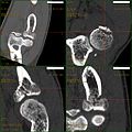
Treatment

Radial head fracture treatment is informed by the Mason-Johnston classification, patient symptoms, and fracture stability. An unstable fracture will involve fracture displacement, fractures to adjacent structures and injury to other associated soft tissues. A stable type 1 radial head fracture is typically managed with conservative measures including joint aspiration, immobilization in a sling for a few days and followed by early range of motion exercises. [2] [6] If range of motion is still limited after joint aspiration it may indicate a mechanical block which is treated surgically. [5] Stable type 2 radial head fractures may be treated as a type 1 if the displacement is minimal. Unstable type 2 - 4 fractures generally warrant surgery. Surgical correction can include fracture fragment excision, radial head reconstruction, open reduction and internal fixation, and radial head excision with artificial replacement. [6] Associated structures that were damaged during the injury may also need to be repaired.

Rehabilitation exercises are recommended and tailored to fracture and treatment type. It is recommended to wait 6 weeks before resuming load bearing with a stable type 1 fracture and 10-12 weeks following surgery for unstable type 2-4 fractures. [7]

Prognosis and Complications

Stable type 1 and 2 radial head fractures often have good outcomes with most cases regaining complete range of motion and having minimal residual stiffness or pain. [5] Outcomes for unstable type 2-4 radial head fractures vary greatly depending on the severity of the injury and the surgical intervention. [5] [6] Some of the more common complications of unstable radial head fractures includes stiffness, poor bone healing, nerve damage, and pain/prominent hardware. [6]

Related Research Articles

<span class="mw-page-title-main">Shoulder problem</span> Medical condition

Shoulder problems including pain, are one of the more common reasons for physician visits for musculoskeletal symptoms. The shoulder is the most movable joint in the body. However, it is an unstable joint because of the range of motion allowed. This instability increases the likelihood of joint injury, often leading to a degenerative process in which tissues break down and no longer function well.

<span class="mw-page-title-main">Joint dislocation</span> Medical injury

A joint dislocation, also called luxation, occurs when there is an abnormal separation in the joint, where two or more bones meet. A partial dislocation is referred to as a subluxation. Dislocations are often caused by sudden trauma on the joint like an impact or fall. A joint dislocation can cause damage to the surrounding ligaments, tendons, muscles, and nerves. Dislocations can occur in any major joint or minor joint. The most common joint dislocation is a shoulder dislocation.

<span class="mw-page-title-main">Rotator cuff tear</span> Medical condition

A rotator cuff tear is an injury where one or more of the tendons or muscles of the rotator cuff of the shoulder get torn. Symptoms may include shoulder pain, which is often worse with movement, limited range of motion, or weakness. This may limit people's ability to brush their hair or put on clothing. Clicking may also occur with movement of the arm.

<span class="mw-page-title-main">Distal radius fracture</span> Fracture of the radius bone near the wrist

A distal radius fracture, also known as wrist fracture, is a break of the part of the radius bone which is close to the wrist. Symptoms include pain, bruising, and rapid-onset swelling. The ulna bone may also be broken.

<span class="mw-page-title-main">Clavicle fracture</span> Medical condition

A clavicle fracture, also known as a broken collarbone, is a bone fracture of the clavicle. Symptoms typically include pain at the site of the break and a decreased ability to move the affected arm. Complications can include a collection of air in the pleural space surrounding the lung (pneumothorax), injury to the nerves or blood vessels in the area, and an unpleasant appearance.

<span class="mw-page-title-main">Ankle fracture</span> Medical condition

An ankle fracture is a break of one or more of the bones that make up the ankle joint. Symptoms may include pain, swelling, bruising, and an inability to walk on the injured leg. Complications may include an associated high ankle sprain, compartment syndrome, stiffness, malunion, and post-traumatic arthritis.

<span class="mw-page-title-main">Osteochondritis dissecans</span> Ischemic bone disease

Osteochondritis dissecans is a joint disorder primarily of the subchondral bone in which cracks form in the articular cartilage and the underlying subchondral bone. OCD usually causes pain during and after sports. In later stages of the disorder there will be swelling of the affected joint which catches and locks during movement. Physical examination in the early stages does only show pain as symptom, in later stages there could be an effusion, tenderness, and a crackling sound with joint movement.

<span class="mw-page-title-main">SLAP tear</span> Medical condition

A SLAP tear or SLAP lesion is an injury to the glenoid labrum. SLAP is an acronym for "superior labral tear from anterior to posterior".

<span class="mw-page-title-main">Pelvic fracture</span> Broken bone in nonacetabular portions of pelvis

A pelvic fracture is a break of the bony structure of the pelvis. This includes any break of the sacrum, hip bones, or tailbone. Symptoms include pain, particularly with movement. Complications may include internal bleeding, injury to the bladder, or vaginal trauma.

<span class="mw-page-title-main">Monteggia fracture</span> Medical condition

The Monteggia fracture is a fracture of the proximal third of the ulna with dislocation of the proximal head of the radius. It is named after Giovanni Battista Monteggia.

<span class="mw-page-title-main">Calcaneal fracture</span> Medical condition

A calcaneal fracture is a break of the calcaneus. Symptoms may include pain, bruising, trouble walking, and deformity of the heel. It may be associated with breaks of the hip or back.

<span class="mw-page-title-main">Humerus fracture</span> Medical condition

A humerus fracture is a break of the humerus bone in the upper arm. Symptoms may include pain, swelling, and bruising. There may be a decreased ability to move the arm and the person may present holding their elbow. Complications may include injury to an artery or nerve, and compartment syndrome.

<span class="mw-page-title-main">Radial tunnel syndrome</span> Medical condition

Radial tunnel syndrome (RTS) is caused by increased pressure on the radial nerve as it travels from the upper arm to the hand and wrist.

<span class="mw-page-title-main">Jefferson fracture</span> Medical condition

A Jefferson fracture is a bone fracture of the anterior and posterior arches of the C1 vertebra, though it may also appear as a three- or two-part fracture. The fracture may result from an axial load on the back of the head or hyperextension of the neck, causing a posterior break, and may be accompanied by a break in other parts of the cervical spine.

<span class="mw-page-title-main">Nasal fracture</span> Medical condition

A nasal fracture, commonly referred to as a broken nose, is a fracture of one of the bones of the nose. Symptoms may include bleeding, swelling, bruising, and an inability to breathe through the nose. They may be complicated by other facial fractures or a septal hematoma.

<span class="mw-page-title-main">Supracondylar humerus fracture</span> Medical condition

A supracondylar humerus fracture is a fracture of the distal humerus just above the elbow joint. The fracture is usually transverse or oblique and above the medial and lateral condyles and epicondyles. This fracture pattern is relatively rare in adults, but is the most common type of elbow fracture in children. In children, many of these fractures are non-displaced and can be treated with casting. Some are angulated or displaced and are best treated with surgery. In children, most of these fractures can be treated effectively with expectation for full recovery. Some of these injuries can be complicated by poor healing or by associated blood vessel or nerve injuries with serious complications.

<span class="mw-page-title-main">Internal fixation</span> Orthopedic operation to fix bone

Internal fixation is an operation in orthopedics that involves the surgical implementation of implants for the purpose of repairing a bone, a concept that dates to the mid-nineteenth century and was made applicable for routine treatment in the mid-twentieth century. An internal fixator may be made of stainless steel, titanium alloy, or cobalt-chrome alloy. or plastics.

Cardiac fibroma, also known as cardiac fibromatosis, cardiac fibrous hamartoma, fibroelastic hamartoma of heart and fibroma of heart is the second highest type of primary cardiac tumor seen in infants and children. This benign tumor made by connective tissue and fibroblast is largely observed in the ventricles of the heart. The left ventricle is the most common location of cardiac fibroma and accounts for approximately 57% of cardiac fibroma cases followed by the right ventricle with 27.5% of cases. Symptoms of the disease depend on the size of the tumor, its location relative to the conduction system, and whether it obstructs blood flow. Two-thirds of children with this tumor are asymptomatic, showing no signs and symptoms. Therefore the cause of cardiac fibroma is unexplained but has been associated with Gorlin Syndrome. Echocardiography is the primarily diagnostic method used to detect if an individual has cardiac fibroma. Resection of the tumor is recommended however heart transplantation is done if surgery is not possible. Overall prognosis of resection is favorable and the chance of recurrence is scarcely reported.

<span class="mw-page-title-main">Wrist osteoarthritis</span> Medical condition

Wrist osteoarthritis is a group of mechanical abnormalities resulting in joint destruction, which can occur in the wrist. These abnormalities include degeneration of cartilage and hypertrophic bone changes, which can lead to pain, swelling and loss of function. Osteoarthritis of the wrist is one of the most common conditions seen by hand surgeons.

<span class="mw-page-title-main">Proximal humerus fracture</span> Break of the upper part of the bone of the arm

A proximal humerus fracture is a break of the upper part of the bone of the arm (humerus). Symptoms include pain, swelling, and a decreased ability to move the shoulder. Complications may include axillary nerve or axillary artery injury.

References

  1. Daniel K Nishijima. "Elbow Fracture" . Retrieved 9 December 2014.
  2. 1 2 3 4 5 Bennett, James B. (September 1993). "Radial head fractures: Diagnosis and management". Journal of Shoulder and Elbow Surgery. 2 (5): 264–273. doi:10.1016/S1058-2746(09)80088-8. PMID   22959508.
  3. 1 2 Kaas, Laurens; van Riet, Roger P.; Vroemen, Jos P.A.M.; Eygendaal, Denise (June 2010). "The epidemiology of radial head fractures". Journal of Shoulder and Elbow Surgery. 19 (4): 520–523. doi:10.1016/j.jse.2009.10.015. PMID   20149972.
  4. 1 2 Pavić, Roman; Margetić, Petra; Hnatešen, Dijana (November 2015). "Diagnosis of occult radial head and neck fracture in adults". Injury. 46: S119–S124. doi:10.1016/j.injury.2015.10.050. PMID   26584731.
  5. 1 2 3 4 5 Duckworth, A. D.; McQueen, M. M.; Ring, D. (February 2013). "Fractures of the radial head". The Bone & Joint Journal. 95-B (2): 151–159. doi:10.1302/0301-620X.95B2.29877. ISSN   2049-4394. PMID   23365021.
  6. 1 2 3 4 5 Rosenblatt, Yishai; Athwal, George S.; Faber, Kenneth J. (April 2008). "Current Recommendations for the Treatment of Radial Head Fractures". Orthopedic Clinics of North America. 39 (2): 173–185. doi:10.1016/j.ocl.2007.12.008. PMID   18374808.
  7. Hackl, Michael; Leschinger, Tim; Uschok, Stephan; Müller, Lars Peter; Wegmann, Kilian (December 2017). "Rehabilitation of elbow fractures and dislocations". Obere Extremität. 12 (4): 201–207. doi:10.1007/s11678-017-0425-1. ISSN   1862-6599. S2CID   79629978.