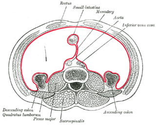
The mesentery is a contiguous set of tissues that attaches the intestines to the posterior abdominal wall in humans and is formed by the double fold of peritoneum. It helps in storing fat and allowing blood vessels, lymphatics, and nerves to supply the intestines, among other functions.

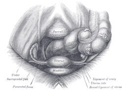
The recto-uterine pouch, also known by various other names, is the extension of the peritoneal cavity between the rectum and the posterior wall of the uterus in the female human body.

The thyroarytenoid muscle is a broad, thin muscle that forms the body of the vocal fold and that supports the wall of the ventricle and its appendix. It functions to relax the vocal folds.
The anterior or lingual surface of the epiglottis is curved forward, and covered on its upper, free part by mucous membrane which is reflected on to the sides and root of the tongue, forming a median and two lateral glossoepiglottic folds; the lateral folds are partly attached to the wall of the pharynx.

The base of the cartilaginous portion of the auditory tube lies directly under the mucous membrane of the nasal part of the pharynx, where it forms an elevation, the torus tubarius, the torus of the auditory tube, or cushion, behind the pharyngeal orifice of the tube. The torus tubarius is very close to the tubal tonsil, which is sometimes also called the tonsil of (the) torus tubarius. Equating the torus with its tonsil however might be seen as incorrect or imprecise.

The greater omentum is a large apron-like fold of visceral peritoneum that hangs down from the stomach. It extends from the greater curvature of the stomach, passing in front of the small intestines and doubles back to ascend to the transverse colon before reaching to the posterior abdominal wall. The greater omentum is larger than the lesser omentum, which hangs down from the liver to the lesser curvature. The common anatomical term "epiploic" derives from "epiploon", from the Greek epipleein, meaning to float or sail on, since the greater omentum appears to float on the surface of the intestines. It is the first structure observed when the abdominal cavity is opened anteriorly.

The broad ligament of the uterus is the wide fold of peritoneum that connects the sides of the uterus to the walls and floor of the pelvis.

The lateral umbilical fold overlies the inferior epigastric artery and its accompanying veins. Unlike the median and medial umbilical folds, the contents of the lateral umbilical fold remain functional after birth. It originates just medial to the deep inguinal ring to the arcuate line on the posterior surface of the anterior abdominal wall.
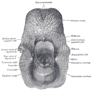
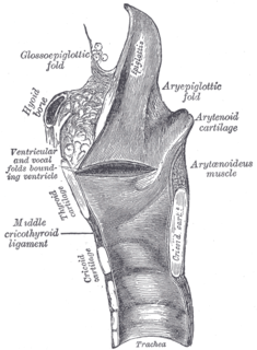
The Aryepiglottic folds are triangular folds of mucous membrane enclosing ligamentous and muscular fibres. They are located at the entrance of the larynx, extending from the lateral borders of the epiglottis to the arytenoid cartilages, hence the name 'aryepiglottic'. They contain the aryepiglottic muscles and form the upper borders of the quadrangular membrane.

The circular folds are large valvular flaps projecting into the lumen of the small intestine.
The median umbilical ligament is a structure in human anatomy. It is a shrivelled piece of tissue that represents the remnant of the embryonic urachus.

The medial umbilical ligament is a paired structure found in human anatomy. It is on the deep surface of the anterior abdominal wall, and is covered by the medial umbilical folds. It should not be confused with the median umbilical ligament, a different structure that represents the remnant of the embryonic urachus.

The transverse folds of rectum are semi-lunar transverse folds of the rectal wall that protrude into the rectum, not the anal canal as that lies below the rectum. Their use seems to be to support the weight of fecal matter, and prevent its urging toward the anus, which would produce a strong urge to defecate. Although the term rectum means straight, these transverse folds overlap each other during the empty state of the intestine to such an extent that, as Houston remarked, they require considerable maneuvering to conduct an instrument along the canal, as often occurs in sigmoidoscopy and colonoscopy.
Culdocentesis is a medical procedure involving the extraction of fluid from the pouch of Douglas through a needle. It can be one diagnostic technique used in identifying pelvic inflammatory disease and ruptured ectopic pregnancies that cause hemoperitoneum.

The urethral crest is an anatomical feature present in the urinary system of both males and females.
Related to the urinary bladder, anteriorly there are the following folds:

The uterosacral ligaments belong to the major ligaments of uterus.

The vestibular fold is one of two thick folds of mucous membrane, each enclosing a narrow band of fibrous tissue, the vestibular ligament, which is attached in front to the angle of the thyroid cartilage immediately below the attachment of the epiglottis, and behind to the antero-lateral surface of the arytenoid cartilage, a short distance above the vocal process.
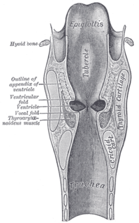
The laryngeal ventricle, is a fusiform fossa, situated between the vestibular and vocal folds on either side, and extending nearly their entire length. There is also a sinus of Morgagni in the pharynx.
The laryngeal cavity extends from the laryngeal inlet downwards to the lower border of the cricoid cartilage where it is continuous with that of the trachea.
The public domain consists of all the creative works to which no exclusive intellectual property rights apply. Those rights may have expired, been forfeited, expressly waived, or may be inapplicable.

Gray's Anatomy is an English language textbook of human anatomy originally written by Henry Gray and illustrated by Henry Vandyke Carter. Earlier editions were called Anatomy: Descriptive and Surgical, Anatomy of the Human Body and Gray's Anatomy: Descriptive and Applied, but the book's name is commonly shortened to, and later editions are titled, Gray's Anatomy. The book is widely regarded as an extremely influential work on the subject, and has continued to be revised and republished from its initial publication in 1858 to the present day. The latest edition of the book, the 41st, was published in September 2015.