
The uterus or womb is a major female hormone-responsive secondary sex organ of the reproductive system in humans and most other mammals. Things occurring in the uterus are described with the term in utero. In the human, the lower end of the uterus, the cervix, opens into the vagina, while the upper end, the fundus, is connected to the fallopian tubes. It is within the uterus that the fetus develops during gestation. In the human embryo, the uterus develops from the paramesonephric ducts which fuse into the single organ known as a simplex uterus. The uterus has different forms in many other animals and in some it exists as two separate uteri known as a duplex uterus.

The peritoneum is the serous membrane forming the lining of the abdominal cavity or coelom in amniotes and some invertebrates, such as annelids. It covers most of the intra-abdominal organs, and is composed of a layer of mesothelium supported by a thin layer of connective tissue. This peritoneal lining of the cavity supports many of the abdominal organs and serves as a conduit for their blood vessels, lymphatic vessels, and nerves.
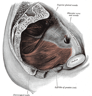
The levator ani is a broad, thin muscle group, situated on either side of the pelvis. It is formed from three muscle components: the pubococcygeus, the iliococcygeus, and the puborectalis.
In medicine, prolapse is a condition in which organs fall down or slip out of place. It is used for organs protruding through the vagina, rectum, or for the misalignment of the valves of the heart. A spinal disc herniation is also sometimes called "disc prolapse". Prolapse means "to fall out of place", from the Latin prolabi meaning "to fall out".

The pelvic floor or pelvic diaphragm is composed of muscle fibers of the levator ani, the coccygeus muscle, and associated connective tissue which span the area underneath the pelvis. The pelvic diaphragm is a muscular partition formed by the levatores ani and coccygei, with which may be included the parietal pelvic fascia on their upper and lower aspects. The pelvic floor separates the pelvic cavity above from the perineal region below. Both males and females have a pelvic floor. To accommodate the birth canal, a female's pelvic cavity is larger than a male's.

Gynecologic ultrasonography or gynecologic sonography refers to the application of medical ultrasonography to the female pelvic organs as well as the bladder, the adnexa, and the recto-uterine pouch. The procedure may lead to other medically relevant findings in the pelvis.

The broad ligament of the uterus is the wide fold of peritoneum that connects the sides of the uterus to the walls and floor of the pelvis.
The suspensory ligament of the ovary, also infundibulopelvic ligament, is a fold of peritoneum that extends out from the ovary to the wall of the pelvis.

The mesovarium is the portion of the broad ligament of the uterus that suspends the ovaries. The ovary is not covered by the mesovarium; rather, it is covered by germinal epithelium.

The pelvic cavity is a body cavity that is bounded by the bones of the pelvis. Its oblique roof is the pelvic inlet. Its lower boundary is the pelvic floor.

The perimetrium is the outer serosal layer of the uterus, derived from the peritoneum overlying the uterine fundus, and can be considered a visceral peritoneum. It consists of a superficial layer of mesothelium, and a thin layer of loose connective tissue beneath it.
The paracolic gutters are spaces between the colon and the abdominal wall.

The fornices of the vagina are the superior portions of the vagina, extending into the recesses created by the vaginal portion of cervix. The word "fornix" is Latin for "arch".

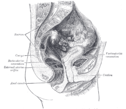
In the male, the peritoneum encircles the sigmoid colon, from which it is reflected to the posterior wall of the pelvis as a fold, the sigmoid mesocolon. It then leaves the sides and, finally, the front of the rectum, and is continued on to the upper ends of the seminal vesicles and the bladder; on either side of the rectum it forms a fossa, the pararectal fossa, which varies in size with the distension of the rectum.

In human female anatomy, the vesico-uterine pouch, also known by various names, is a fold of peritoneum over the uterus and the urinary bladder. Like the recto-uterine pouch, it is a female pelvic recess. However, it is a shallower pouch close to the anterior fornix of the vagina.

The recto-vesical pouch is the pocket that lies between the rectum and the urinary bladder in human males and other male mammals.

In human anatomy, the omental foramen, is the passage of communication, or foramen, between the greater sac, and the lesser sac.
Culdoscopy is an endoscopic procedure performed to examine the rectouterine pouch and pelvic viscera by the introduction of a culdoscope through the posterior vaginal wall. The word culdoscopy is derived from the term cul-de-sac, which means literally in French "bottom of a sac", and refers to the rectouterine pouch.
An obstetric labor complication is a difficulty or abnormality that arises during the process of labor or delivery.

The vaginal support structures are those muscles, bones, ligaments, tendons, membranes and fascia, of the pelvic floor that maintain the position of the vagina within the pelvic cavity and allow the normal functioning of the vagina and other reproductive structures in the female. Defects or injuries to these support structures in the pelvic floor leads to pelvic organ prolapse. Anatomical and congenital variations of vaginal support structures can predispose a woman to further dysfunction and prolapse later in life. The urethra is part of the anterior wall of the vagina and damage to the support structures there can lead to incontinence and urinary retention.


