Related Research Articles

The lumbar vertebrae are, in human anatomy, the five vertebrae between the rib cage and the pelvis. They are the largest segments of the vertebral column and are characterized by the absence of the foramen transversarium within the transverse process and by the absence of facets on the sides of the body. They are designated L1 to L5, starting at the top. The lumbar vertebrae help support the weight of the body, and permit movement.

The internal carotid artery is a major paired artery, one on each side of the head and neck, in human anatomy. They arise from the common carotid arteries where these bifurcate into the internal and external carotid arteries at cervical vertebral level 3 or 4; the internal carotid artery supplies the brain, while the external carotid nourishes other portions of the head, such as face, scalp, skull, and meninges.

The abdominal aorta is the largest artery in the abdominal cavity. As part of the aorta, it is a direct continuation of the descending aorta.

In human anatomy, the basilar artery is one of the arteries that supplies the brain with oxygen-rich blood.

The celiacartery, also known as the celiac trunk, or truncus coeliacus, is the first major branch of the abdominal aorta. It is 1.25 cm in length. Branching from the aorta at thoracic vertebra 12 (T12) in humans, it is one of three anterior/ midline branches of the abdominal aorta.

In human anatomy, the superior mesenteric artery (SMA) arises from the anterior surface of the abdominal aorta, just inferior to the origin of the celiac trunk, and supplies the intestine from the lower part of the duodenum through two-thirds of the transverse colon, as well as the pancreas.

The anterior cerebral artery (ACA) is one of a pair of arteries on the brain that supplies oxygenated blood to most midline portions of the frontal lobes and superior medial parietal lobes. The two anterior cerebral arteries arise from the internal carotid artery and are part of the circle of Willis. The left and right anterior cerebral arteries are connected by the anterior communicating artery.

In human anatomy, the inferior mesenteric artery, often abbreviated as IMA, is the third main branch of the abdominal aorta and arises at the level of L3, supplying the large intestine from the left colic flexure to the upper part of the rectum, which includes the descending colon, the sigmoid colon, and part of the rectum. Proximally, its territory of distribution overlaps with the middle colic artery, and therefore the superior mesenteric artery. The SMA and IMA anastomose via the marginal artery of the colon and via Riolan's arcade. The territory of distribution of the IMA is more or less equivalent to the embryonic hindgut.

The external iliac arteries are two major arteries which bifurcate off the common iliac arteries anterior to the sacroiliac joint of the pelvis. They proceed anterior and inferior along the medial border of the psoas major muscles. They exit the pelvic girdle posterior and inferior to the inguinal ligament about one third laterally from the insertion point of the inguinal ligament on the pubic tubercle at which point they are referred to as the femoral arteries. The external iliac artery is usually the artery used to attach the renal artery to the recipient of a kidney transplant.
The middle cerebral artery (MCA) is one of the three major paired arteries that supply blood to the cerebrum. The MCA arises from the internal carotid and continues into the lateral sulcus where it then branches and projects to many parts of the lateral cerebral cortex. It also supplies blood to the anterior temporal lobes and the insular cortices.

The internal iliac artery is the main artery of the pelvis.
The cystic artery supplies oxygenated blood to the gallbladder and cystic duct.

The superior thyroid artery arises from the external carotid artery just below the level of the greater cornu of the hyoid bone and ends in the thyroid gland.
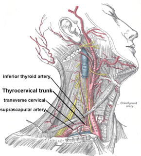
The inferior thyroid artery is an artery in the neck. It arises from the thyrocervical trunk and passes upward, in front of the vertebral artery and longus colli muscle. It then turns medially behind the carotid sheath and its contents, and also behind the sympathetic trunk, the middle cervical ganglion resting upon the vessel.

The superior pancreaticoduodenal artery is an artery that supplies blood to the duodenum and pancreas.
The inferior pancreaticoduodenal artery branches from the superior mesenteric artery or from its first intestinal branch, opposite the upper border of the inferior part of the duodenum.

A bronchopulmonary segment is a portion of lung supplied by a specific tertiary bronchus and arteries. These arteries branch from the pulmonary and bronchial arteries, and run together through the center of the segment. Veins and lymphatic vessels drain along the edges of the segment. The segments are separated from each other by layers of connective tissue. Each bronchopulmonary segment is a discrete anatomical and functional unit, and this separation means that a bronchopulmonary segment can be surgically removed without affecting the function of the others.

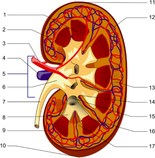
The renal hilum or renal pedicle is the hilum of the kidney, that is, its recessed central fissure where its vessels, nerves and ureter pass. The medial border of the kidney is concave in the center and convex toward either extremity; it is directed forward and a little downward. Its central part presents a deep longitudinal fissure, bounded by prominent overhanging anterior and posterior lips. This fissure is a hilum that transmits the vessels, nerves, and ureter. From anterior to posterior, the renal vein exits, the renal artery enters, and the renal pelvis exits the kidney.
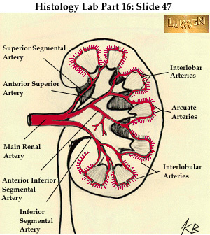
The interlobar arteries are vessels of the renal circulation which supply the renal lobes. The interlobar arteries branch from the lobar arteries which branch from the segmental arteries, from the renal artery.

The following outline is provided as an overview of and topical guide to human anatomy:
References
This article incorporates text in the public domain from the 20th edition of Gray's Anatomy (1918)
The public domain consists of all the creative works to which no exclusive intellectual property rights apply. Those rights may have expired, been forfeited, expressly waived, or may be inapplicable.

Gray's Anatomy is an English language textbook of human anatomy originally written by Henry Gray and illustrated by Henry Vandyke Carter. Earlier editions were called Anatomy: Descriptive and Surgical and Gray's Anatomy: Descriptive and Applied, but the book's name is commonly shortened to, and later editions are titled, Gray's Anatomy. The book is widely regarded as an extremely influential work on the subject, and has continued to be revised and republished from its initial publication in 1858 to the present day. The latest edition of the book, the 41st, was published in September 2015.