Suberites

Last updated

Suberites
Suberites domuncula.jpg
Suberites domuncula
Scientific classification Red Pencil Icon.png
Kingdom: Animalia
Phylum: Porifera
Class: Demospongiae
Order: Suberitida
Family: Suberitidae
Genus: Suberites
Nardo, 1833
Species

See text

Synonyms
List
  • CarnleiaBurton, 1930
  • ChoanitesMantell, 1822 sensu De Laubenfels, 1936
  • FiculinaGray, 1867
  • LaxosuberellaBurton, 1930
  • LitamenaNardo, 1833
  • LithumenaRenier, 1828
  • Raspailia (Syringella) sensu Schmidt, 1868
  • SuberanthusLendenfeld, 1898
  • SuberellaThiele, 1905
  • SuberellaBurton, 1929
  • SyringellaSchmidt, 1868

Suberites is a genus of sea sponges in the family Suberitidae. [1] Sponges, known scientifically as Porifera, are the oldest metazoans and are used to elucidate the basics of multicellular evolution. [2] These living fossils are ideal for studying the principal features of metazoans, such as extracellular matrix interactions, signal-receptor systems, nervous or sensory systems, and primitive immune systems. Thus, sponges are useful tools with which to study early animal evolution. They appeared approximately 580 million years ago, in the Ediacaran. [2]

Contents

Evolutionary significance

As members of the oldest phylum of metazoans, Suberites serve as model organisms to elucidate features of the earliest animals. [2] [3] [4] Suberites and their relatives are used to determine the structure of the first metazoans [2] and have been studied to determine how totipotency has replaced by pluripotency in most higher animals. [5] Among other things, Suberites show that tyrosine-phosphorylation machinery evolved in animals independently from other eukaryotes. [2] Suberites are also used as models to elucidate the evolution of transmembrane receptors and cell-junction proteins. [6] A combination of stem cell and apoptosis factors studies is used as a model for studies of development in higher animals. [7]

Ecology

Suberites are a global genus. One species, Suberites zeteki, is found in Hawaii. S. zeteki associates with many fungi. [8] Another, S. japonicas, is native to the waters around Japan. [9] S. aurantiacus is found in the Caribbean sea. [10] S.carnosus lives in the Indian Ocean and in the Mediterranean Sea and can also be found in Irish waters. [10] [11] S. diversicolor can be found in Indonesia. [12] Due to Suberites’ ability to efficiently filter water, many microbes, especially fungal species, are filtered through. If these microbes escape digestion, they can deposit on the sponge and reside there indefinitely. [8] Symbiotic bacteria produce toxins, such as okadaic acid, which defend them from colonization by parasitic annelids. [13] [14] Expression of various enzymes by Suberites influences the growth of their symbiotic bacteria. [14] Suberites often live on the shells on the mollusk Hexaplex trunculus . [13] Suberites have mechanisms of defense against predation, such as the toxic chemicals found below. [15]

Physiology

Suberites display neuronal communications, but neuronal networks are mysteriously missing. [16] However, they do have many of the same sensory receptors and signals found in higher animals. [17] Researchers in China and Germany have found that sponge spicules contribute to their neural communication. [18] In effect, the silicaceous structures act as fiber optic cables to convey light signals generated from the protein luciferase. [17] [18] The sponges generate light from luciferin, after it is acted upon by luciferase. [17] [19] Suberites have also been shown to produce light in response to tactile stimulation. [19] Suberites consist mostly of cells, in contrast with other Porifera (such as the class Hexactinellida) which are syncytial. [2] As a result, Suberites have slower reaction times in their neural communication. Suberites utilized many Ras-like GTPases which are used for signaling and affect development. [20] According to comparative studies, Suberites have some of the most simple indicator proteins, such as collagen, of known animals. [2] Like all sponges, Suberites are filter-feeders. They are extremely efficient and can process thousands of liters of water per day. [8] [21] S. domuncula has been used for study of graft rejection. Researchers have discovered that apoptotic factors are induced in the tissue that is rejected. [22]

Development

Suberites consist of many telomerase-positive cells, which means the cells are essentially immortal, barring cell death signal. [2] In most cases, the signal is a lack of connection either to the extracellular matrix or other cells. [2] [7] Their apoptotic cells are similar to homologous to mammalian. However, maintenance of long-lived cells involves proteins such as SDLAGL that are highly similar to yeast and human homologs. [2] Certain inorganic materials, such as iron and selenium, influence the growth of Suberites, including the primmorph growth and spicule formation. [23] [24] [25] Suberites undergo cell differentiation through a variety of mechanisms based on cell-cell communication. [26]

Morphology

Suberites are key examples of the importance of the extracellular matrix in animals. In sponges, it is mediated by proteoglycans. [2] Spicule formation is also important for Suberites. Spicules are structural support of sponges, similar to skeletons in higher animals. They are normally hollow structures that are formed by lamellar growth. [27] [28] [29] Whereas higher animal skeletons are largely calcium-based, sponge spicules consist mostly of silica, a silicon dioxide polymer. [30] These inorganic structures provide support for the animals. [17] [31] The spicules are biologically-formed silica structures, also known as biosilica. [30] [31] [32] [33] Silica deposition begins intracellularly and is carried out by the enzyme silicatein. [27] [28] [30] [31] [34] Silicateins are modulated by a group of proteins called silintaphins. [35] The process occurs in specialized cells known as sclerocytes. [27] [28] [31] Biosilica formation in Suberites differs from other species that utilize biosilica in this regard. Most other species, such as certain plants and diatoms, simply deposit a supersaturated biosilica solution. [17] The network of silica found in sponges mediates much of the sponges’ neural communications.

Immunity and defense

Suberites show the cytokine-like molecule allograft inflammatory factor one (AIF-1), which is similar to vertebrate AIF-1. [2] [36] Immune response relies on phosphorylation cascades involving the p38 kinase. [36] S. domuncula was the first demonstrated immune response of invertebrate species (1). These sponges also have similar graft-response inflammation to vertebrates. [2] Their immune systems are much simpler than vertebrates; they consist of only innate immunity. [2] Because they filter thousands of liters of water per day, and their environment contains a high concentration of bacteria and viruses, Suberites have developed a highly potent system of immunity. [21] Despite the efficiency of their immune systems, Suberites can be susceptible to infection which often stimulates cell death through apoptotic pathways. [21]

Suberites, namely S. domuncula, defend themselves from macroscopic threats with a neurotoxin known as suberitine. [37] It was the first known protein discovered in a sponge. [37] The neurotoxic properties of suberitine arise from its ability to block action potentials. [38] It additionally has hemolytic properties, which do not originate from phospholipase A activity. [38] It has some antibacterial activity; however, the extent of the activity due solely to suberitine is not currently defined. [39] The sponge itself neutralizes the toxin through a pathway that is not fully understood, but involves retinal, a β-carotene metabolite. [40] S. japonicas also produces several cytotoxic compounds, seragamides A-F. The seragamides act by interfering with cytoskeleton activity, specifically the actin microfilaments. [9] The activity of the seragamides is a possible route for anti-cancer drugs, similar to existing drugs which target microtubules. [9] Suberites also produce cytotoxic compounds known as nakijinamines, which resemble other toxins found in Suberites, but the role of the nakijinamines has not yet been found. [41] Many of the bioactive compounds found on Suberites are microbial in nature. [11]

Species

The following species are recognised in the genus Suberites: [1]

Related Research Articles

<span class="mw-page-title-main">Sponge</span> Animals of the phylum Porifera

Sponges, the members of the phylum Porifera, are a basal animal clade as a sister of the diploblasts. They are multicellular organisms that have bodies full of pores and channels allowing water to circulate through them, consisting of jelly-like mesohyl sandwiched between two thin layers of cells.

<span class="mw-page-title-main">Choanoflagellate</span> Group of eukaryotes considered the closest living relatives of animals

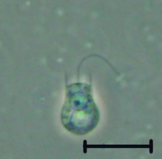
The choanoflagellates are a group of free-living unicellular and colonial flagellate eukaryotes considered to be the closest living relatives of the animals. Choanoflagellates are collared flagellates, having a funnel shaped collar of interconnected microvilli at the base of a flagellum. Choanoflagellates are capable of both asexual and sexual reproduction. They have a distinctive cell morphology characterized by an ovoid or spherical cell body 3–10 µm in diameter with a single apical flagellum surrounded by a collar of 30–40 microvilli. Movement of the flagellum creates water currents that can propel free-swimming choanoflagellates through the water column and trap bacteria and detritus against the collar of microvilli, where these foodstuffs are engulfed. This feeding provides a critical link within the global carbon cycle, linking trophic levels. In addition to their critical ecological roles, choanoflagellates are of particular interest to evolutionary biologists studying the origins of multicellularity in animals. As the closest living relatives of animals, choanoflagellates serve as a useful model for reconstructions of the last unicellular ancestor of animals.

<span class="mw-page-title-main">Demosponge</span> Class of sponges

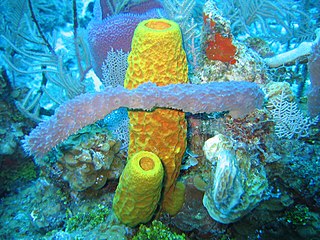
Demosponges (Demospongiae) are the most diverse class in the phylum Porifera. They include 76.2% of all species of sponges with nearly 8,800 species worldwide. They are sponges with a soft body that covers a hard, often massive skeleton made of calcium carbonate, either aragonite or calcite. They are predominantly leuconoid in structure. Their "skeletons" are made of spicules consisting of fibers of the protein spongin, the mineral silica, or both. Where spicules of silica are present, they have a different shape from those in the otherwise similar glass sponges. Some species, in particular from the Antarctic, obtain the silica for spicule building from the ingestion of siliceous diatoms.

<i>Grantia</i> Genus of sponges

Grantia is a genus of calcareous sponges belonging to the family Grantiidae. Species of the genus Grantia contain spicules and spongin fibers.

<i>Suberites domuncula</i> Species of sponge

Suberites domuncula is a species of sea sponge belonging to the family Suberitidae.

<i>Polymastia</i> (sponge) Genus of sponges

Polymastia is a genus of sea sponges containing about 30 species. These are small to large encrusting or dome-shaped sponges with a smooth surface having many teat-shaped projections (papillae). In areas of strong wave action, this genus does not grow the teat structures, but instead grows in a corrugated form.

<i>Chondrocladia</i> Genus of sponges

Chondrocladia is a genus of carnivorous demosponges of the family Cladorhizidae. Neocladia was long considered a junior synonym, but has recently become accepted as a distinct genus.

<i>Axinella</i> Genus of sponges

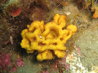
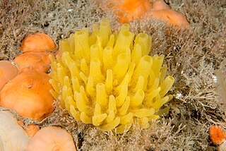
Axinella is a genus of sponges in the family Axinellidae first described in 1862 by Eduard Oscar Schmidt. Species of Axinella occur in the Indian and Pacific Oceans. Most of these sponges are smaller than 20 cm, and have a yellow or orange colour.

<span class="mw-page-title-main">Sponge spicule</span> Structural element of sea sponges

Spicules are structural elements found in most sponges. The meshing of many spicules serves as the sponge's skeleton and thus it provides structural support and potentially defense against predators.

Monorhaphis is a monotypic genus of siliceous deep sea Hexactinellid sponges. The single species is the type species Monorhaphis chuni, a sponge known for creating a single giant basal spicule (G.B.S.) to anchor the sponge in the sediments. The species was described by Franz Eilhard Schulze in 1904 from specimens collected by the German Deep Sea Expedition in 1898-1899. Monorhaphis is also the only genus in the monotypic family Monorhaphididae.

Homaxinella is a genus of sea sponges in the family Suberitidae. The type species is Homaxinella balfourensis.

Homaxinella balfourensis is a species of sea sponge in the family Suberitidae. It is found in the seas around Antarctica and can grow in two forms, either branching out in one plane like a fan or forming an upright club-like structure.

<span class="mw-page-title-main">Precambrian body plans</span> Structure and development of early multicellular organisms

Until the late 1950s, the Precambrian was not believed to have hosted multicellular organisms. However, with radiometric dating techniques, it has been found that fossils initially found in the Ediacara Hills in Southern Australia date back to the late Precambrian. These fossils are body impressions of organisms shaped like disks, fronds and some with ribbon patterns that were most likely tentacles.

<i>Gelliodes</i> Genus of sponges

Gelliodes is a genus of sponges in the family Niphatidae.

Amphilectus is a genus of demosponges, comprising around 20 species found in oceans around the world.

<span class="mw-page-title-main">Silicatein</span>

Silicateins are enzymes which catalyse the formation of biosilica from monomeric silicon compounds extracted from the natural environment. Environmental silicates are absorbed by specific biota, including diatoms, radiolaria, silicoflagellates, and siliceous sponges; silicateins have so far only been found in sponges. Silicateins are homologous to the cysteine protease cathepsin.

Hamacantha is a genus of sponges in the family Hamacanthidae. This species in this genus differ from those in the other genera in this family through the presence of diancistras, distinctive microscleres. These are thought to aid in framing the skeleton by joining monactine megascleres. This genus contains 30 species in three subgenera.

<i>Phorbas</i> (sponge) Genus of sponges

Phorbas is a genus of demosponges belonging to the family Hymedesmiidae.

<i>Latrunculia</i> Genus of demosponges

Latrunculia is a genus of demosponges. It is well known for the diverse array of chemical compounds found in its species, including the latrunculins, which are named after this genus. Many of these are medically important, including anti-cancer compounds such as discorhabdins.

Tetilla is a genus of demosponges in the family Tetillidae. It is widely distributed. They are mainly found in deeper habitats.

References

  1. 1 2 "WoRMS - World Register of Marine Species - Suberites Nardo, 1833". www.marinespecies.org. Retrieved 15 November 2010.
  2. 1 2 3 4 5 6 7 8 9 10 11 12 13 14 Müller, Werner E.G (June 2001). "Review: How was metazoan threshold crossed? The hypothetical Urmetazoa". Comparative Biochemistry and Physiology Part A: Molecular & Integrative Physiology. 129 (2–3): 433–460. doi:10.1016/s1095-6433(00)00360-3.
  3. Wiens, Matthias; Mangoni, Alfonso; D’Esposito, Monica; Fattorusso, Ernesto; Korchagina, Natalia; Schröder, Heinz C.; Grebenjuk, Vladislav A.; Krasko, Anatoli; Batel, Renato; Müller, Isabel M.; Müller, Werner E. G. (August 2003). "The Molecular Basis for the Evolution of the Metazoan Bodyplan: Extracellular Matrix-Mediated Morphogenesis in Marine Demosponges". Journal of Molecular Evolution . 57 (S1): S60–S75. doi:10.1007/s00239-003-0008-1.
  4. Müller, Werner E. G.; Müller, Isabel M.; Schröder, Heinz C. (September 2006). "Evolutionary relationship of Porifera within the eukaryotes". Hydrobiologia . 568 (S1): 167–176. doi:10.1007/s10750-006-0318-6.
  5. Müller, Werner E.G; Korzhev, Michael; Le Pennec, Gaël; Müller, Isabel M; Schröder, Heinz C (July 2003). "Origin of metazoan stem cell system in sponges: first approach to establish the model (Suberites domuncula)" (PDF). Biomolecular Engineering. 20 (4–6): 369–379. doi:10.1016/S1389-0344(03)00055-8.
  6. Adell, Teresa; Gamulin, Vera; Perović-Ottstadt, Sanja; Wiens, Matthias; Korzhev, Michael; Müller, Isabel M.; Müller, Werner E. G. (July 2004). "Evolution of Metazoan Cell Junction Proteins: The Scaffold Protein MAGI and the Transmembrane Receptor Tetraspanin in the Demosponge Suberites domuncula". Journal of Molecular Evolution. 59 (1): 41–50. doi:10.1007/s00239-004-2602-2.
  7. 1 2 Luthringer, B; Isbert, S; Müller, W E G; Zilberberg, C; Thakur, N L; Wörheide, G; Stauber, R H; Kelve, M; Wiens, M (February 2011). "Poriferan survivin exhibits a conserved regulatory role in the interconnected pathways of cell cycle and apoptosis". Cell Death & Differentiation. 18 (2): 201–213. doi: 10.1038/cdd.2010.87 . PMID   20651742.
  8. 1 2 3 G. Zheng, L. Binglin, Z. Chengchao, W. Guangyi, Molecular Detection of Fungal Communities in the Hawaiian Marine Sponges Suberites zeteki and Mycale armata. Applied & Environmental Microbiology 74, 6091 (2008).
  9. 1 2 3 C. Tanaka, J. Tanaka, R. F. Bolland, G. Marriott, T. Higa, Seragamides A–F, new actin-targeting depsipeptides from the sponge Suberites japonicus Thiele. Tetrahedron 62, 3536 (2006).
  10. 1 2 L. P. Ponomarenko, O. A. Vanteeva, S. A. Rod'kina, V. B. Krasokhin, S. S. Afiyatullov, Metabolites of the marine sponge Suberites cf. aurantiacus. Chemistry of Natural Compounds 46, 335 (2010).
  11. 1 2 B. Flemer et al., Diversity and antimicrobial activities of microbes from two Irish marine sponges, Suberites carnosus and Leucosolenia sp. Journal of Applied Microbiology 112, 289 (2012).
  12. D. F. R. Cleary et al., Habitat- and host-related variation in sponge bacterial symbiont communities in Indonesian waters. FEMS Microbiology Ecology 85, 465 (2013).
  13. 1 2 H. C. Schröder et al., Okadaic Acid, an Apoptogenic Toxin for Symbiotic/Parasitic Annelids in the Demosponge Suberites domuncula. Applied & Environmental Microbiology 72, 4907 (2006).
  14. 1 2 W. E. G. Müller et al., Oxygen-Controlled Bacterial Growth in the Sponge Suberites domuncula: toward a Molecular Understanding of the Symbiotic Relationships between Sponge and Bacteria. Applied & Environmental Microbiology 70, 2332 (2004).
  15. W. E. G. Müller et al., Molecular/chemical ecology in sponges: evidence for an adaptive antibacterial response in Suberites domuncula. Marine Biology 144, 19 (2004).
  16. W. E. G. Müller et al., Matrix-mediated canal formation in primmorphs from the sponge Suberites domuncula involves the expression of a CD36 receptor-ligand system. Journal of Cell Science 117, 2579 (2004).
  17. 1 2 3 4 5 X. Wang, X. Fan, H. Schröder, W. Müller, Flashing light in sponges through their siliceous fiber network: A new strategy of 'neuronal transmission' in animals. Chinese Science Bulletin 57, 3300 (2012).
  18. 1 2 W. E. G. Müller et al., Luciferase a light source for the silica-based optical waveguides (spicules) in the demosponge Suberites domuncula. Cellular and Molecular Life Sciences 66, 537 (2009).
  19. 1 2 W. E. G. Müller et al., A cryptochrome-based photosensory system in the siliceous sponge Suberites domuncula (Demospongiae). FEBS Journal 277, 1182 (2010).
  20. H. Cetkovic, A. Mikoc, W. E. G. Müller, V. Gamulin, Ras-like Small GTPases Form a Large Family of Proteins in the Marine Sponge Suberites domuncula. Journal of Molecular Evolution 64, 332 (2007).
  21. 1 2 3 Wiens, Matthias; Korzhev, Michael; Krasko, Anatoli; Thakur, Narsinh L.; Perović-Ottstadt, Sanja; Breter, Hans J.; Ushijima, Hiroshi; Diehl-Seifert, Bärbel; Müller, Isabel M.; Müller, Werner E.G. (July 2005). "Innate Immune Defense of the Sponge Suberites domuncula against Bacteria Involves a MyD88-dependent Signaling Pathway". Journal of Biological Chemistry . 280 (30): 27949–27959. doi: 10.1074/jbc.M504049200 .
  22. Wiens, Matthias; Perović-Ottstadt, Sanja; Müller, Isabel M.; Müller, Werner E. G. (November 2004). "Allograft rejection in the mixed cell reaction system of the demosponge Suberites domuncula is controlled by differential expression of apoptotic genes". Immunogenetics. 56 (8): 597–610. doi:10.1007/s00251-004-0718-6. PMID   15517243.
  23. L. Valisano, G. Bavestrello, M. Giovine, A. Arillo, C. Cerrano, Effect of iron and dissolved silica on primmorphs of Petrosia ficiformis (Poiret, 1789). Chemistry & Ecology 23, 233 (2007).
  24. A. Krasko et al., Iron Induces Proliferation and Morphogenesis in Primmorphs from the Marine Sponge Suberites domuncula. DNA & Cell Biology 21, 67 (2002).
  25. W. E. G. Müller et al., Selenium affects biosilica formation in the demosponge Suberites domuncula. FEBS Journal 272, 3838 (2005).
  26. H. C. Schröder et al., Differentiation capacity of epithelial cells in the sponge Suberites domuncula. Cell & Tissue Research 316, 271 (2004).
  27. 1 2 3 H. C. Schröder et al., Biosilica formation in spicules of the sponge Suberites domuncula: Synchronous expression of a gene cluster. Genomics 85, 666 (2005).
  28. 1 2 3 H. C. Schröder et al., Apposition of silica lamellae during growth of spicules in the demosponge Suberites domuncula: Biological/biochemical studies and chemical/biomimetical confirmation. Journal of Structural Biology 159, 325 (2007).
  29. F. Natalio et al., Silicatein-mediated incorporation of titanium into spicules from the demosponge Suberites domuncula. Cell & Tissue Research 339, 429 (2010).
  30. 1 2 3 W. Xiaohong et al., Evagination of Cells Controls Bio-Silica Formation and Maturation during Spicule Formation in Sponges. PLoS ONE 6, 1 (2011).
  31. 1 2 3 4 X. Wang et al., Silicateins, silicatein interactors and cellular interplay in sponge skeletogenesis: formation of glass fiber-like spicules. FEBS Journal 279, 1721 (2012).
  32. W. E. G. Müller et al., Hardening of bio-silica in sponge spicules involves an aging process after its enzymatic polycondensation: Evidence for an aquaporin-mediated water absorption. BBA − General Subjects 1810, 713 (2011).
  33. W. E. G. Müller et al., Silicateins, the major biosilica forming enzymes present in demosponges: Protein analysis and phylogenetic relationship. Gene 395, 62 (2007).
  34. W. E. G. Müller et al., Identification of a silicatein(-related) protease in the giant spicules of the deep-sea hexactinellid Monorhaphis chuni. Journal of Experimental Biology 211, 300 (2008)
  35. W. E. G. Müller et al., The silicatein propeptide acts as inhibitor/modulator of self-organization during spicule axial filament formation. FEBS Journal 280, 1693 (2013).
  36. 1 2 H. C. Schröder et al., Functional Molecular Biodiversity: Assessing the Immune Status of Two Sponge Populations ( Suberites domuncula) on the Molecular Level. Marine Ecology 25, 93 (2004).
  37. 1 2 L. Cariello, L. Zanetti, Suberitine, the toxic protein from the marine sponge suberites domuncula. Comparative Biochemistry and Physiology 64C, 15 (1979).
  38. 1 2 L. Cariello, E. Tosti, L. Zanetti, The hemolytic activity of suberitine. Comparative Biochemistry and Physiology 73C, 91 (1981).
  39. N. L. Thakur et al., Antibacterial activity of the sponge suberites domuncula and its primmorphs: potential basis for epibacterial chemical defense. Aquatic Microbial Ecology 31, 77 (2003).
  40. W. Muller et al., Differential expression of the desmosponge (suberites domuncula) carotenoid oxygenases in response to light: protection mechanism against the self-produced toxic protein (suberitine). Marine Drugs 10, 177 (2012).
  41. Y. Takahashi et al., Heteroaromatic alkaloids, nakijinamines, from a sponge Suberites sp. Tetrahedron 68, 8545 (2012).