Ursodoxicoltaurine

Last updated

Ursodoxicoltaurine
Tauroursodeoxycholic acid.svg
Names
IUPAC name
2-(3α,7β-Dihydroxy-5β-cholan-24-amido)ethane-1-sulfonic acid
Systematic IUPAC name
2-{(4R)-4-[(1R,3aS,3bR,4S,5aS,7R,9aS,9bS,11aR)-4,7-Dihydroxy-9a,11a-dimethylhexadecahydro-1H-cyclopenta[a]phenanthren-1-yl]pentanamido}ethane-1-sulfonic acid
Other names
Tauroursodeoxycholic acid; TUDCA; 3α,7β-Dihydroxy-5β-cholanoyltaurine; UR 906; Ursodeoxycholyltaurine; Taurursodiol
Identifiers
3D model (JSmol)
ChEBI
ChEMBL
ChemSpider
KEGG
PubChem CID
UNII
  • InChI=1S/C26H45NO6S/c1-16(4-7-23(30)27-12-13-34(31,32)33)19-5-6-20-24-21(9-11-26(19,20)3)25(2)10-8-18(28)14-17(25)15-22(24)29/h16-22,24,28-29H,4-15H2,1-3H3,(H,27,30)(H,31,32,33)/t16-,17+,18-,19-,20+,21+,22+,24+,25+,26-/m1/s1 Yes check.svgY
    Key: BHTRKEVKTKCXOH-LBSADWJPSA-N Yes check.svgY
  • InChI=1/C26H45NO6S/c1-16(4-7-23(30)27-12-13-34(31,32)33)19-5-6-20-24-21(9-11-26(19,20)3)25(2)10-8-18(28)14-17(25)15-22(24)29/h16-22,24,28-29H,4-15H2,1-3H3,(H,27,30)(H,31,32,33)/t16-,17+,18-,19-,20+,21+,22+,24+,25+,26-/m1/s1
    Key: BHTRKEVKTKCXOH-LBSADWJPBX
  • C[C@H](CCC( = O)NCCS( = O)( = O)O)[C@H]1CC[C@@H]2[C@@]1(CC[C@H]3[C@H]2[C@H](C[C@H]4[C@@]3(CC[C@H](C4)O)C)O)C
Properties
C26H45NO6S
Molar mass 499.71 g·mol−1
Pharmacology
A05AA05 ( WHO )
Except where otherwise noted, data are given for materials in their standard state (at 25 °C [77 °F], 100 kPa).
X mark.svgN  verify  (what is  Yes check.svgYX mark.svgN ?)

Ursodoxicoltaurine is the international nonproprietary name (INN) for the pharmaceutical form of tauroursodeoxycholic acid (TUDCA). It is also known as taurursodiol. Tauroursodeoxycholic acid is a naturally occurring hydrophilic bile acid which is the taurine conjugated form of ursodeoxycholic acid (UDCA). Humans have only trace amounts of tauroursodeoxycholic acid but bears have large amounts of tauroursodeoxycholic acid and ursodeoxycholic acid in their bile. [1]

Contents

Synthesis

Bile acids are naturally synthesized from cholesterol in the liver and are conjugated with specific amino-acids, specifically taurine. Bear bile contains several bile acids including taurochenodeoxycholic acid, ursodeoxycholic acid, and chenodeoxycholic acid. [2] UDCA and its taurine conjugates comprise about 47% of the bile in American black bears and up to 76% in Asiatic bears. [1] [3] Ursodeoxycholic acid and tauroursodeoxycholic acid were first chemically synthesized in 1954 in Japan. [1] Ursodeoxycholic acid is produced in several countries for the treatment of gallstones and primary biliary cholangitis. [4]

Medical uses

In Canada and the United States, ursodoxicoltaurine, in combination with sodium phenylbutyrate, was indicated for the treatment of amyotrophic lateral sclerosis (ALS). [5] [6] Following failed results from the phase 3 PHOENIX trial (NCT05021536) It has been removed from the market in April 2024. Amylyx Pharmaceuticals has announced that effective immediately Relyvrio will no longer be available. [7]

Cellular mechanisms

Apoptosis is largely influenced by the mitochondria. If the mitochondria are distressed, they release cytochrome C (cyC) and calcium which activate caspases to propagate a cascade of cellular mechanisms to cause apoptosis. Tauroursodeoxycholic acid prevents apoptosis with its role in the BAX pathway. [8] Tauroursodeoxycholic acid prevents BAX from being transported to the mitochondria which protects the mitochondria from perturbation and the activation of caspases. [9] Many effects of tauroursodeoxycholic acid appear to be dependent on the activation of the cell membrane receptors TGR5, S1PR2 and α5β1-Integrin. [9]

Tauroursodeoxycholic acid also acts as a chemical chaperone to help maintain the stability and correct folding of proteins. [10]

Research

Ursodoxicoltaurine has been shown to reduce apoptosis and to have protective effects in neurodegenerative diseases and the eye, particularly for retinal degenerative disorders. [10] [11]

Studies have shown that tauroursodeoxycholic acid has neuroprotective actions based on its potent ability to inhibit apoptosis, attenuate oxidative stress, and reduce endoplasmic reticulum stress in different experimental models of these illnesses. [9]

Studies have shown protective effects of tauroursodeoxycholic acid in eye diseases. [11]

Photoreceptor cells

A study examined the effects of tauroursodeoxycholic acid on cones, in relation to retinitis pigmentosa (RP), a disease in which retinal rods and cones undergo apoptosis. Mice models were used, a wild-type and a mutant RP model, rd10. Both models were injected with tauroursodeoxycholic acid every 3 days from post-natal day 6 (p6) to p30 and compared to the vehicle. Electroretinography (ERG), photoreceptor cell counts, cone photoreceptor nuclei counts, and TUNEL labeling were all analyzed to show the effects of ursodoxicoltaurine. The dark-adapted and light-adapted ERG responses were greater in the ursodoxicoltaurine treated mouse than the vehicle treated mouse. Ursodoxicoltaurine treated mice also had more photoreceptor counts, yet non-altered retinal morphology or function. Even at P30, a stage where rod and cone function is usually greatly diminished in the rd10 mouse model, the photoreceptor function was protected. [12]

Another study, from the Department of Ophthalmology at Johns Hopkins University, in Baltimore, Maryland, saw similar effects in two components of bile, bilirubin and ursodoxicoltaurine, in relation to RP. Oxidative stress and prolonged light exposure were studied in rd10 mice and albino mice. In rd10 mice, intraperitoneal injections of bilirubin or ursodoxicoltaurine were given every 3 days starting at P6. This caused a considerable preservation in cone cell amount and function at P50, and a modest rod cell amount at P30. In the albino mice models, intraperitoneal injections of bilirubin or ursodoxicoltaurine were given prior to prolonged light exposure. Both treatments had positive effects on the health of the mouse retina, including a reduced accumulation of superoxide radicals, rod cell death, and disruption of cone inner and outer segments. The findings of the study are elucidating optimized conditions for RP treatment. [13]

Choroidal neovascularization

A study done at the Department of Ophthalmology at Seoul National University College of Medicine examined the effects of ursodoxicoltaurine and UDCA on laser-treated choroids of rat models. Argon lasers were used to induce choroidal neovascularization (CNV) in rat models. Ursodoxicoltaurine and UDCA were injected intraperitoneally 24 hours before and daily after the laser treatment. Fourteen days after laser-treatment, the eyes were examined for effects. Fluorescein angiography showed lower leakage from the CNV in UDCA and ursodoxicoltaurine treated groups than the control group. Additionally, vascular endothelial growth factor (VEGF) levels in the retina were examined and showed lower levels in the ursodoxicoltaurine treated group compared to the control group, whereas no effect in the UDCA treated group. ursodoxicoltaurine and UDCA may suppress CNV formation, which may be associated with its anti-inflammatory effects. [14]

Synaptic connectivity

A study from the Department of Physiology in University of Alicante, in Alicante, Spain, shows the effects of ursodoxicoltaurine in the P23H transgenic rat, a model of autosomal dominant retinitis pigmentosa. The transgenic rats were injected with ursodoxicoltaurine once a week starting from P21 until P120, along with vehicle-administered controls. At P120, the functionality of the retina was examined via ERG and immunoflourescent microscopy. The amplitude of the a- and b- waves were considerably higher in ursodoxicoltaurine treated rats, compared to the control group. Photoreceptor density in the center of the retina was three-fold greater in ursodoxicoltaurine treated rats. Also, TUNEL results showed smaller amounts of TUNEL-positive cells. The synaptic contacts amongst photoreceptor cells, bipolar cells, and horizontal cells were preserved in the ursodoxicoltaurine treated P23H rats. Additionally, the synaptic terminals in the outer plexiform layer were of greater density that in control rats. The neuroprotective effects of ursodoxicoltaurine are not only preserving retinal morphology and function, but also its synaptic contacts, a potentially useful aspect in delaying RP. [15]

Other diseases

Tauroursodeoxycholic acid has also been suggested to have potential application in heart disease, Huntington's disease, Parkinson's disease, amyotrophic lateral sclerosis and stroke in view of the drug's ability to reduce apoptotic effects. [10] [9] [16] [8]

Related Research Articles

<span class="mw-page-title-main">Retina</span> Part of the eye

The retina is the innermost, light-sensitive layer of tissue of the eye of most vertebrates and some molluscs. The optics of the eye create a focused two-dimensional image of the visual world on the retina, which then processes that image within the retina and sends nerve impulses along the optic nerve to the visual cortex to create visual perception. The retina serves a function which is in many ways analogous to that of the film or image sensor in a camera.

<span class="mw-page-title-main">Gallstone</span> Disease where stones form in the gallbladder

A gallstone is a stone formed within the gallbladder from precipitated bile components. The term cholelithiasis may refer to the presence of gallstones or to any disease caused by gallstones, and choledocholithiasis refers to the presence of migrated gallstones within bile ducts.

<span class="mw-page-title-main">Retinitis pigmentosa</span> Gradual retinal degeneration leading to progressive sight loss

Retinitis pigmentosa (RP) is a genetic disorder of the eyes that causes loss of vision. Symptoms include trouble seeing at night and decreasing peripheral vision. As peripheral vision worsens, people may experience "tunnel vision". Complete blindness is uncommon. Onset of symptoms is generally gradual and often begins in childhood.

<span class="mw-page-title-main">Photoreceptor cell</span> Type of neuroepithelial cell

A photoreceptor cell is a specialized type of neuroepithelial cell found in the retina that is capable of visual phototransduction. The great biological importance of photoreceptors is that they convert light into signals that can stimulate biological processes. To be more specific, photoreceptor proteins in the cell absorb photons, triggering a change in the cell's membrane potential.

<span class="mw-page-title-main">Primary biliary cholangitis</span> Autoimmune disease of the liver

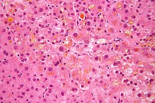
Primary biliary cholangitis (PBC), previously known as primary biliary cirrhosis, is an autoimmune disease of the liver. It results from a slow, progressive destruction of the small bile ducts of the liver, causing bile and other toxins to build up in the liver, a condition called cholestasis. Further slow damage to the liver tissue can lead to scarring, fibrosis, and eventually cirrhosis.

<span class="mw-page-title-main">Ursodeoxycholic acid</span> Medication and metabolite of cholesterol

Ursodeoxycholic acid (UDCA), also known as ursodiol, is a secondary bile acid, produced in humans and most other species from metabolism by intestinal bacteria. It is synthesized in the liver in some species, and was first identified in bile of bears of genus Ursus, from which its name derived. In purified form, it has been used to treat or prevent several diseases of the liver or bile ducts.

Retinal hemorrhage is a disorder of the eye in which bleeding occurs in the retina, the light sensitive tissue, located on the back wall of the eye. There are photoreceptor cells in the retina called rods and cones, which transduce light energy into nerve signals that can be processed by the brain to form visual images. Retinal hemorrhage is strongly associated with child abuse in infants and young children and often leaves such abused infants permanently blind. In older children and adults, retinal hemorrhage can be caused by several medical conditions such as hypertension, retinal vein occlusion, anemia, leukemia or diabetes.

<span class="mw-page-title-main">Lipofuscin</span> Lipid-containing residue associated with aging

Lipofuscin is the name given to fine yellow-brown pigment granules composed of lipid-containing residues of lysosomal digestion. It is considered to be one of the aging or "wear-and-tear" pigments, found in the liver, kidney, heart muscle, retina, adrenals, nerve cells, and ganglion cells.

<span class="mw-page-title-main">Retina horizontal cell</span>

Horizontal cells are the laterally interconnecting neurons having cell bodies in the inner nuclear layer of the retina of vertebrate eyes. They help integrate and regulate the input from multiple photoreceptor cells. Among their functions, horizontal cells are believed to be responsible for increasing contrast via lateral inhibition and adapting both to bright and dim light conditions. Horizontal cells provide inhibitory feedback to rod and cone photoreceptors. They are thought to be important for the antagonistic center-surround property of the receptive fields of many types of retinal ganglion cells.

Intrinsically photosensitive retinal ganglion cells (ipRGCs), also called photosensitive retinal ganglion cells (pRGC), or melanopsin-containing retinal ganglion cells (mRGCs), are a type of neuron in the retina of the mammalian eye. The presence of an additional photoreceptor was first suspected in 1927 when mice lacking rods and cones still responded to changing light levels through pupil constriction; this suggested that rods and cones are not the only light-sensitive tissue. However, it was unclear whether this light sensitivity arose from an additional retinal photoreceptor or elsewhere in the body. Recent research has shown that these retinal ganglion cells, unlike other retinal ganglion cells, are intrinsically photosensitive due to the presence of melanopsin, a light-sensitive protein. Therefore, they constitute a third class of photoreceptors, in addition to rod and cone cells.

<span class="mw-page-title-main">Cholestasis</span> Medical condition

Cholestasis is a condition where the flow of bile from the liver to the duodenum is impaired. The two basic distinctions are:

<span class="mw-page-title-main">Bile acid</span> Steroid acid found predominantly in the bile of mammals and other vertebrates

Bile acids are steroid acids found predominantly in the bile of mammals and other vertebrates. Diverse bile acids are synthesized in the liver. Bile acids are conjugated with taurine or glycine residues to give anions called bile salts.

<span class="mw-page-title-main">Müller glia</span> Glial cell type in the retina

Müller glia, or Müller cells, are a type of retinal glial cells, first recognized and described by Heinrich Müller. They are found in the vertebrate retina, where they serve as support cells for the neurons, as all glial cells do. They are the most common type of glial cell found in the retina. While their cell bodies are located in the inner nuclear layer of the retina, they span across the entire retina.

<span class="mw-page-title-main">PDE6B</span> Protein-coding gene in the species Homo sapiens

Rod cGMP-specific 3',5'-cyclic phosphodiesterase subunit beta is the beta subunit of the protein complex PDE6 that is encoded by the PDE6B gene. PDE6 is crucial in transmission and amplification of visual signal. The existence of this beta subunit is essential for normal PDE6 functioning. Mutations in this subunit are responsible for retinal degeneration such as retinitis pigmentosa or congenital stationary night blindness.

<span class="mw-page-title-main">Disc shedding</span>

Disc shedding is the process by which photoreceptor cells in the retina are renewed. The disc formations in the outer segment of photoreceptors, which contain the photosensitive opsins, are completely renewed every ten days.

<span class="mw-page-title-main">Hyodeoxycholic acid</span> Chemical compound

Hyodeoxycholic acid, also known as 3α,6α-Dihydroxy-5β-cholan-24-oic acid or HDCA, is a secondary bile acid, one of the metabolic byproducts of intestinal bacteria. It differs from deoxycholic acid in that the 6α-hydroxyl is in the 12 position in the former. The 6α-hydroxyl group makes HDCA a hydrophilic acid, a property it shares with hyocholic acid. HDCA is present in mammalian species in different proportions. It is the main acid constituent of hog bile, and for this reason it was used industrially as precursor for steroid synthesis before total synthesis became practical.

Gene therapy using lentiviral vectors was being explored in early stage trials as of 2009.

Retinal gene therapy holds a promise in treating different forms of non-inherited and inherited blindness.

<i>S</i>-Nitrosoglutathione Chemical compound

S-Nitrosoglutathione (GSNO) is an endogenous S-nitrosothiol (SNO) that plays a critical role in nitric oxide (NO) signaling and is a source of bioavailable NO. NO coexists in cells with SNOs that serve as endogenous NO carriers and donors. SNOs spontaneously release NO at different rates and can be powerful terminators of free radical chain propagation reactions, by reacting directly with ROO• radicals, yielding nitro derivatives as end products. NO is generated intracellularly by the nitric oxide synthase (NOS) family of enzymes: nNOS, eNOS and iNOS while the in vivo source of many of the SNOs is unknown. In oxygenated buffers, however, formation of SNOs is due to oxidation of NO to dinitrogen trioxide (N2O3). Some evidence suggests that both exogenous NO and endogenously derived NO from nitric oxide synthases can react with glutathione to form GSNO.

<span class="mw-page-title-main">Retinal regeneration</span>

Retinal regeneration refers to the restoration of vision in vertebrates that have suffered retinal lesions or retinal degeneration.

References

  1. 1 2 3 Hagey LR, Crombie DL, Espinosa E, Carey MC, Igimi H, Hofmann AF (November 1993). "Ursodeoxycholic acid in the Ursidae: biliary bile acids of bears, pandas, and related carnivores". Journal of Lipid Research. 34 (11): 1911–7. doi: 10.1016/S0022-2275(20)35109-9 . PMID   8263415.
  2. Luo Q, Chen Q, Wu Y, Jiang M, Chen Z, Zhang X, Chen H (2010). "[Chemical constituents of bear bile]". Zhongguo Zhong Yao Za Zhi. 35 (18): 2416–2419. PMID   21141490.
  3. Boatright JH, Nickerson JM, Moring AG, Pardue MT (September 2009). "Bile acids in treatment of ocular disease". Journal of Ocular Biology, Diseases, and Informatics. 2 (3): 149–159. doi:10.1007/s12177-009-9030-x. PMC   2798994 . PMID   20046852.
  4. Carey EJ, Ali AH, Lindor KD (October 2015). "Primary biliary cirrhosis". Lancet. 386 (10003): 1565–75. doi:10.1016/S0140-6736(15)00154-3. PMID   26364546. S2CID   44811681.
  5. "Albrioza monograph" (PDF). 1 June 2022. Archived (PDF) from the original on 14 June 2022. Retrieved 14 June 2022.
  6. "Relyvrio- sodium phenylbutyrate/taurursodiol powder, for suspension". DailyMed. 30 September 2022. Retrieved 3 January 2023.
  7. "Failed clinical trial of Sodium phenylbutyrate/ursodoxicoltaurine".
  8. 1 2 Rivard AL, Steer CJ, Kren BT, Rodrigues CM, Castro RE, Bianco RW, Low WC (2007). "Administration of tauroursodeoxycholic acid (TUDCA) reduces apoptosis following myocardial infarction in rat". The American Journal of Chinese Medicine. 35 (2): 279–95. doi:10.1142/S0192415X07004813. PMID   17436368.
  9. 1 2 3 4 Zangerolamo L, Vettorazzi JF, Rosa LR, Carneiro EM, Barbosa HC (May 2021). "The bile acid TUDCA and neurodegenerative disorders: An overview". Life Sciences. 272: 119252. doi:10.1016/j.lfs.2021.119252. PMID   33636170. S2CID   232066323.
  10. 1 2 3 Khalaf K, Tornese P, Cocco A, Albanese A (June 2022). "Tauroursodeoxycholic acid: a potential therapeutic tool in neurodegenerative diseases". Translational Neurodegeneration. 11 (1): 33. doi: 10.1186/s40035-022-00307-z . PMC   9166453 . PMID   35659112.
  11. 1 2 Daruich A, Picard E, Boatright JH, Behar-Cohen F (2019). "Review: The bile acids urso- and tauroursodeoxycholic acid as neuroprotective therapies in retinal disease". Molecular Vision. 25: 610–624. PMC   6817734 . PMID   31700226.
  12. Phillips Joe M; Walker Tiffany A; Choi Hee-Young; Faulkner Amanda E; Kim Moon K; Sidney Sheree S; Boyd Amber P; Nickerson John M; Boatright Jeffrey H; Pardue Machelle T (2008). "Tauroursodeoxycholic acid preservation of photoreceptor structure and function in the rd10 mouse through postnatal day 30". Invest Ophthalmol Vis Sci. 49 (5): 2148–2155. doi:10.1167/iovs.07-1012. PMC   2626193 . PMID   18436848.
  13. Oveson BC, Iwase T, Hackett SF, Lee SY, Usui S, Sedlak TW, Snyder SH, Campochiaro PA, Sung JU (January 2011). "Constituents of bile, bilirubin and TUDCA, protect against oxidative stress-induced retinal degeneration". Journal of Neurochemistry. 116 (1): 144–53. doi:10.1111/j.1471-4159.2010.07092.x. PMC   4083853 . PMID   21054389.
  14. Woo SJ, Kim JH, Yu HG (2010). "Ursodeoxycholic acid and tauroursodeoxycholic acid suppress choroidal neovascularization in a laser-treated rat model". J Ocul Pharmacol Ther. 26 (3): 223–229. doi:10.1089/jop.2010.0012. PMID   20565307.
  15. Fernández-Sánchez L, Lax P, Pinilla I, Martín-Nieto J, Cuenca N (2011). "Tauroursodeoxycholic Acid (TUDCA) Prevents Retinal Degeneration in Transgenic P23H Rats". Invest Ophthalmol Vis Sci. 52 (8): 4998–5008. doi:10.1167/iovs.11-7496. PMID   21508111.
  16. Vang S, Longley K, Steer CJ, Low WC (May 2014). "The Unexpected Uses of Urso- and Tauroursodeoxycholic Acid in the Treatment of Non-liver Diseases". Global Advances in Health and Medicine. 3 (3): 58–69. doi:10.7453/gahmj.2014.017. PMC   4030606 . PMID   24891994.