WNT4 deficiency

Last updated

WNT4 deficiency is a rare genetic disorder that affects females and it results in the underdevelopment and sometimes absence of the uterus and vagina. WNT4 deficiency is caused by mutations of the WNT4 gene. Abnormally high androgen levels are found in the blood and can initiate and promote the development of male sex characteristics. This is seen as male pattern of hair growth on the chest and face. Those with this genetic defect develop breasts but do not have their period. Mayer–Rokitansky–Küster–Hauser syndrome is a related but distinct syndrome. Some women who have an initial diagnosis of MRKH have later been found to have WNT4 deficiency. Most women with MRKH syndrome do not have genetic mutations of the WNT4 gene. The failure to begin the menstrual cycle may be the initial clinical sign of WNT4 deficiency. WNT4 deficiency can cause significant psychological challenges and counseling is recommended. [1] [2]

Contents

Signs and symptoms

The case reports for WNT4 deficiency are few and far between making it difficult to assess the full list of signs and symptoms. More cases ought to be identified in order to achieve a better, and well-rounded, potential spectrum of associated symptoms. [3] However, from the cases that have been identified and given the mechanism of action, there have been symptoms that have been characterized to explain WNT4 deficiency.

Ovary, the female reproductive organ. Ovary nih.jpg
Ovary, the female reproductive organ.

Causes

WNT4 deficiency is an extremely rare disorder that affects females. The exact cause is still unknown due to very limited presented cases worldwide. However, what is known is that it is caused by mutations in the WNT4 gene. WNT4 is a secreted protein that is encoded by the WNT4 gene which is found on chromosome 1. [4] The main function of this gene is to promote female sex development and repress male sex development. The WNT4 gene provides instructions for producing a specific protein that is responsible for the formation of the female reproductive system, the kidneys, and several hormone producing glands.

The WNT4 protein is important in regulating the formation of the Müllerian ducts, also known as the paramesonephric duct, which are the structures in the embryo that eventually will develop into the uterus, fallopian tubes, cervix, and the upper part of the vagina. Consequently, this protein is essential in development of the ovaries and development of oocytes, or female egg cells. [5]

As said, this disorder can be caused by a mutation in the gene due to a random spontaneous event, or it can be inherited as an autosomal dominant trait. Dominant genetic disorders occur when only a single copy of an abnormal gene is necessary to portray its phenotypic qualities. [3] Based on the laws of normal genetics, the abnormal gene can be inherited from either parent and the risk of passing the gene from affected parent to future offspring is 50% regardless of the sex of the offspring given its dominant properties. However, in this case it is not possible to inherit the gene from a mother because they are infertile therefore meaning that they cannot have children. It is still unknown if this mutation can be inherited from the father or if it is caused by new mutations in the gene. [6]

Mechanism/pathophysiology

Human Chromosome 1 Ideogram human chromosome 1.svg
Human Chromosome 1

WNT4 is a secreted protein that is encoded by the WNT4 gene which is found on chromosome 1. [4] Investigators have found that the gene is located on the short arm (p) of chromosome one, which is denoted as (1p35). 1 denotes the chromosome number, p denotes the arm, and 35 denotes the band that the mutation is located on the chromosome. The exact functions of the protein of the WNT4 gene are not fully understood and more research ought to be obtained in order to determine the underlying mechanism of action that causes the symptoms associated with this disease. [3]

However, what is known is that WNT4 is a secreted molecule that binds to the members of the frizzled family of receptors which results in the transcriptional regulation of target genes. Frizzled is a family of atypical G protein-coupled receptors that serve as receptors in the Wnt signaling pathway and other signaling pathways. WNT4 is produced in the ovarian somatic cells and it up-regulates the Dax1 gene known to aggravate the nuclear-receptor steroidogenic factor1 which ultimately will inhibit steroidogenic enzymes. WNT4 increases follistatin expression which ultimately inhibits the formation of anti-testis action and supports ovarian germ cell survival. [7]

Diagnosis

WNT4 deficiency is present at birth meaning that it is congenital, but can be left undiagnosed or unidentified until adolescence when female puberty begins. The start of the females menstrual cycle commences her puberty years, however primary amenorrhea or lack of menstruation, may be what brought a patient in to see a physician and that is when diagnosis begins. Due to this condition being extremely rare, a diagnosis of WNT4 deficiency may be suspected only after having done extensive clinical evaluation, detailed patient history, and the identification of characteristic symptoms. These symptoms include an absent or underdeveloped uterus and/or vagina occurring in conjunction with normal external genitalia. [3]

Specialized imaging techniques such as ultrasounds or magnetic resonance imaging (MRI) can be used to see images of the internal organs (uterus, ovaries, and kidney) to further confirm WNT4 deficiency. In addition, due to modern science, molecular genetic analysis can reveal any mutations of the WNT4 gene to confirm this diagnosis. Karyotyping can be performed as well to rule out any other conditions because it examines the integrity of chromosome in a sample of cells. A female presenting with WNT4 deficiency will have a normal 46, XX karyotype, however, abnormalities on those chromosomes can be seen. [3]

WNT4 deficiency can sometimes be misdiagnosed because there are other disorders that elicit very similar symptoms. One other rare disorder is known as Mayer-Rokitansky-Kuster-Hauser (MRKH) syndrome. It is said that women who have WNT4 deficiency have been misdiagnosed with MRKH syndrome due to very similar signs and symptoms, however, the main difference is that in MRKH syndrome, there is no mutation present on the WNT4 gene even though there is an underdevelopment of the uterus/vagina and amenorrhea while still maintaining normal ovarian function. [3] MRKH syndrome is typically the first diagnosis given, but upon extensive genetic testing and imaging analysis, WNT4 deficiency is the correct diagnosis.

Additionally, complete androgen insensitivity syndrome is a rare disorder in which individuals are genetically male (46, XY) but they do not respond to androgens. This syndrome demonstrates intersex because phenotypically the person is female and displays all external phenotypes of a female, but genetically male because their karyotype denotes 46, XY. [3]

Prevention/treatment

Since WNT4 is a congenital disease, and it is autosomal dominantly inherited, there is no prevention. Since one of the main signs of WNT4 deficiency is having an absent uterus, conceiving is not an option. However, other forms of conception may be available to them since they have functional ovaries but since this disease has an autosomal dominant pattern of inheritance, genetic testing might be a suggestion in order to prevent the passing of the gene.

Treatment, on the other hand, is still in the works considering there are not many cases reported and of those cases, severity of diagnosis differs. However, generally speaking based on what is known, treatment of WNT4 deficiency is directed toward specific symptoms that are apparent in each individual meaning that they arise on a case by case basis. Nonetheless, treatment will require efforts from a team of specialists such as pediatricians (given a diagnosis under age 18), internists, gynecologists, nephrologists, endocrinologists, plastic surgeons, urologists, psychiatrists, and more. [3]

Prognosis

Due to how rare this disorder is, evidence on prognosis is not yet identified. However, based on what is known, a woman who is diagnosed with WNT4 deficiency may have the potential to live a long life. However, this diagnosis does lead to infertility, difficulty engaging in intercourse, the reoccurrence of kidney stones, etc. which may mean preserving a good quality of life may be difficult. Therefore, difficulty accepting this diagnosis may require extensive counseling and group therapy in order to accept and adapt to a different lifestyle.

Epidemiology

WNT4 deficiency does not have any concrete evidence of epidemiology due to its rare nature. The most concrete evidence that is identified lies in the hands of genetics due to its autosomal dominant pattern of inheritance and the mutation present on chromosome 1. However, there is no evidence to show population favorability, distribution patterns, risk factors, environmental factors, etc.

Current research/research directions

WNT4 deficiency is an extremely rare genetic disorder and not much research can be found in relation to the disease itself. The knowledge behind the gene and how it acts in the body is present however, with such minimal cases to compare to, it is hard to develop research on the disease. A future perspective to consider, on the other hand, regarding the WNT4 gene can be used to evaluate conditions such as polycystic ovary disease/syndrome (PCOS) which is a very common disease that affects many females. PCOS also displays hyperandrogenic characteristics and a better characterization of the milder form of WNT4 and its signaling pathways in ovarian tissues can help to further explain its contribution to the hyperandrogenic states seen in patients with PCOS. [2]

Research regarding the WNT4 gene itself is in the works in relation to other conditions that stem from the properties related to this gene. That being said, although this disorder is extremely rare, the WNT4 gene is very widely used in other biological functions. For instance, in 2016, a group of researchers studied how the WNT4 gene mediates estrogen receptor signaling and endocrine resistance in invasive lobular carcinoma cell lines. Since WNT4 is a critical signaling molecule in mammary gland development regulated by a progesterone receptor, its pathway was used in breast cancer research in order to modulate the endocrine response in invasive lobular carcinoma. [8] Additionally, WNT4 gene is used in research regarding kidney function. In 2013, DiRocco et al. published a research article regarding WNT4 and beta-catenin signaling in medullary kidney fibroblasts and discovered that WNT4 expression in renal fibrosis is important in the proliferation process. [9]

Related Research Articles

<span class="mw-page-title-main">Androgen insensitivity syndrome</span> Medical condition

Androgen insensitivity syndrome (AIS) is a difference in sex development involving hormonal resistance due to androgen receptor dysfunction.

<span class="mw-page-title-main">Leigh syndrome</span> Mitochondrial metabolism disease characterized by progressive loss of mental and movement abilities

Leigh syndrome is an inherited neurometabolic disorder that affects the central nervous system. It is named after Archibald Denis Leigh, a British neuropsychiatrist who first described the condition in 1951. Normal levels of thiamine, thiamine monophosphate, and thiamine diphosphate are commonly found, but there is a reduced or absent level of thiamine triphosphate. This is thought to be caused by a blockage in the enzyme thiamine-diphosphate kinase, and therefore treatment in some patients would be to take thiamine triphosphate daily.

<span class="mw-page-title-main">Lipoid congenital adrenal hyperplasia</span> Medical condition

Lipoid congenital adrenal hyperplasia is an endocrine disorder that is an uncommon and potentially lethal form of congenital adrenal hyperplasia (CAH). It arises from defects in the earliest stages of steroid hormone synthesis: the transport of cholesterol into the mitochondria and the conversion of cholesterol to pregnenolone—the first step in the synthesis of all steroid hormones. Lipoid CAH causes mineralocorticoid deficiency in affected infants and children. Male infants are severely undervirilized causing their external genitalia to look feminine. The adrenals are large and filled with lipid globules derived from cholesterol.

<span class="mw-page-title-main">Persistent Müllerian duct syndrome</span> Medical condition

Persistent Müllerian duct syndrome (PMDS) is the presence of Müllerian duct derivatives in what would be considered a genetically and otherwise physically normal male animal by typical human based standards. In humans, PMDS typically is due to an autosomal recessive congenital disorder and is considered by some to be a form of pseudohermaphroditism due to the presence of Müllerian derivatives.

Müllerian agenesis, also known as Müllerian aplasia, vaginal agenesis, or Mayer–Rokitansky–Küster–Hauser syndrome, is a congenital malformation characterized by a failure of the Müllerian ducts to develop, resulting in a missing uterus and variable degrees of vaginal hypoplasia of its upper portion. Müllerian agenesis is the cause in 15% of cases of primary amenorrhoea. Because most of the vagina does not develop from the Müllerian duct, instead developing from the urogenital sinus, along with the bladder and urethra, it is present even when the Müllerian duct is completely absent. Because ovaries do not develop from the Müllerian ducts, affected people might have normal secondary sexual characteristics but are infertile due to the lack of a functional uterus. However, biological motherhood is possible through uterus transplantation or use of gestational surrogates.

<span class="mw-page-title-main">Coffin–Lowry syndrome</span> Medical condition

Coffin–Lowry syndrome is a genetic disorder that is X-linked dominant and which causes severe mental problems sometimes associated with abnormalities of growth, cardiac abnormalities, kyphoscoliosis, as well as auditory and visual abnormalities.

<span class="mw-page-title-main">Birt–Hogg–Dubé syndrome</span> Rare autosomal dominant cancer syndrome

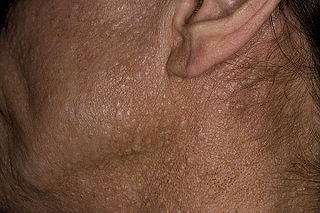
Birt–Hogg–Dubé syndrome (BHD), also Hornstein–Birt–Hogg–Dubé syndrome, Hornstein–Knickenberg syndrome, and fibrofolliculomas with trichodiscomas and acrochordons is a human, adult onset, autosomal dominant genetic disorder caused by the FLCN gene. It can cause susceptibility to kidney cancer, renal and pulmonary cysts, and noncancerous tumors of the hair follicles, called fibrofolliculomas. The symptoms seen in each family are unique, and can include any combination of the three symptoms. Fibrofolliculomas are the most common manifestation, found on the face and upper trunk in over 80% of people with BHD over the age of 40. Pulmonary cysts are equally common (84%) and 24% of people with BHD eventually experience a collapsed lung. Kidney tumors, both cancerous and benign, occur in 14–34% of people with BHD; the associated kidney cancers are often rare hybrid tumors.

Vaginal atresia is a condition in which the vagina is abnormally closed or absent. The main causes can either be complete vaginal hypoplasia, or a vaginal obstruction, often caused by an imperforate hymen or, less commonly, a transverse vaginal septum. It results in uterovaginal outflow tract obstruction. This condition does not usually occur by itself within an individual, but coupled with other developmental disorders within the female. The disorders that are usually coupled with a female who has vaginal atresia are Mayer-Rokitansky-Küster-Hauser syndrome, Bardet-Biedl syndrome, or Fraser syndrome. One out of every 5,000 women have this abnormality.

<span class="mw-page-title-main">Larsen syndrome</span> Medical condition

Larsen syndrome (LS) is a congenital disorder discovered in 1950 by Larsen and associates when they observed dislocation of the large joints and face anomalies in six of their patients. Patients with Larsen syndrome normally present with a variety of symptoms, including congenital anterior dislocation of the knees, dislocation of the hips and elbows, flattened facial appearance, prominent foreheads, and depressed nasal bridges. Larsen syndrome can also cause a variety of cardiovascular and orthopedic abnormalities. This rare disorder is caused by a genetic defect in the gene encoding filamin B, a cytoplasmic protein that is important in regulating the structure and activity of the cytoskeleton. The gene that influences the emergence of Larsen syndrome is found in chromosome region, 3p21.1-14.1, a region containing human type VII collagen gene. Larsen syndrome has recently been described as a mesenchyme disorder that affects the connective tissue of an individual. Autosomal dominant and recessive forms of the disorder have been reported, although most cases are autosomal dominant. Reports have found that in Western societies, Larsen syndrome can be found in one in every 100,000 births, but this is most likely an underestimate because the disorder is frequently unrecognized or misdiagnosed.

<span class="mw-page-title-main">Alström syndrome</span> Rare genetic disorder involving childhood obesity and multiple organ dysfunction

Alström syndrome (AS), also called Alström–Hallgren syndrome, is a very rare autosomal recessive genetic disorder characterised by childhood obesity and multiple organ dysfunction. Symptoms include early-onset type 2 diabetes, cone-rod dystrophy resulting in blindness, sensorineural hearing loss and dilated cardiomyopathy. Endocrine disorders typically also occur, such as hypergonadotrophic hypogonadism and hypothyroidism, as well as acanthosis nigricans resulting from hyperinsulinemia. Developmental delay is seen in almost half of people with Alström syndrome.

XX gonadal dysgenesis is a type of female hypogonadism in which the ovaries do not function to induce puberty in an otherwise normal girl whose karyotype is found to be 46,XX. With nonfunctional streak ovaries, she is low in estrogen levels (hypoestrogenic) and has high levels of FSH and LH. Estrogen and progesterone therapy is usually then commenced. Some cases are considered a severe version of premature ovarian failure where the ovaries fail before puberty.

<span class="mw-page-title-main">Disorders of sex development</span> Medical conditions involving the development of the reproductive system

Disorders of sex development (DSDs), also known as differences in sex development, diverse sex development and variations in sex characteristics (VSC), are congenital conditions affecting the reproductive system, in which development of chromosomal, gonadal, or anatomical sex is atypical.

<span class="mw-page-title-main">WNT4</span> Protein-coding gene in the species Homo sapiens

WNT4 is a secreted protein that, in humans, is encoded by the WNT4 gene, found on chromosome 1. It promotes female sex development and represses male sex development. Loss of function may have consequences, such as female to male sex reversal.

<span class="mw-page-title-main">Complete androgen insensitivity syndrome</span> Intersex condition that results in a phenotypic female

Complete androgen insensitivity syndrome (CAIS) is an AIS condition that results in the complete inability of the cell to respond to androgens. As such, the insensitivity to androgens is only clinically significant when it occurs in individuals who are exposed to significant amounts of testosterone at some point in their lives. The unresponsiveness of the cell to the presence of androgenic hormones prevents the masculinization of male genitalia in the developing fetus, as well as the development of male secondary sexual characteristics at puberty, but does allow, without significant impairment, female genital and sexual development in those with the condition.

<span class="mw-page-title-main">Leydig cell hypoplasia</span> Medical condition

Leydig cell hypoplasia (LCH), also known as Leydig cell agenesis, is a rare autosomal recessive genetic and endocrine syndrome affecting an estimated 1 in 1,000,000 genetic males. It is characterized by an inability of the body to respond to luteinizing hormone (LH), a gonadotropin which is normally responsible for signaling Leydig cells of the testicles to produce testosterone and other androgen sex hormones. The condition manifests itself as pseudohermaphroditism, hypergonadotropic hypogonadism, reduced or absent puberty, and infertility.

Müllerian duct anomalies are those structural anomalies caused by errors in Müllerian duct development during embryonic morphogenesis. Factors that precipitate include genetics, and maternal exposure to teratogens.

Acromesomelic dysplasia is a rare skeletal disorder that causes abnormal bone and cartilage development, leading to shortening of the forearms, lower legs, hands, feet, fingers, and toes. Five different genetic mutations have been implicated in the disorder. Treatment is individualized but is generally aimed at palliating symptoms, for example, treatment of kyphosis and lumbar hyperlordosis.

<span class="mw-page-title-main">Vaginal anomalies</span> Congenital defect; abnormal or absent vagina

Vaginal anomalies are abnormal structures that are formed during the prenatal development of the female reproductive system and are rare congenital defects that result in an abnormal or absent vagina.

<span class="mw-page-title-main">SRD5A3-CDG</span> Medical condition

SRD5A3-CDG is a rare, non X-linked congenital disorder of glycosylation (CDG) due to a mutation in the steroid 5 alpha reductase type 3 gene. It is one of over 150 documented types of Congenital disorders of Glycosylation. Like many other CDGs, SRD5A3 is ultra-rare, with around 38 documented cases in the world.

<span class="mw-page-title-main">17q12 microdeletion syndrome</span> Rare genetic anomaly in humans

17q12 microdeletion syndrome, also known as 17q12 deletion syndrome, is a rare chromosomal anomaly caused by the deletion of a small amount of material from a region in the long arm of chromosome 17. It is typified by deletion of the HNF1B gene, resulting in kidney abnormalities and renal cysts and diabetes syndrome. It also has neurocognitive effects, and has been implicated as a genetic factor for autism and schizophrenia.

References

  1. "WNT4 Deficiency - NORD (National Organization for Rare Disorders)". NORD (National Organization for Rare Disorders). Retrieved 2018-01-22.
  2. 1 2 Biason-Lauber, A.; De Filippo, G.; Konrad, D.; Scarano, G.; Nazzaro, A.; Schoenle, E. J. (January 2007). "WNT4 deficiency--a clinical phenotype distinct from the classic Mayer-Rokitansky-Kuster-Hauser syndrome: a case report". Human Reproduction (Oxford, England). 22 (1): 224–229. doi: 10.1093/humrep/del360 . ISSN   0268-1161. PMID   16959810.
  3. 1 2 3 4 5 6 7 8 "WNT4 Deficiency". NORD (National Organization for Rare Disorders). Retrieved 2020-11-11.
  4. 1 2 "WNT4", Wikipedia, 2020-09-23, retrieved 2020-11-11
  5. "WNT4 gene: MedlinePlus Genetics". medlineplus.gov. Retrieved 2020-11-11.
  6. "Müllerian aplasia and hyperandrogenism: MedlinePlus Genetics". medlineplus.gov. Retrieved 2020-11-11.
  7. Biason-Lauber, Konrad, A, D (2008). "WNT4 and sex development" (PDF). Sexual Development. 2 (4–5): 210–218. doi:10.1159/000152037. PMID   18987495. S2CID   35718787.{{cite journal}}: CS1 maint: multiple names: authors list (link)
  8. Sikora, Matthew J.; Jacobsen, Britta M.; Levine, Kevin; Chen, Jian; Davidson, Nancy E.; Lee, Adrian V.; Alexander, Caroline M.; Oesterreich, Steffi (2016-09-20). "WNT4 mediates estrogen receptor signaling and endocrine resistance in invasive lobular carcinoma cell lines". Breast Cancer Research. 18 (1): 92. doi: 10.1186/s13058-016-0748-7 . ISSN   1465-542X. PMC   5028957 . PMID   27650553.
  9. DiRocco, Derek P.; Kobayashi, Akio; Taketo, Makoto M.; McMahon, Andrew P.; Humphreys, Benjamin D. (2013-09-01). "Wnt4/β−Catenin Signaling in Medullary Kidney Myofibroblasts". Journal of the American Society of Nephrology. 24 (9): 1399–1412. doi: 10.1681/ASN.2012050512 . ISSN   1046-6673. PMC   3752942 . PMID   23766539.