Deamidation

Last updated
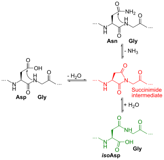
Deamidation reaction of Asn-Gly (top right) to Asp-Gly (at left) or iso(Asp)-Gly (in green at bottom right) Deamidation Asn Gly.svg
Deamidation reaction of Asn-Gly (top right) to Asp-Gly (at left) or iso(Asp)-Gly (in green at bottom right)

Deamidation is a chemical reaction in which an amide functional group in the side chain of the amino acids asparagine or glutamine is removed or converted to another functional group. Typically, asparagine is converted to aspartic acid or isoaspartic acid. Glutamine is converted to glutamic acid or pyroglutamic acid (5-oxoproline). In a protein or peptide, these reactions are important because they may alter its structure, stability or function and may lead to protein degradation. The net chemical change is the addition of a water group and removal of an ammonia group, which corresponds to a +1 (0.98402) Da mass increase. Although deamidation occurs on glutamine, glycosylated asparagine and other amides, these are negligible under typical proteolysis conditions. [1]

Contents

In the deamidation of an asparagine residue under physiological conditions, the side chain is attacked by the nitrogen atom of the following peptide group (in black at top right of Figure), forming an asymmetric succinimide intermediate (in red). The asymmetry of the intermediate results in two products of its hydrolysis, either aspartic acid (in black at left) or isoaspartic acid, which is a beta amino acid (in green at bottom right). However, there is a concern that aspartic acid can be isomerized after deamidation. [2] The deamidation of a glutamine residue may proceed via the same mechanism but at a much slower rate since formation of the six-member-ring glutarimide intermediate is less favoured than the succinimide intermediate for asparagine. In general, deamidation can be eliminated by proteolysis at an acidic pH or at a slightly basic pH (4.5 and 8.0, respectively) using the endoprotease, Glu-C. [2]

The rates of deamidation depend on multiple factors, including the primary sequences and higher-order structures of the proteins, pH, temperature, and components in the solutions. Most potential deamidation sites are stabilized by higher order structure. Asn-Gly (NG),is the most flexible and since it is acidic, it is most prone to deamidation with a half-life around 24 h under physiological conditions (pH 7.4, 37 °C). [3]

As a free amino acid, or as the N-terminal residue of a peptide or protein, glutamine deamidates readily to form pyroglutamic acid (5-oxoproline). The reaction proceeds via nucleophilic attack of the α-amino group on the side-chain amide to form a γ-lactam with the elimination of ammonia from the side-chain.

Analytical method

Protein deamidation has been commonly analyzed by reverse-phase liquid chromatography (RPLC) through peptide mapping. Recently reported novel ERLIC-MS/MS method would enhance the separation of deamidated and non-deamidated peptides with increased identification and quantitation quantification. [4]

Mass spectrometry is commonly used to characterize deamidation states of proteins, including therapeutic monoclonal antibodies. [5] The technique is especially useful for deamidation analysis due to its high sensitivity, speed, and specificity. This allows site-specific deamidation analysis. [6]

A major challenge of using mass spectrometry is the formation of deamidation artifacts during sample preparation. These artifacts significantly skew results because they suggest greater rates of spontaneous deamidation than what is truly observed. This can prove problematic in the case of therapeutic proteins which can be mischaracterized in QC protocols if a large percentage of detected deamidation is due to artifacts. Recent studies indicate that lower pH can reduce the rate of deamidation artifacts. [2]

Kinetics of deamidation

Deamidation reactions have been conjectured to be one of the factors that limit the useful lifetime of proteins. [1]

Deamidation proceeds much more quickly if the susceptible amino acid is followed by a small, flexible residue such as glycine whose low steric hindrance leaves the peptide group open for attack. Deamidation reactions also proceed much more quickly at elevated pH (>10) and temperature.

The endoprotease, Glu-C, has shown specificity to only glutamic acid when in specific pH conditions (4.5 and 8.0) and cleaved the C-terminal side when in a solution with Tris-HCl, bicarbonate, or acetate.

See also

Related Research Articles

<span class="mw-page-title-main">Protein primary structure</span> Linear sequence of amino acids in a peptide or protein

Protein primary structure is the linear sequence of amino acids in a peptide or protein. By convention, the primary structure of a protein is reported starting from the amino-terminal (N) end to the carboxyl-terminal (C) end. Protein biosynthesis is most commonly performed by ribosomes in cells. Peptides can also be synthesized in the laboratory. Protein primary structures can be directly sequenced, or inferred from DNA sequencess.

<span class="mw-page-title-main">Proteolysis</span> Breakdown of proteins into smaller polypeptides or amino acids

Proteolysis is the breakdown of proteins into smaller polypeptides or amino acids. Uncatalysed, the hydrolysis of peptide bonds is extremely slow, taking hundreds of years. Proteolysis is typically catalysed by cellular enzymes called proteases, but may also occur by intra-molecular digestion.

<span class="mw-page-title-main">Protease</span> Enzyme that cleaves other proteins into smaller peptides

A protease is an enzyme that catalyzes proteolysis, breaking down proteins into smaller polypeptides or single amino acids, and spurring the formation of new protein products. They do this by cleaving the peptide bonds within proteins by hydrolysis, a reaction where water breaks bonds. Proteases are involved in many biological functions, including digestion of ingested proteins, protein catabolism, and cell signaling.

<span class="mw-page-title-main">Aspartic acid</span> Amino acid

Aspartic acid (symbol Asp or D; the ionic form is known as aspartate), is an α-amino acid that is used in the biosynthesis of proteins. Like all other amino acids, it contains an amino group and a carboxylic acid. Its α-amino group is in the protonated –NH+
3
form under physiological conditions, while its α-carboxylic acid group is deprotonated −COO under physiological conditions. Aspartic acid has an acidic side chain (CH2COOH) which reacts with other amino acids, enzymes and proteins in the body. Under physiological conditions (pH 7.4) in proteins the side chain usually occurs as the negatively charged aspartate form, −COO. It is a non-essential amino acid in humans, meaning the body can synthesize it as needed. It is encoded by the codons GAU and GAC.

<span class="mw-page-title-main">Asparagine</span> Chemical compound

Asparagine is an α-amino acid that is used in the biosynthesis of proteins. It contains an α-amino group, an α-carboxylic acid group, and a side chain carboxamide, classifying it as a polar, aliphatic amino acid. It is non-essential in humans, meaning the body can synthesize it. It is encoded by the codons AAU and AAC.

<span class="mw-page-title-main">Post-translational modification</span> Biological processes

Post-translational modification (PTM) is the covalent and generally enzymatic modification of proteins following protein biosynthesis. This process often occurs in the endoplasmic reticulum and the golgi apparatus. Proteins are synthesized by ribosomes translating mRNA into polypeptide chains, which may then undergo PTM to form the mature protein product. PTMs are important components in cell signaling, as for example when prohormones are converted to hormones.

<span class="mw-page-title-main">Proteinogenic amino acid</span> Amino acid that is incorporated biosynthetically into proteins during translation

Proteinogenic amino acids are amino acids that are incorporated biosynthetically into proteins during translation. The word "proteinogenic" means "protein creating". Throughout known life, there are 22 genetically encoded (proteinogenic) amino acids, 20 in the standard genetic code and an additional 2 that can be incorporated by special translation mechanisms.

<span class="mw-page-title-main">Serine protease</span> Class of enzymes

Serine proteases are enzymes that cleave peptide bonds in proteins. Serine serves as the nucleophilic amino acid at the (enzyme's) active site. They are found ubiquitously in both eukaryotes and prokaryotes. Serine proteases fall into two broad categories based on their structure: chymotrypsin-like (trypsin-like) or subtilisin-like.

Edman degradation, developed by Pehr Edman, is a method of sequencing amino acids in a peptide. In this method, the amino-terminal residue is labeled and cleaved from the peptide without disrupting the peptide bonds between other amino acid residues.

<span class="mw-page-title-main">Protein sequencing</span> Sequencing of amino acid arrangement in a protein

Protein sequencing is the practical process of determining the amino acid sequence of all or part of a protein or peptide. This may serve to identify the protein or characterize its post-translational modifications. Typically, partial sequencing of a protein provides sufficient information to identify it with reference to databases of protein sequences derived from the conceptual translation of genes.

<span class="mw-page-title-main">Peptide mass fingerprinting</span>

Peptide mass fingerprinting (PMF) is an analytical technique for protein identification in which the unknown protein of interest is first cleaved into smaller peptides, whose absolute masses can be accurately measured with a mass spectrometer such as MALDI-TOF or ESI-TOF. The method was developed in 1993 by several groups independently. The peptide masses are compared to either a database containing known protein sequences or even the genome. This is achieved by using computer programs that translate the known genome of the organism into proteins, then theoretically cut the proteins into peptides, and calculate the absolute masses of the peptides from each protein. They then compare the masses of the peptides of the unknown protein to the theoretical peptide masses of each protein encoded in the genome. The results are statistically analyzed to find the best match.

<span class="mw-page-title-main">Citrullination</span> Biological process

Citrullination or deimination is the conversion of the amino acid arginine in a protein into the amino acid citrulline. Citrulline is not one of the 20 standard amino acids encoded by DNA in the genetic code. Instead, it is the result of a post-translational modification. Citrullination is distinct from the formation of the free amino acid citrulline as part of the urea cycle or as a byproduct of enzymes of the nitric oxide synthase family.

In molecular biology, the Signal Peptide Peptidase (SPP) is a type of protein that specifically cleaves parts of other proteins. It is an intramembrane aspartyl protease with the conserved active site motifs 'YD' and 'GxGD' in adjacent transmembrane domains (TMDs). Its sequences is highly conserved in different vertebrate species. SPP cleaves remnant signal peptides left behind in membrane by the action of signal peptidase and also plays key roles in immune surveillance and the maturation of certain viral proteins.

<span class="mw-page-title-main">Isoaspartate</span>

Isoaspartic acid is an aspartic acid residue isomeric to the typical α peptide linkage. It is a β-amino acid, with the side chain carboxyl moved to the backbone. Such a change is caused by a chemical reaction in which the nitrogen atom on the N+1 following peptide bond nucleophilically attacks the γ-carbon of the side chain of an asparagine or aspartic acid residue, forming a succinimide intermediate. Hydrolysis of the intermediate results in two products, either aspartic acid or isoaspartic acid, which is a β-amino acid. The reaction also results in the deamidation of the asparagine residue. Racemization may occur leading to the formation of D-aminoacids.

<span class="mw-page-title-main">Triticeae glutens</span> Seed storage protein in mature wheat seeds

Gluten is the seed storage protein in mature wheat seeds. It is the sticky substance in bread wheat which allows dough to rise and retain its shape during baking. The same, or very similar, proteins are also found in related grasses within the tribe Triticeae. Seed glutens of some non-Triticeae plants have similar properties, but none can perform on a par with those of the Triticeae taxa, particularly the Triticum species. What distinguishes bread wheat from these other grass seeds is the quantity of these proteins and the level of subcomponents, with bread wheat having the highest protein content and a complex mixture of proteins derived from three grass species.

<span class="mw-page-title-main">Carboxypeptidase A</span>

Carboxypeptidase A usually refers to the pancreatic exopeptidase that hydrolyzes peptide bonds of C-terminal residues with aromatic or aliphatic side-chains. Most scientists in the field now refer to this enzyme as CPA1, and to a related pancreatic carboxypeptidase as CPA2.

Protein footprinting is a term used to refer to a method of biochemical analysis that investigates protein structure, assembly, and interactions within a larger macromolecular assembly. It was originally coined in reference to the use of limited proteolysis to investigate contact sites within a monoclonal antibody - protein antigen complex and a year later to examine the protection from hydroxyl radical cleavage conferred by a protein bound to DNA within a DNA-protein complex. In DNA footprinting the protein is envisioned to make an imprint at a particular point of interaction. This latter method was adapted through the direct treatment of proteins and their complexes with hydroxyl radicals and can be generally denoted RP-MS akin to the designation used for Hydrogen-deuterium exchange Mass Spectrometry.

Peptidyl-Asp metalloendopeptidase is an enzyme. This enzyme catalyses the following chemical reaction

<span class="mw-page-title-main">Glutamic protease</span>

Glutamic proteases are a group of proteolytic enzymes containing a glutamic acid residue within the active site. This type of protease was first described in 2004 and became the sixth catalytic type of protease. Members of this group of protease had been previously assumed to be an aspartate protease, but structural determination showed it to belong to a novel protease family. The first structure of this group of protease was scytalidoglutamic peptidase, the active site of which contains a catalytic dyad, glutamic acid (E) and glutamine (Q), which give rise to the name eqolisin. This group of proteases are found primarily in pathogenic fungi affecting plant and human.

Asparagine peptide lyase are one of the seven groups in which proteases, also termed proteolytic enzymes, peptidases, or proteinases, are classified according to their catalytic residue. The catalytic mechanism of the asparagine peptide lyases involves an asparagine residue acting as nucleophile to perform a nucleophilic elimination reaction, rather than hydrolysis, to catalyse the breaking of a peptide bond.

References

  1. 1 2 Clarke, S (2003). "Aging as war between chemical and biochemical processes: protein methylation and the recognition of age-damaged proteins for repair". Ageing Res Rev. 2 (3): 263–285. doi:10.1016/S1568-1637(03)00011-4. PMID   12726775. S2CID   18135051.
  2. 1 2 3 Liu, Shanshan; Moulton, Kevin Ryan; Auclair, Jared Robert; Zhou, Zhaohui Sunny (2016-04-01). "Mildly acidic conditions eliminate deamidation artifact during proteolysis: digestion with endoprotease Glu-C at pH 4.5". Amino Acids. 48 (4): 1059–1067. doi:10.1007/s00726-015-2166-z. ISSN   1438-2199. PMC   4795971 . PMID   26748652.
  3. Tyler-Cross R, Schirch V (1991) Effects of amino acid sequence, buffers, and ionic strength on the rate and mechanism of deamidation of asparagine residues in small peptides. J Biol Chem 266:22549–22556
  4. Zhen, Jing (2018). "Antibody characterization using novel ERLIC-MS/MS-based peptide mapping". mAbs. 10 (7): 1–9. doi:10.1080/19420862.2018.1505179. PMC   6204790 . PMID   30130443.
  5. Wang, Weijie; Meeler, Andrea R.; Bergerud, Luke T.; Hesselberg, Mark; Byrne, Michael; Wu, Zhuchun (2012). "Quantification and characterization of antibody deamidation by peptide mapping with mass spectrometry". International Journal of Mass Spectrometry. 312: 107–113. Bibcode:2012IJMSp.312..107W. doi:10.1016/j.ijms.2011.06.006.
  6. Hao, Piliang; Adav, Sunil S.; Gallart-Palau, Xavier; Sze, Siu Kwan (November 2017). "Recent advances in mass spectrometric analysis of protein deamidation". Mass Spectrometry Reviews. 36 (6): 677–692. Bibcode:2017MSRv...36..677H. doi:10.1002/mas.21491. ISSN   1098-2787. PMID   26763661.