In human anatomy, the ulnar nerve is a nerve that runs near the ulna bone. The ulnar collateral ligament of elbow joint is in relation with the ulnar nerve. The nerve is the largest unprotected nerve in the human body, so injury is common. This nerve is directly connected to the little finger, and the adjacent half of the ring finger, innervating the palmar aspect of these fingers, including both front and back of the tips, perhaps as far back as the fingernail beds.

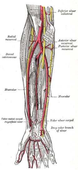
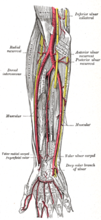
The ulnar artery is the main blood vessel, with oxygenated blood, of the medial aspect of the forearm. It arises from the brachial artery and terminates in the superficial palmar arch, which joins with the superficial branch of the radial artery. It is palpable on the anterior and medial aspect of the wrist.

The opponens digiti minimi is a muscle in the hand. It is of a triangular form, and placed immediately beneath the palmaris brevis, abductor digiti minimi and flexor digiti minimi brevis. It is one of the three hypothenar muscles that control the little finger.
The opponens pollicis is a small, triangular muscle in the hand, which functions to oppose the thumb. It is one of the three thenar muscles, lying deep to the abductor pollicis brevis and lateral to the flexor pollicis brevis.
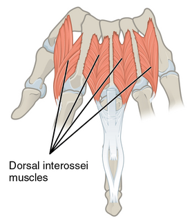
In human anatomy, the dorsal interossei (DI) are four muscles in the back of the hand that act to abduct (spread) the index, middle, and ring fingers away from hand's midline and assist in flexion at the metacarpophalangeal joints and extension at the interphalangeal joints of the index, middle and ring fingers.

In human anatomy, the abductor digiti minimi is a skeletal muscle situated on the ulnar border of the palm of the hand. It forms the ulnar border of the palm and its spindle-like shape defines the hypothenar eminence of the palm together with the skin, connective tissue, and fat surrounding it. Its main function is to pull the little finger away from the other fingers.

The flexor digiti minimi brevis is a hypothenar muscle in the hand that flexes the little finger at the metacarpophalangeal joint. It lies lateral to the abductor digiti minimi when the hand is in anatomical position.
The superficial palmar arch is formed predominantly by the ulnar artery, with a contribution from the superficial palmar branch of the radial artery. However, in some individuals the contribution from the radial artery might be absent, and instead anastomoses with either the princeps pollicis artery, the radialis indicis artery, or the median artery, the former two of which are branches from the radial artery.
The deep palmar arch is an arterial network found in the palm. It is usually formed mainly from the terminal part of the radial artery, with the ulnar artery contributing via its deep palmar branch, by an anastomosis. This is in contrast to the superficial palmar arch, which is formed predominantly by the ulnar artery.

The superficial branch of the radial nerve passes along the front of the radial side of the forearm to the commencement of its lower third. It is a sensory nerve.

The lateral plantar nerve is a branch of the tibial nerve, in turn a branch of the sciatic nerve and supplies the skin of the fifth toe and lateral half of the fourth, as well as most of the deep muscles, its distribution being similar to that of the ulnar nerve in the hand.
The palmar metacarpal arteries, three or four in number, arise from the convexity of the deep volar arch

The dorsal carpal arch is an anatomical term for the combination (anastomosis) of dorsal carpal branch of the radial artery and the dorsal carpal branch of the ulnar artery near the back of the wrist.
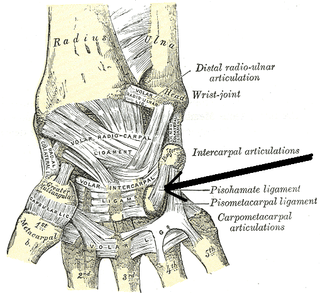
The pisohamate ligament is a ligament in the hand. It is the volar ligament that connects the pisiform to the hamate. It is a prolongation of the tendon of the flexor carpi ulnaris.
In anatomy, arterial tree is used to refer to all arteries and/or the branching pattern of the arteries. This article regards the human arterial tree. Starting from the aorta:
The palmar carpal arch is the combination (anastomosis) of two arteries: the palmar carpal branch of radial artery and the palmar carpal branch of ulnar artery.
The proper palmar digital arteries travel along the sides of the phalanges, each artery lying just below its corresponding digital nerve.

The deep branch of the ulnar nerve is a terminal, primarily motor branch of the ulnar nerve. It is accompanied by the deep palmar branch of ulnar artery.

The cervical spinal nerve 8 (C8) is a spinal nerve of the cervical segment.
The public domain consists of all the creative works to which no exclusive intellectual property rights apply. Those rights may have expired, been forfeited, expressly waived, or may be inapplicable.

Gray's Anatomy is an English language textbook of human anatomy originally written by Henry Gray and illustrated by Henry Vandyke Carter. Earlier editions were called Anatomy: Descriptive and Surgical, Anatomy of the Human Body and Gray's Anatomy: Descriptive and Applied, but the book's name is commonly shortened to, and later editions are titled, Gray's Anatomy. The book is widely regarded as an extremely influential work on the subject, and has continued to be revised and republished from its initial publication in 1858 to the present day. The latest edition of the book, the 41st, was published in September 2015.