Exophiala dermatitidis

Last updated

Exophiala dermatitidis
Wangiella dermatitidis PAS stain PHIL 3781 lores.jpg
histopathologic changes in phaeohyphomycosis due to Exophiala dermatitidis, formerly Wangiella dermatitidis using PAS stain
Scientific classification OOjs UI icon edit-ltr.svg
Domain: Eukaryota
Kingdom: Fungi
Division: Ascomycota
Class: Eurotiomycetes
Order: Chaetothyriales
Family: Herpotrichiellaceae
Genus: Exophiala
Species:
E. dermatitidis
Binomial name
Exophiala dermatitidis
(Kano) de Hoog (1977)
Synonyms [1]
  • Hormiscium dermatitidisKano (1934) [2]
  • Fonsecaea dermatitidis(Kano) Carrion (1950) [3]
  • Hormodendrum dermatitidis(Kano) Conant (1954) [4]
  • Phialophora dermatitidis(Kano) C.W.Emmons (1963) [5]
  • Rhinocladiella dermatitidisSchol-Schwarz (1968) [6]
  • Wangiella dermatitidis(Kano) McGinnis (1977) [7]

Exophiala dermatitidis is a thermophilic black yeast, and a member of the Herpotrichiellaceae. [8] [9] While the species is only found at low abundance in nature, metabolically active strains are commonly isolated in saunas, steam baths, and dish washers. [9] [10] Exophiala dermatitidis only rarely causes infection in humans, however cases have been reported around the world. In East Asia, the species has caused lethal brain infections in young and otherwise healthy individuals. [11] The fungus has been known to cause cutaneous and subcutaneous phaeohyphomycosis, [12] [13] and as a lung colonist in people with cystic fibrosis in Europe. [14] In 2002, an outbreak of systemic E. dermatitidis infection occurred in women who had received contaminated steroid injections at North Carolina hospitals. [15] [16]

Contents

Appearance and general description

Exophiala dermatitidis forms slow growing, brown or black colonies. [11] [12] As is common amongst black yeasts, E. dermatitidis is an anamorphic fungus with multiple conidial forms. [17] [18] [19] This morphological plasticity has complicated taxonomic determination based solely on physical appearance. [19] Young colonies are described as waxy, [11] mucoid, [17] smooth, [11] [12] and yeastlike. [12] [13] Over time pigmented aerial hyphae develop, and older colonies are described as appearing filamentous and velvety. [12] [13] [17] [20] Conidia are between globular and elliptical in shape, and form at the mouth of short annellidic conidiogenous cells. Annellations can only be observed by electron microscopy. [12] [13] [17] Prior to analysis by scanning electron microscope, the conidiogenous cells were observed to form from non-annelated phialides and phialides without collarettes. [13] These observations caused the species to be erroneously provided with its own monotypic genus, Wangiella. [13] The species name Wangiella dermatitidis is still commonly used in the scientific literature.

The black fungus also takes on diverse morphologies in vivo. Infected tissues contain mixtures of ovoid yeast-like cells, short septate hyphae that may be branched or unbranched, toruloid hyphae, as well as isotopically enlarged sclerotic (muriform-like) cells that resemble those found in chromoblastomycosis. [21] [22] The muriform-like cells isolated from E. dermatitidis infections have thinner walls than those found in chromoblastomycosis, and cells are divided along a single plane. [21]

A sexual form of E. dermatitidis has not been observed. [8] However, the occurrence of two mating type idiomorphs in approximately equal numbers among clinical and environmental isolates suggests that E. dermatitidis reproduces sexually. [23] Sequencing of rDNA has shown that clinical isolates of E. dermatitidis are very closely related to Capronia mansonii, and It has been predicted that the ascospores and ascomata of an E. dermatitidis teleomorph would therefore resemble those of C. mansonii. [8]

Ideal growth conditions for E. dermatitidis occur between 40 °C (104 °F) and 42 °C (108 °F), [11] [13] [24] however E. dermatitidis can tolerate temperatures as high as 47 °C (117 °F). [10] Metabolically active fungus is highly abundant in Turkish steam baths that routinely reach temperatures of over 60 °C (140 °F), but is typically not found in more hot and dry sauna facilities, or in cool environments surrounding steam baths. [24] It is thought that extracellular polysaccharides may protect the species from stress in hot and moist environments, as this feature is typical of strains isolated from steam baths. [9] [24] Exophiala dermatitidis has carotenoid pigments in its cell wall that may serve a role in protecting E. dermatitidis from UV damage. [19]

Thermophilicity, negative potassium nitrate assimilation, negative melezitose assimilation, and an ability to decompose tyrosine are used to distinguish E. dermatitidis from other black yeasts. [11] [12] [19] [20] An exoantigen test is useful in species determination, and DNA analysis can also be performed. [19] The fungus has been selectively isolated using high temperature incubation (at 40 °C) on media containing cycloheximide. [9]

Geographic distribution

Exophiala dermatitidis has been isolated around the world in low abundance from a variety of environmental sources, including soil, decaying timber, and wasp nests. [9] [13] [21] The thermophilicity and acid tolerance of E. dermatitidis suggests passage through warm-blooded animals, and it is hypothesized that its ecological niche might be associated with tropical, frugivorous bird and bat species. [14] [25] An ability to utilize nutrients in diverse environments, to adhere to fruit surfaces, and progress through different morphological phases are considered to provide further evidence for this theory. [25] Clinical isolates tend to harbor strains that are found only rarely in nature. [14] It may be the case that traits linked to halotolerance have predisposed these strains towards infecting humans. [14]

While E. dermatitidis has been found only in low abundance in nature, the species is well suited to survive in a number of warm and wet man made niches. Metabolically active strains are isolated in high abundance from surfaces inside saunas, steam baths and humidifiers. [9] [24] E. dermatitidis is one of the most common fungal species to inhabit dishwashers, and has been found in dishwashers around the world. [10]

Pathophysiology

Exophiala dermatitidis is typically considered a human opportunistic pathogen, as those affected by E. dermatitidis often have underlying health conditions. [11] [12] An exception to this is the neurotropic clinical presentation, which is typically found in young and otherwise healthy individuals. [11]

While over 100 fungal species can cause phaeohyphomycosis, E. dermatitidis is one of the two fungi most frequently implicated, along with another Exophiala species, E. jeanselmei . [26] [27] Exophiala dermatitidis is considered to be one of the most pathogenic fungi in the genus Exophiala, [14] and is highly deadly, with a fatality rate of over 40%. [19] [28] The high fatality rate is primarily due to an ability to form systemic and neurotropic infections, which represent approximately half of reported E. dermatitidis cases. [19]

Local and superficial infections

Exophiala dermatitidis forms cutaneous and subcutaneous phaeohyphomycosis, which most commonly affect the face and neck. [11] [12] [13] [17] [19] [21] Indeed, the fungus was originally isolated from the skin of a patient with lesions on their cheek, neck, and ear. [19] Cells isolated from cutaneous infections are often spherical, and may form toruloid or moniliform chains. [19] Exophiala dermatitidis has been implicated various superficial infections including onychomycosis, otitis externa, and eye infections causing keratitis. [11] [19] [27]

In Europe E. dermatitidis tends to be associated with cystic fibrosis, [14] and is frequently found to have colonized the lungs of CF patients. [17] In one study, E. dermatitidis could be isolated from 6.2% of cystic fibrosis patients using erythritol-chloramphenicol agar culture dishes. [29] Exophiala dermatitidis has also been reported as the etiological agent of lung infections causing pneumonia. [11]

Systemic infections

Exophiala dermatitidis forms neurotrophic infections, and is the black yeast that most commonly causes life-threatening phaeohyphomycosis. [11] [12] [21] Conditions that might predispose people towards an invasive opportunistic infection include diabetes mellitus, lymphocytic leukemia, bronchiectasis, rheumatoid arthritis, and catheterization. [30] Systemic infections are often reported to be without cutaneous or subcutaneous involvement. [21]

Systemic E. dermatitidis infections can include cerebral metastases. [11] [12] The fatality rate for such infections is reported to be over 90%. [28] Central nervous system phaeohyphomycosis is rare, and for unknown reasons primarily arise in East Asia, despite a cosmopolitan distribution of the fungus. [9] Within East Asian populations, young and otherwise healthy people have developed cerebral infections. [9] Lung infections in European CF patients and neurotropic mycosis in East Asia are caused by E. dermatitidis strains that are genetically similar, and host factors such as immunological differences may be responsible for the different infection patterns. [31] Exophiala dermatitidis occurs at very high frequency in both Asian and European saunas, and absence of neurotrophic mycosis in Europe isn't explained by reduced exposure to the fungus. [24]

In 2002 a small outbreak of systemic E. dermatitidis infection occurred in North Carolina hospitals, involving five women who received steroid injections for pain management. [15] [16] In one of women the infection wasn't evident until 152 days after injection of the contaminated solution. [16] Isolates from these patients were found by the FDA to be susceptible to all of voriconazole, itraconazole, and amphotericin B. In one patient the infection caused sacroiliitis, while the remaining four developed meningitis. [16] Meningitis eventually caused death in one patient, while voriconazole was successful in treating infection in the four other patients. [16] The outbreak was traced back to a single compounding pharmacy, which was found by the FDA to have inadequately controlled for sterility of its products. [15] [16]

Immune response

Exophiala dermatitidis typically causes a non-specific and granulomatous inflammatory response. [21] Lymphocytes, histiocytes, multinucleated giant cells and neutrophils are recruited. [21] The host responses are highly variable, often include cyst formation, and vary from weak reaction to an intense inflammatory response that results in tissue necrosis. [21]

As with other black yeasts that cause phaeohyphomycosis, melanin appears to have defensive purpose, and helps protect E. dermatitidis from death within human neutrophils. [9] [26] Pathogenic strains of E. dermatitidis contain five times more melanin than saprophytic E. dermatitidis, while melanin deficient mutants of pathogenic strains have dramatically reduced virulence. [19]

Treatment

A diagnosis of E. dermatitidis infection of the CNS can only be reliably achieved following biopsy. [21] For systemic infections there are few treatment options, and E. dermatitidis is described as "notoriously resistant" to antifungal drugs. [19] [21] During the North Carolina outbreak, treatment with voriconazole was effective in four out of the five patients, and all of voriconazole, itraconazole, and amphotericin B were found to be effective in vitro. [16] Terbinafine has also been found to be effective in vitro, and combinations of antifungal drugs can have a synergistic effect against E. dermatitidis. [32] A 2012 article found that of reported cases, 44% of patients responded to amphotericin B treatment, 50% responded to voriconazole treatment, and 71.4% responded to itraconazole therapy. [28]

For small and local infections surgery may be an option. [19] As E. dermatitidis infections are believed to be caused by traumatic implantation of the fungus, surgeons must be exceedingly careful to not re-introduce infection during operation. [19] Despite the high heat tolerance of E. dermatitidis, heat treatment of cutaneous lesions have been effective. [19]

Related Research Articles

<i>Talaromyces marneffei</i> Species of fungus

Talaromyces marneffei, formerly called Penicillium marneffei, was identified in 1956. The organism is endemic to southeast Asia where it is an important cause of opportunistic infections in those with HIV/AIDS-related immunodeficiency. Incidence of T. marneffei infections has increased due to a rise in HIV infection rates in the region.

<span class="mw-page-title-main">Sporotrichosis</span> Medical condition

Sporotrichosis, also known as rose handler's disease, is a fungal infection that may be localised to skin, lungs, bone and joint, or become systemic. It presents with firm painless nodules that later ulcerate. Following initial exposure to Sporothrix schenckii, the disease typically progresses over a period of a week to several months. Serious complications may develop in people who have a weakened immune system.

<i>Aspergillus terreus</i> Species of fungus

Aspergillus terreus, also known as Aspergillus terrestris, is a fungus (mold) found worldwide in soil. Although thought to be strictly asexual until recently, A. terreus is now known to be capable of sexual reproduction. This saprotrophic fungus is prevalent in warmer climates such as tropical and subtropical regions. Aside from being located in soil, A. terreus has also been found in habitats such as decomposing vegetation and dust. A. terreus is commonly used in industry to produce important organic acids, such as itaconic acid and cis-aconitic acid, as well as enzymes, like xylanase. It was also the initial source for the drug mevinolin (lovastatin), a drug for lowering serum cholesterol.

Exophiala jeanselmei is a saprotrophic fungus in the family Herpotrichiellaceae. Four varieties have been discovered: Exophiala jeanselmei var. heteromorpha, E. jeanselmei var. lecanii-corni, E. jeanselmei var. jeanselmei, and E. jeanselmei var. castellanii. Other species in the genus Exophiala such as E. dermatitidis and E. spinifera have been reported to have similar annellidic conidiogenesis and may therefore be difficult to differentiate.

<i>Blastomyces dermatitidis</i> Species of fungus

Blastomyces dermatitidis is a dimorphic fungus that causes blastomycosis, an invasive and often serious fungal infection found occasionally in humans and other animals. It lives in soil and wet, decaying wood, often in an area close to a waterway such as a lake, river or stream. Indoor growth may also occur, for example, in accumulated debris in damp sheds or shacks. The fungus is endemic to parts of eastern North America, particularly boreal northern Ontario, southeastern Manitoba, Quebec south of the St. Lawrence River, parts of the U.S. Appalachian mountains and interconnected eastern mountain chains, the west bank of Lake Michigan, the state of Wisconsin, and the entire Mississippi Valley including the valleys of some major tributaries such as the Ohio River. In addition, it occurs rarely in Africa both north and south of the Sahara Desert, as well as in the Arabian Peninsula and the Indian subcontinent. Though it has never been directly observed growing in nature, it is thought to grow there as a cottony white mold, similar to the growth seen in artificial culture at 25 °C (77 °F). In an infected human or animal, however, it converts in growth form and becomes a large-celled budding yeast. Blastomycosis is generally readily treatable with systemic antifungal drugs once it is correctly diagnosed; however, delayed diagnosis is very common except in highly endemic areas.

Black yeasts, sometimes also black fungi, dematiaceous fungi, microcolonial fungi or meristematic fungi is a diverse group of slow-growing microfungi which reproduce mostly asexually. Only few genera reproduce by budding cells, while in others hyphal or meristematic (isodiametric) reproduction is preponderant. Black yeasts share some distinctive characteristics, in particular a dark colouration (melanisation) of their cell wall. Morphological plasticity, incrustation of the cell wall with melanins and presence of other protective substances like carotenoids and mycosporines represent passive physiological adaptations which enable black fungi to be highly resistant against environmental stresses. The term "polyextremotolerance" has been introduced to describe this phenotype, an example of which is the species Aureobasidium pullulans. Presence of 1,8-dihydroxynaphthalene melanin in the cell wall confers to the microfungi their characteristic olivaceous to dark brown/black colour.

Exophiala hongkongensis is an ascomycete fungus species. Described as new to science in 2013, it was discovered on the toenail clipping of a patient with the fungal disease onychomycosis.

<i>Cladophialophora bantiana</i> Species of fungus

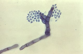
Cladophialophora bantiana is a melanin producing mold known to cause brain abscesses in humans. It is one of the most common causes of systemic phaeohyphomycosis in mammals. Cladophialophora bantiana is a member of the ascomycota and has been isolated from soil samples from around the world.

Phaeohyphomycosis is a diverse group of fungal infections, caused by dematiaceous fungi whose morphologic characteristics in tissue include hyphae, yeast-like cells, or a combination of these. It can be associated an array of melanistic filamentous fungi including Alternaria species,Exophiala jeanselmei, and Rhinocladiella mackenziei.

Phialemonium curvatum is a pathogenic fungus in the phylum Ascomycota. The genus was created to accommodate taxa intermediate to Acremonium and Phialophora. This genus is characterized by its abundance of adelophialides and few discrete phialides with no signs of collarettes. Specifically, P. curvatum is characterized by its grayish white colonies and its allantoid conidia. Phialemonium curvatum is typically found in a variety of environments including air, soil, industrial water and sewage. Furthermore, P. curvatum affects mainly immunocompromised and is rarely seen in immunocompetent people. The species has been known to cause peritonitis, endocarditis, endovascular infections, osteomyelitis as well as cutaneous infections of wounds and burns.

Scedosporiosis is the general name for any mycosis - i.e., fungal infection - caused by a fungus from the genus Scedosporium. Current population-based studies suggest Scedosporium prolificans and Scedosporium apiospermum to be among the most common infecting agents from the genus, although infections caused by other members thereof are not unheard of. The latter is an asexual form (anamorph) of another fungus, Pseudallescheria boydii. The former is a “black yeast”, currently not characterized as well, although both of them have been described as saprophytes.

<i>Exophiala phaeomuriformis</i> Species of fungus

Exophiala phaeomuriformis is thermophilic fungus belonging to the genus Exophiala and the family Herpotrichiellaceae. it is a member of the group of fungi known as black yeasts, and is typically found in hot and humid locations, such as saunas, bathrooms, and dishwashers. This species can cause skin infections and is typically classified as a Biosafety Risk Group 2 agent.

<i>Fonsecaea compacta</i> Species of fungus

Fonsecaea compacta is a saprophytic fungal species found in the family Herpotrichiellaceae. It is a rare etiological agent of chromoblastomycosis, with low rates of correspondence observed from reports. The main active components of F. compacta are glycolipids, yet very little is known about its composition. F. compacta is widely regarded as a dysplastic variety of Fonsecaea pedrosoi, its morphological precursor. The genus Fonsecaea presently contains two species, F. pedrosoi and F. compacta. Over 100 strains of F. pedrosoi have been isolated but only two of F. compacta.

<i>Rhinocladiella mackenziei</i> Species of fungus

Rhinocladiella mackenziei is a deeply pigmented mold that is a common cause of human cerebral phaeohyphomycosis. Rhinocladiella mackenziei was believed to be endemic solely to the Middle East, due to the first cases of infection being limited to the region. However, cases of R. mackenziei infection are increasingly reported from regions outside the Middle East. This pathogen is unique in that the majority of cases have been reported from immunologically normal people.

<i>Phialophora verrucosa</i> Species of fungus

Phialophora verrucosa is a pathogenic, dematiaceous fungus that is a common cause of chromoblastomycosis. It has also been reported to cause subcutaneous phaeohyphomycosis and mycetoma in very rare cases. In the natural environment, it can be found in rotting wood, soil, wasp nests, and plant debris. P. verrucosa is sometimes referred to as Phialophora americana, a closely related environmental species which, along with P. verrucosa, is also categorized in the P. carrionii clade.

Cladosporium oxysporum is an airborne fungus that is commonly found outdoors and is distributed throughout the tropical and subtropical region, it is mostly located In Asia and Africa. It spreads through airborne spores and is often extremely abundant in outdoor air during the spring and summer seasons. It mainly feeds on decomposing organic matter in warmer climates, but can also be parasitic and feed on living plants. The airborne spores can occasionally cause cutaneous infections in humans, and the high prevalence of C. oxysporum in outdoor air during warm seasons contributes to its importance as an etiological agent of allergic disease and possibly human cutaneous phaeohyphomycosis in tropical regions.

Exophiala pisciphila is a mesophilic black yeast and member of the dark septate endophytes. This saprotrophic fungus is found commonly in marine and soil environments. It is abundant in harsh environments like soil contaminated with heavy metals. E. pisciphila forms symbiotic relationships with various plants by colonizing on roots, conferring resistance to drought and heavy metal stress. It is an opportunistic pathogen that commonly causes infections in captive fish and amphibians, while rarely causing disease in humans. Secondary metabolites produced by this species have potential clinical antibiotic and antiretroviral applications.

<span class="mw-page-title-main">Trichosporon asteroides</span> Fungus of the genus Trichosporon

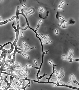
Trichosporon asteroides is an asexual basidiomycetous fungus first described from human skin but now mainly isolated from blood and urine. T. asteroides is a hyphal fungus with a characteristically yeast-like appearance due to the presence of slimy arthroconidia. Infections by this species usually respond to treatment with azoles and amphotericin B.

Arthrographis kalrae is an ascomycetous fungus responsible for human nail infections described in 1938 by Cochet as A. langeronii. A. kalrae is considered a weak pathogen of animals including human restricted to the outermost keratinized layers of tissue. Infections caused by this species are normally responsive to commonly used antifungal drugs with only very rare exceptions.

Cladophialophora arxii is a black yeast shaped dematiaceous fungus that is able to cause serious phaeohyphomycotic infections. C. arxii was first discovered in 1995 in Germany from a 22-year-old female patient suffering multiple granulomatous tracheal tumours. It is a clinical strain that is typically found in humans and is also capable of acting as an opportunistic fungus of other vertebrates Human cases caused by C. arxii have been reported from all parts of the world such as Germany and Australia.

References

  1. "Exophiala dermatitidis (Kano) de Hoog 1977". MycoBank. International Mycological Association. Retrieved 22 August 2012.
  2. Kano K. (1934). "A new pathogenic Hormiscium Kunze causing chromoblastomycosis". Aichi Igakkai Zasshi (in Japanese). 41: 1657–73.
  3. Carrión AL. (1950). "Yeastlike dematiaceous fungi infecting the human skin: Special reference to so-called Hormiscium dermatitidis". Archives of Dermatology and Syphilology. 61 (6): 996–1009. doi:10.1001/archderm.1950.01530130114017. PMID   15419816.
  4. Conant NF, Smith DT, Baker RD, Callaway JL, Martin DS (1954). Manual of Clinical Mycology (2 ed.). Philadelphia, Pennsylvania: WB Saunders. p. 276.
  5. Emmons CW, Binford CH, Utz JP (1963). "Medical Mycology". London, UK: Henry Kimpton: 291.{{cite journal}}: Cite journal requires |journal= (help)
  6. Schol-Schwarz, Marie Beatrice (1968). "Rhinocladiella, its synonym Fonsecaea and its relation to Phialophora". Antonie van Leeuwenhoek. 34 (2): 119–152. doi:10.1007/BF02046424. PMID   5301320. S2CID   43876060.
  7. McGinnis MR. (1977). "Wangiella dermatitidis, a correction". Mycotaxon. 6 (2): 367–9.
  8. 1 2 3 Untereiner, Wendy A.; Naveau, Francoise A. (1999). "Molecular Systematics of the Herpotrichiellaceae with an Assessment of the Phylogenetic Positions of Exophiala dermatitidis and Phialophora americana". Mycologia. 91 (1): 67–83. doi:10.2307/3761194. JSTOR   3761194.
  9. 1 2 3 4 5 6 7 8 9 Reiss E, Shadomy HJ (2011). Fundamentals of Medical Mycology. John Wiley & Sons. p. 500. ISBN   978-0-470-17791-4.
  10. 1 2 3 Zalar, P.; Novak, M.; de Hoog, G.S.; Gunde-Cimerman, N. (2011). "Dishwashers – A man-made ecological niche accommodating human opportunistic fungal pathogens". Fungal Biology. 115 (10): 997–1007. doi:10.1016/j.funbio.2011.04.007. PMID   21944212.
  11. 1 2 3 4 5 6 7 8 9 10 11 12 13 de Hoog, G. S.; Guarro, J.; Gené, J.; Figueras, M. J. (2000). Atlas of clinical fungi (2. ed.). Utrecht: Centraalbureau voor Schimmelcultures [u.a.] ISBN   90-70351-43-9.
  12. 1 2 3 4 5 6 7 8 9 10 11 Howard, Dexter H. (2003). Pathogenic fungi in humans and animals (2nd ed.). New York: Marcel Dekker. ISBN   0203909100.
  13. 1 2 3 4 5 6 7 8 9 Kwon-Chung, K.J.; Bennett, John E. (1992). Medical mycology. Philadelphia: Lea & Febiger. ISBN   0-8121-1463-9.
  14. 1 2 3 4 5 6 Gunde-Cimerman, Nina; Plemenitaš, Ana; Oren, Aharon (2005). Adaptation to life at high salt concentrations in archaea, bacteria, and eukarya. Dordrecht: Springer. pp. 384–388. ISBN   1402036329.
  15. 1 2 3 Richardson, Malcolm; Warnock, David W. (2010). Fungal infection : diagnosis and management (4th ed.). Oxford: Wiley-Blackwell. p. 387. ISBN   978-1405170567 . Retrieved 8 November 2014.
  16. 1 2 3 4 5 6 7 Centers for Disease Control and Prevention (13 December 2002). "Exophiala infection from contaminated injectable steroids prepared by a compounding pharmacy – United States, July–November 2002". Morbidity and Mortality Weekly Report. 51 (49): 1109–12. PMID   12530707 . Retrieved 8 November 2014.
  17. 1 2 3 4 5 6 Anaissie, Elias J.; McGinnis, Michael R.; Pfaller, Michael A. (2009). Clinical Mycology (2nd ed.). Edinburgh: Churchill Livingstone. p. 334. ISBN   978-1416056805 . Retrieved 11 October 2014.
  18. Georgiev, Vassil St. (1998). Infectious diseases in immunocompromised hosts. Boca Raton: CRC Press. p. 909. ISBN   0849385539 . Retrieved 16 October 2014.
  19. 1 2 3 4 5 6 7 8 9 10 11 12 13 14 15 16 Matsumoto, T; Matsuda, T; McGinnis, MR; Ajello, L (1993). "Clinical and mycological spectra of Wangiella dermatitidis infections". Mycoses. 36 (5–6): 145–55. doi:10.1111/j.1439-0507.1993.tb00743.x. PMID   8264710. S2CID   27029853.
  20. 1 2 Tille, Patricia (2013). Bailey & Scott's Diagnostic Microbiology. Elsevier Health Sciences. p. 766.
  21. 1 2 3 4 5 6 7 8 9 10 11 Matsumoto, T.; Ajello, L.; Matsuda, T.; Szaniszlo, P.J.; Walsh, T.J. (1994). "Developments in hyalohyphomycosis and phaeohyphomycosis". Medical Mycology. 32 (s1): 329–349. doi:10.1080/02681219480000951. PMID   7722796.
  22. "Wangiella (Exophiala) dermatitidis". Broad Institute. Retrieved 12 November 2014.
  23. Metin B, Döğen A, Yıldırım E, de Hoog GS, Heitman J, Ilkit M. Mating type (MAT) locus and possible sexuality of the opportunistic pathogen Exophiala dermatitidis. Fungal Genet Biol. 2019 Mar;124:29–38. doi: 10.1016/j.fgb.2018.12.011. Epub 2019 Jan 3. PMID 30611834
  24. 1 2 3 4 5 Matos, T.; de Hoog, G. S.; de Boer, A. G.; de Crom, I.; Haase, G. (November 2002). "High prevalence of the neurotrope and related oligotrophic black yeasts in sauna facilities". Mycoses. 45 (9–10): 373–377. doi: 10.1046/j.1439-0507.2002.00779.x . PMID   12421284. S2CID   4842988.
  25. 1 2 Sudhadham, M.; Prakitsin, S.; Sivichai, S.; Chaiyarat, R.; Dorrestein, G.M; Menken, S.B.J.; de Hoog, G.S. (2008). "The neurotropic black yeast Exophiala dermatitidis has a possible origin in the tropical rain forest". Studies in Mycology. 61: 145–155. doi:10.3114/sim.2008.61.15. PMC   2610309 . PMID   19287537.
  26. 1 2 Johnson, Malcolm D.; Richardson, Elizabeth M. (2006). The pocket guide to fungal infection (2nd ed.). Malden, Mass.: Blackwell Pub. pp. 134–138. ISBN   9780470757734.
  27. 1 2 Domer, J. E. (2004). Human fungal pathogens : with 31 tables (2nd ed.). Berlin [u.a.]: Springer. pp. 78–79. ISBN   9783540426295 . Retrieved 8 November 2014.
  28. 1 2 3 Patel, Atul K.; Patel, Ketan K.; Darji, Prakash; Singh, Rachna; Shivaprakash, M. R.; Chakrabarti, Arunaloke (May 2013). "endocarditis on native aortic valve in a postrenal transplant patient and review of literature on infections". Mycoses. 56 (3): 365–372. doi:10.1111/myc.12009. PMID   23013169. S2CID   40764387.
  29. Horré, R.; Schaal, K.P.; Siekmeier, R.; Sterzik, B.; de Hoog, G.S.; Schnitzler, N. (2004). "Isolation of Fungi, Especially Exophiala dermatitidis, in Patients Suffering from Cystic Fibrosis". Respiration. 71 (4): 360–366. doi:10.1159/000079640. PMID   15316209. S2CID   4832528.
  30. Szaniszlo PJ. (2002). "Molecular genetic studies of the model dematiaceous pathogen Wangiella dermatitidis". International Journal of Medical Microbiology. 292 (5–6): 381–90. doi:10.1078/1438-4221-00221. PMID   12452284.
  31. Uijthof, J. M. J.; Hoog, G. S.; Cock, A. W. A. M.; Takeo, K.; Nishimura, K. (24 April 2009). "Pathogenicity of strains of the black yeast Exophiala (Wangiella) dermatitidis: an evaluation based on polymerase chain reaction". Mycoses. 37 (7–8): 235–242. doi:10.1111/j.1439-0507.1994.tb00419.x. PMID   7739652. S2CID   20385551.
  32. Acton, Ashton Q. (2012). Azole Antifungals—Advances in Research and Application: 2012 Edition: ScholarlyBrief. ScholarlyEditions. pp. 14–15. ISBN   978-1481612265 . Retrieved 9 November 2014.