Related Research Articles

A fascial compartment is a section within the body that contains muscles and nerves and is surrounded by deep fascia. In the human body, the limbs can each be divided into two segments – the upper limb can be divided into the arm and the forearm and the sectional compartments of both of these – the fascial compartments of the arm and the fascial compartments of the forearm contain an anterior and a posterior compartment. Likewise, the lower limbs can be divided into two segments – the leg and the thigh – and these contain the fascial compartments of the leg and the fascial compartments of the thigh.

The spermatic cord is the cord-like structure in males formed by the vas deferens and surrounding tissue that runs from the deep inguinal ring down to each testicle. Its serosal covering, the tunica vaginalis, is an extension of the peritoneum that passes through the transversalis fascia. Each testicle develops in the lower thoracic and upper lumbar region and migrates into the scrotum. During its descent it carries along with it the vas deferens, its vessels, nerves etc. There is one on each side.

The inguinal canal is a passage in the anterior abdominal wall on each side of the body, which in males, convey the spermatic cords and in females, the round ligament of the uterus. The inguinal canals are larger and more prominent in males.

The retroperitoneal space (retroperitoneum) is the anatomical space behind (retro) the peritoneum. It has no specific delineating anatomical structures. Organs are retroperitoneal if they have peritoneum on their anterior side only. Structures that are not suspended by mesentery in the abdominal cavity and that lie between the parietal peritoneum and abdominal wall are classified as retroperitoneal.

A fascia is a generic term for macroscopic membranous bodily structures. Fasciae are classified as superficial, visceral or deep, and further designated according to their anatomical location.

The mesentery is an organ that attaches the intestines to the posterior abdominal wall and is formed by the double fold of peritoneum. It helps in storing fat and allowing blood vessels, lymphatics, and nerves to supply the intestines, among other functions.
Deep fascia is a fascia, a layer of dense connective tissue that can surround individual muscles and groups of muscles to separate into fascial compartments.

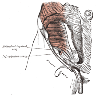
In human anatomy, the inferior epigastric artery is an artery that arises from the external iliac artery. It is accompanied by the inferior epigastric vein; inferiorly, these two inferior epigastric vessels together travel within the lateral umbilical fold The inferior epigastric artery then traverses the arcuate line of rectus sheath to enter the rectus sheath, then anastomoses with the superior epigastric artery within the rectus sheath.

In anatomy, the abdominal wall represents the boundaries of the abdominal cavity. The abdominal wall is split into the anterolateral and posterior walls.

The transversalis fascia is the fascial lining of the anterolateral abdominal wall situated between the inner surface of the transverse abdominal muscle, and the preperitoneal fascia. It is directly continuous with the iliac fascia, the internal spermatic fascia, and pelvic fascia.

The fascia of Camper is a thick superficial layer of the anterior abdominal wall.

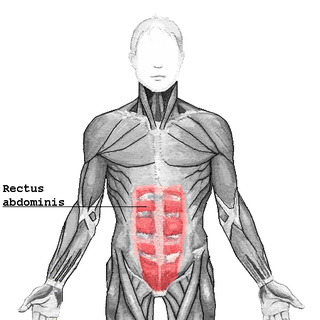
The rectus sheath is a tough fibrous compartment formed by the aponeuroses of the transverse abdominal muscle, and the internal and external oblique muscles. It contains the rectus abdominis and pyramidalis muscles, as well as vessels and nerves.

The bare area of the liver is a large triangular area on the diaphragmatic surface of the liver. It is the only part of the liver with no peritoneal covering, although it is still covered by Glisson's capsule. It is attached directly to the diaphragm by loose connective tissue. The bare area of the liver is relevant to the portacaval anastomosis, encloses the right extraperitoneal subphrenic space, and can be a site of spread of infection from the abdominal cavity to the thoracic cavity
The extraperitoneal space is the portion of the abdomen and pelvis which does not lie within the peritoneum.

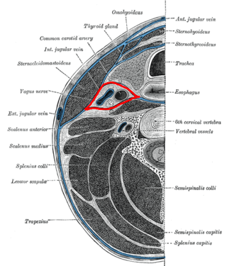
The buccopharyngeal fascia is a fascia of the pharynx. It represents the posterior portion of the pretracheal fascia. It covers the superior pharyngeal constrictor muscles, and buccinator muscle.
The presacral fascia lines the anterior aspect of the sacrum, enclosing the sacral vessels and nerves. It continues anteriorly as the pelvic parietal fascia, covering the entire pelvic cavity.
The retroinguinal space is the extraperitoneal space situated deep to the inguinal ligament. It's limited by the fascia transversalis anteriorly, the peritoneum posteriorly and the iliac fascia laterally. This preperitoneal space communicates with prevesical space of Retzius. It is divided into two compartments. The medial compartment contains vasculature including the femoral artery and vein. The lateral compartment allows for passage of the iliopsoas, allowing attachment to the femur, along with the femoral nerve.
Toldt's fascia, is a discrete layer of connective tissue containing lymphatic channels. It is found between the two mesothelial layers that separate the mesocolon from the underlying retroperitoneum. It was first described by the Austrian anatomist Carl Toldt (1840–1920) as a fascial plane formed by the fusion of the visceral peritoneum with the parietal peritoneum. This was later called Toldt's fascia.

The vaginal support structures are those muscles, bones, ligaments, tendons, membranes and fascia, of the pelvic floor that maintain the position of the vagina within the pelvic cavity and allow the normal functioning of the vagina and other reproductive structures in the female. Defects or injuries to these support structures in the pelvic floor leads to pelvic organ prolapse. Anatomical and congenital variations of vaginal support structures can predispose a woman to further dysfunction and prolapse later in life. The urethra is part of the anterior wall of the vagina and damage to the support structures there can lead to incontinence and urinary retention.
The temporoparietal fascia is a superficial fascia of the side of the head over the area of the temporal fossa situated superficial to the (deep) temporal fascia, and deep to the skin and subcutaneous tissue of the region.
References
- 1 2 3 4 "extraperitoneal fascia". TheFreeDictionary.com. Retrieved 2023-05-17.
- 1 2 Arregui, M. E. (1997-07-01). "Surgical anatomy of the preperitoneal fasciae and posterior transversalis fasciae in the inguinal region". Hernia. 1 (2): 101–110. doi:10.1007/BF02427673. ISSN 1248-9204.
- 1 2 3 4 Kingsnorth, Andrew N.; Skandalakis, Panagiotis N.; Colborn, Gene L.; Weidman, Thomas A.; Skandalakis, Lee J.; Skandalakis, John E. (2000-02-01). "EMBRYOLOGY, ANATOMY, AND SURGICAL APPLICATIONS OF THE PREPERITONEAL SPACE". Surgical Clinics of North America. 80 (1): 1–24. doi:10.1016/S0039-6109(05)70394-7. ISSN 0039-6109.
- ↑ Memon, Muhammed Ashraf; Quinn, Thomas H.; Cahill, Donald R. (June 1999). "Transversalis Fascia: Historical Aspects and its Place in Contemporary Inguinal Herniorrhaphy". Journal of Laparoendoscopic & Advanced Surgical Techniques. 9 (3): 267–272. doi:10.1089/lap.1999.9.267. ISSN 1092-6429.