A glycome is the entire complement or complete set of all sugars, whether free or chemically bound in more complex molecules, of an organism. An alternative definition is the entirety of carbohydrates in a cell. The glycome may in fact be one of the most complex entities in nature. "Glycomics, analogous to genomics and proteomics, is the systematic study of all glycan structures of a given cell type or organism" and is a subset of glycobiology.[1]
"Carbohydrate", "glycan", "saccharide", and "sugar" are generic terms used interchangeably in this context and includes monosaccharides, oligosaccharides, polysaccharides, and derivatives of these compounds. Carbohydrates consist of "hydrated carbon", i.e. [CH2O]n. Monosaccharides are a carbohydrate that cannot be hydrolyzed into a simpler carbohydrate and are the building blocks of oligosaccharides and polysaccharides. Oligosaccharides are linear or branched chains of monosaccharides attached to one another via glycosidic linkages. The number of monosaccharide units can vary. Polysaccharides are glycans composed of repeating monosaccharides, generally greater than ten monosaccharide units in length.[2]
The glycome exceeds the complexity of the proteome as a result of the even greater diversity of the glycome's constituent carbohydrates and is further complicated by the sheer multiplicity of possibilities in the combination and interaction of the carbohydrates with each other and with proteins. "The spectrum of all glycan structures — the glycome — is immense. In humans, its size is orders of magnitude greater than the number of proteins that are encoded by the genome, one percent of which encodes proteins that make, modify, localize or bind sugar chains, which are known as glycans."[3]
The outer surface of the cell is a sea of lipids with a fleet of sugar molecules, many of which are attached to proteins, fats or both, that interact with molecules outside the cell and are critical for the communication between cells and the stickiness of a cell. "Glycans are nature's biologic modifiers," says Jamey Marth, a Howard Hughes Medical Institute investigator at the University of California San Diego."Glycans generally don't turn physiologic processes on and off, rather they modify the behavior of the cell by responding to external stimuli."[4]
Tools used for research on glycome
The following are examples of the commonly used techniques in glycan analysis:[5]
High-resolution mass spectrometry (MS) and high-performance liquid chromatography (HPLC)
The most commonly applied methods are MS and HPLC, in which the glycan part is cleaved either enzymatically or chemically from the target and subjected to analysis.[6] In case of glycolipids, they can be analyzed directly without separation of the lipid component.
N-glycans from glycoproteins are analyzed routinely by high-performance-liquid-chromatography (reversed phase, normal phase and ion exchange HPLC) after tagging the reducing end of the sugars with a fluorescent compound (reductive labeling).[7] A large variety of different labels were introduced in the recent years, where 2-aminobenzamide (AB), anthranilic acid (AA), 2-aminopyridin (PA), 2-aminoacridone (AMAC) and 3-(acetylamino)-6-aminoacridine (AA-Ac) are just a few of them.[8]
O-glycans are usually analysed without any tags, due to the chemical release conditions preventing them to be labeled.
Fractionated glycans from high-performance liquid chromatography (HPLC) instruments can be further analyzed by MALDI-TOF-MS(MS) to get further information about structure and purity. Sometimes glycan pools are analyzed directly by mass spectrometry without prefractionation, although a discrimination between isobaric glycan structures is more challenging or even not always possible. Anyway, direct MALDI-TOF-MS analysis can lead to a fast and straightforward illustration of the glycan pool.[9]
In recent years, high performance liquid chromatography online coupled to mass spectrometry became very popular. By choosing porous graphitic carbon as a stationary phase for liquid chromatography, even non derivatized glycans can be analyzed. Detection is here done by mass spectrometry, but in instead of MALDI-MS, electrospray ionisation (ESI) is more frequently used.[10][11][12]
Multiple Reaction Monitoring (MRM)
Although MRM has been used extensively in metabolomics and proteomics, its high sensitivity and linear response over a wide dynamic range make it especially suited for glycan biomarker research and discovery. MRM is performed on a triple quadrupole (QqQ) instrument, which is set to detect a predetermined precursor ion in the first quadrupole, a fragmented in the collision quadrupole, and a predetermined fragment ion in the third quadrupole. It is a non-scanning technique, wherein each transition is detected individually and the detection of multiple transitions occurs concurrently in duty cycles. This technique is being used to characterize the immune glycome.[13][14]
Table 1:Advantages and disadvantages of mass spectrometry in glycan analysis
Advantages
Disadvantages
Applicable for small sample amounts (lower fmol range)
Useful for complex glycan mixtures (generation of a further analysis dimension).
Attachment sides can be analysed by tandem MS experiments (side specific glycan analysis).
Glycan sequencing by tandem MS experiments.
Destructive method.
Need of a proper experimental design.
Arrays
Lectin and antibody arrays provide high-throughput screening of many samples containing glycans. This method uses either naturally occurring lectins or artificial monoclonal antibodies, where both are immobilized on a certain chip and incubated with a fluorescent glycoprotein sample.
Glycan arrays, like that offered by the Consortium for Functional Glycomics and Z Biotech LLC, contain carbohydrate compounds that can be screened with lectins or antibodies to define carbohydrate specificity and identify ligands.
Metabolic and covalent labeling of glycans
Metabolic labeling of glycans can be used as a way to detect glycan structures. A well known strategy involves the use of azide-labeled sugars which can be reacted using the Staudinger ligation. This method has been used for in vitro and in vivo imaging of glycans.
↑ Hase S, Ikenaka T, Matsushima Y (November 1978). "Structure analyses of oligosaccharides by tagging of the reducing end sugars with a fluorescent compound". Biochem. Biophys. Res. Commun. 85 (1): 257–63. doi:10.1016/S0006-291X(78)80037-0. PMID743278.
↑ Pabst M, Kolarich D, Pöltl G, etal. (January 2009). "Comparison of fluorescent labels for oligosaccharides and introduction of a new postlabeling purification method". Anal. Biochem. 384 (2): 263–73. doi:10.1016/j.ab.2008.09.041. PMID18940176.
↑ Harvey DJ, Bateman RH, Bordoli RS, Tyldesley R (2000). "Ionisation and fragmentation of complex glycans with a quadrupole time-of-flight mass spectrometer fitted with a matrix-assisted laser desorption/ionisation ion source". Rapid Commun. Mass Spectrom. 14 (22): 2135–42. doi:10.1002/1097-0231(20001130)14:22<2135::AID-RCM143>3.0.CO;2-#. PMID11114021.
↑ Schulz, BL; Packer NH, NH; Karlsson, NG (Dec 2002). "Small-scale analysis of O-linked oligosaccharides from glycoproteins and mucins separated by gel electrophoresis". Anal. Chem. 74 (23): 6088–97. doi:10.1021/ac025890a. PMID12498206.
↑ Pabst M, Bondili JS, Stadlmann J, Mach L, Altmann F (July 2007). "Mass + retention time = structure: a strategy for the analysis of N-glycans by carbon LC-ESI-MS and its application to fibrin N-glycans". Anal. Chem. 79 (13): 5051–7. doi:10.1021/ac070363i. PMID17539604.
Glycomics is the comprehensive study of glycomes, including genetic, physiologic, pathologic, and other aspects. Glycomics "is the systematic study of all glycan structures of a given cell type or organism" and is a subset of glycobiology. The term glycomics is derived from the chemical prefix for sweetness or a sugar, "glyco-", and was formed to follow the omics naming convention established by genomics and proteomics.
Glycoproteins are proteins which contain oligosaccharide chains covalently attached to amino acid side-chains. The carbohydrate is attached to the protein in a cotranslational or posttranslational modification. This process is known as glycosylation. Secreted extracellular proteins are often glycosylated.
The Consortium for Functional Glycomics (CFG) is a large research initiative funded in 2001 by a glue grant from the National Institute of General Medical Sciences (NIGMS) to “define paradigms by which protein-carbohydrate interactions mediate cell communication”. To achieve this goal, the CFG studies the functions of:
Defined in the narrowest sense, glycobiology is the study of the structure, biosynthesis, and biology of saccharides that are widely distributed in nature. Sugars or saccharides are essential components of all living things and aspects of the various roles they play in biology are researched in various medical, biochemical and biotechnological fields.
Lectins are carbohydrate-binding proteins that are highly specific for sugar groups that are part of other molecules, so cause agglutination of particular cells or precipitation of glycoconjugates and polysaccharides. Lectins have a role in recognition at the cellular and molecular level and play numerous roles in biological recognition phenomena involving cells, carbohydrates, and proteins. Lectins also mediate attachment and binding of bacteria, viruses, and fungi to their intended targets.
An oligosaccharide is a saccharide polymer containing a small number of monosaccharides. Oligosaccharides can have many functions including cell recognition and cell adhesion.
The terms glycans and polysaccharides are defined by IUPAC as synonyms meaning "compounds consisting of a large number of monosaccharides linked glycosidically". However, in practice the term glycan may also be used to refer to the carbohydrate portion of a glycoconjugate, such as a glycoprotein, glycolipid, or a proteoglycan, even if the carbohydrate is only an oligosaccharide. Glycans usually consist solely of O-glycosidic linkages of monosaccharides. For example, cellulose is a glycan composed of β-1,4-linked D-glucose, and chitin is a glycan composed of β-1,4-linked N-acetyl-D-glucosamine. Glycans can be homo- or heteropolymers of monosaccharide residues, and can be linear or branched.
Glycoproteomics is a branch of proteomics that identifies, catalogs, and characterizes proteins containing carbohydrates as a result of post-translational modifications. Glycosylation is the most common post-translational modification of proteins, but continues to be the least studied on the proteome level. Mass spectrometry (MS) is an analytical technique used to improve the study of these proteins on the proteome level. Glycosylation contributes to several concerted biological mechanisms essential to maintaining physiological function. The study of the glycosylation of proteins is important to understanding certain diseases, like cancer, because a connection between a change in glycosylation and these diseases has been discovered. To study this post-translational modification of proteins, advanced mass spectrometry techniques based on glycoproteomics have been developed to help in terms of therapeutic applications and the discovery of biomarkers.
Protein mass spectrometry refers to the application of mass spectrometry to the study of proteins. Mass spectrometry is an important method for the accurate mass determination and characterization of proteins, and a variety of methods and instrumentations have been developed for its many uses. Its applications include the identification of proteins and their post-translational modifications, the elucidation of protein complexes, their subunits and functional interactions, as well as the global measurement of proteins in proteomics. It can also be used to localize proteins to the various organelles, and determine the interactions between different proteins as well as with membrane lipids.
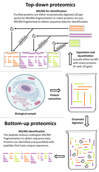
Top-down proteomics is a method of protein identification that either uses an ion trapping mass spectrometer to store an isolated protein ion for mass measurement and tandem mass spectrometry (MS/MS) analysis or other protein purification methods such as two-dimensional gel electrophoresis in conjunction with MS/MS. Top-down proteomics is capable of identifying and quantitating unique proteoforms through the analysis of intact proteins. The name is derived from the similar approach to DNA sequencing. During mass spectrometry intact proteins are typically ionized by electrospray ionization and trapped in a Fourier transform ion cyclotron resonance, quadrupole ion trap or Orbitrap mass spectrometer. Fragmentation for tandem mass spectrometry is accomplished by electron-capture dissociation or electron-transfer dissociation. Effective fractionation is critical for sample handling before mass-spectrometry-based proteomics. Proteome analysis routinely involves digesting intact proteins followed by inferred protein identification using mass spectrometry (MS). Top-down MS (non-gel) proteomics interrogates protein structure through measurement of an intact mass followed by direct ion dissociation in the gas phase.
Anne Dell is an Australian biochemist specialising in the study of glycomics and the carbohydrate structures that modify proteins. Anne's work could be used to figure out how pathogens such as HIV are able to evade termination by the immune system which could be applied toward understanding how this occurs in fetuses. Her research has also led to the development of higher sensitivity mass spectroscopy techniques which have allowed for the better studying of the structure of carbohydrates. Anne also established GlycoTRIC at Imperial College London, a research center that allows for glycobiology to be better understood in biomedical applications. She is currently Professor of Carbohydrate Biochemistry and Head of the Department of Life Sciences at Imperial College London. Dell's other contributions to the study of Glycobiology are the additions she has made to the textbook "Essentials of Glycobiology" Dell was appointed Commander of the Order of the British Empire (CBE) in the 2009 Birthday Honours.
N-linked glycosylation, is the attachment of an oligosaccharide, a carbohydrate consisting of several sugar molecules, sometimes also referred to as glycan, to a nitrogen atom, in a process called N-glycosylation, studied in biochemistry. The resulting protein is called an N-linked glycan, or simply an N-glycan.
Peptide:N-glycosidase F, commonly referred to as PNGase F, is an amidase of the peptide-N4-(N-acetyl-beta-glucosaminyl)asparagine amidase class. PNGase F works by cleaving between the innermost GlcNAc and asparagine residues of high mannose, hybrid, and complex oligosaccharides from N-linked glycoproteins and glycopeptides. This results in a deaminated protein or peptide and a free glycan.
Translational glycobiology or applied glycobiology is the branch of glycobiology and glycochemistry that focuses on developing new pharmaceuticals through glycomics and glycoengineering. Although research in this field presents many difficulties, translational glycobiology presents applications with therapeutic glycoconjugates, with treating various bone diseases, and developing therapeutic cancer vaccines and other targeted therapies. Some mechanisms of action include using the glycan for drug targeting, engineering protein glycosylation for better efficacy, and glycans as drugs themselves.
Carbohydrate Structure Database (CSDB) is a free curated database and service platform in glycoinformatics, launched in 2005 by a group of Russian scientists from N.D. Zelinsky Institute of Organic Chemistry, Russian Academy of Sciences. CSDB stores published structural, taxonomical, bibliographic and NMR-spectroscopic data on natural carbohydrates and carbohydrate-related molecules.
In biochemistry, paucimannosylation is an enzymatic post-translational modification involving the attachment of relatively simple mannose (Man) and N-Acetylglucosamine (GlcNAc) containing carbohydrates (glycans) to proteins. The paucimannosidic glycans may also be modified with other types of monosaccharides including fucose (Fuc) and xylose (Xyl) depending on the species, tissue and cell origin.
The Minimum Information Required About a Glycomics Experiment (MIRAGE) initiative is part of the Minimum Information Standards and specifically applies to guidelines for reporting on a glycomics experiment. The initiative is supported by the Beilstein Institute for the Advancement of Chemical Sciences. The MIRAGE project focuses on the development of publication guidelines for interaction and structural glycomics data as well as the development of data exchange formats. The project was launched in 2011 in Seattle and set off with the description of the aims of the MIRAGE project.
Glycan arrays, like that offered by the Consortium for Functional Glycomics (CFG), National Center for Functional Glycomics (NCFG) and Z Biotech, LLC, contain carbohydrate compounds that can be screened with lectins, antibodies or cell receptors to define carbohydrate specificity and identify ligands. Glycan array screening works in much the same way as other microarray that is used for instance to study gene expression DNA microarrays or protein interaction Protein microarrays.
Ten Feizi is a Turkish Cypriot/British molecular biologist who is Professor and Director of the Glycosciences Laboratory at Imperial College London. Her research considers the structure and function of glycans. She was awarded the Society for Glycobiology Rosalind Kornfeld award in 2014. She was also awarded the Fellowship of the Academy of Medical Sciences in 2021.
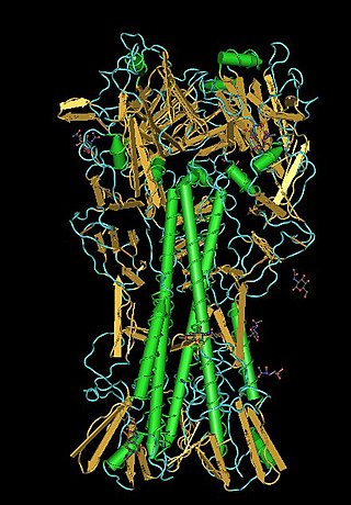
Glycan-Protein interactions represent a class of biomolecular interactions that occur between free or protein-bound glycans and their cognate binding partners. Intramolecular glycan-protein (protein-glycan) interactions occur between glycans and proteins that they are covalently attached to. Together with protein-protein interactions, they form a mechanistic basis for many essential cell processes, especially for cell-cell interactions and host-cell interactions. For instance, SARS-CoV-2, the causative agent of COVID-19, employs its extensively glycosylated spike (S) protein to bind to the ACE2 receptor, allowing it to enter host cells. The spike protein is a trimeric structure, with each subunit containing 22 N-glycosylation sites, making it an attractive target for vaccine search.
This page is based on this Wikipedia article Text is available under the CC BY-SA 4.0 license; additional terms may apply. Images, videos and audio are available under their respective licenses.