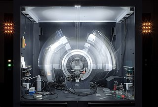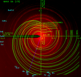
X-ray crystallography is the experimental science determining the atomic and molecular structure of a crystal, in which the crystalline structure causes a beam of incident X-rays to diffract into many specific directions. By measuring the angles and intensities of these diffracted beams, a crystallographer can produce a three-dimensional picture of the density of electrons within the crystal. From this electron density, the mean positions of the atoms in the crystal can be determined, as well as their chemical bonds, their crystallographic disorder, and various other information.
In X-ray crystallography, wide-angle X-ray scattering (WAXS) or wide-angle X-ray diffraction (WAXD) is the analysis of Bragg peaks scattered to wide angles, which are caused by sub-nanometer-sized structures. It is an X-ray-diffraction method and commonly used to determine a range of information about crystalline materials. The term WAXS is commonly used in polymer sciences to differentiate it from SAXS but many scientists doing "WAXS" would describe the measurements as Bragg/X-ray/powder diffraction or crystallography.

Neutron diffraction or elastic neutron scattering is the application of neutron scattering to the determination of the atomic and/or magnetic structure of a material. A sample to be examined is placed in a beam of thermal or cold neutrons to obtain a diffraction pattern that provides information of the structure of the material. The technique is similar to X-ray diffraction but due to their different scattering properties, neutrons and X-rays provide complementary information: X-Rays are suited for superficial analysis, strong x-rays from synchrotron radiation are suited for shallow depths or thin specimens, while neutrons having high penetration depth are suited for bulk samples.
In physics, the phase problem is the problem of loss of information concerning the phase that can occur when making a physical measurement. The name comes from the field of X-ray crystallography, where the phase problem has to be solved for the determination of a structure from diffraction data. The phase problem is also met in the fields of imaging and signal processing. Various approaches of phase retrieval have been developed over the years.
Electron crystallography is a method to determine the arrangement of atoms in solids using a transmission electron microscope (TEM). It can involve the use of high-resolution transmission electron microscopy images, electron diffraction patterns including convergent-beam electron diffraction or combinations of these. It has been successful in determining some bulk structures, and also surface structures. Two related methods are low-energy electron diffraction which has solved the structure of many surfaces, and reflection high-energy electron diffraction which is used to monitor surfaces often during growth.
A diffractometer is a measuring instrument for analyzing the structure of a material from the scattering pattern produced when a beam of radiation or particles interacts with it.

Powder diffraction is a scientific technique using X-ray, neutron, or electron diffraction on powder or microcrystalline samples for structural characterization of materials. An instrument dedicated to performing such powder measurements is called a powder diffractometer.

X-ray reflectivity is a surface-sensitive analytical technique used in chemistry, physics, and materials science to characterize surfaces, thin films and multilayers. It is a form of reflectometry based on the use of X-rays and is related to the techniques of neutron reflectometry and ellipsometry.
Hugo M. Rietveld was a Dutch crystallographer who is famous for his publication on the full profile refinement method in powder diffraction, which became later known as the Rietveld refinement method. The method is used for the characterisation of crystalline materials from X-ray powder diffraction data. The Rietveld refinement uses a least squares approach to refine a theoretical line profile until it matches the measured profile. The introduction of this technique which used the full profile instead of individual reflections was a significant step forward in the diffraction analysis of powder samples.
Multi-wavelength anomalous diffraction is a technique used in X-ray crystallography that facilitates the determination of the three-dimensional structure of biological macromolecules via solution of the phase problem.
Diffraction topography is a imaging technique based on Bragg diffraction. Diffraction topographic images ("topographies") record the intensity profile of a beam of X-rays diffracted by a crystal. A topography thus represents a two-dimensional spatial intensity mapping of reflected X-rays, i.e. the spatial fine structure of a Laue reflection. This intensity mapping reflects the distribution of scattering power inside the crystal; topographs therefore reveal the irregularities in a non-ideal crystal lattice. X-ray diffraction topography is one variant of X-ray imaging, making use of diffraction contrast rather than absorption contrast which is usually used in radiography and computed tomography (CT). Topography is exploited to a lesser extends with neutrons, and has similarities to dark field imaging in the electron microscope community.
Small-angle X-ray scattering (SAXS) is a small-angle scattering technique by which nanoscale density differences in a sample can be quantified. This means that it can determine nanoparticle size distributions, resolve the size and shape of (monodisperse) macromolecules, determine pore sizes, characteristic distances of partially ordered materials, and much more. This is achieved by analyzing the elastic scattering behaviour of X-rays when travelling through the material, recording their scattering at small angles. It belongs to the family of small-angle scattering (SAS) techniques along with small-angle neutron scattering, and is typically done using hard X-rays with a wavelength of 0.07 – 0.2 nm. Depending on the angular range in which a clear scattering signal can be recorded, SAXS is capable of delivering structural information of dimensions between 1 and 100 nm, and of repeat distances in partially ordered systems of up to 150 nm. USAXS can resolve even larger dimensions, as the smaller the recorded angle, the larger the object dimensions that are probed.
Multiple isomorphous replacement (MIR) is historically the most common approach to solving the phase problem in X-ray crystallography studies of proteins. For protein crystals this method is conducted by soaking the crystal of a sample to be analyzed with a heavy atom solution or co-crystallization with the heavy atom. The addition of the heavy atom (or ion) to the structure should not affect the crystal formation or unit cell dimensions in comparison to its native form, hence, they should be isomorphic.

Rigaku Corporation is an international manufacturer and distributor of scientific, analytical and industrial instrumentation specializing in X-ray related technologies, including X-ray crystallography, X-ray diffraction (XRD), X-ray reflectivity, X-ray fluorescence (XRF), automation, cryogenics and X-ray optics.

Miniflex is an X-ray diffraction (XRD) analytical measuring instrument produced by Rigaku. The current instrument is the fourth in a series introduced in 1973.
In crystallography, mosaicity is a measure of the spread of crystal plane orientations. A mosaic crystal is an idealized model of an imperfect crystal, imagined to consist of numerous small perfect crystals (crystallites) that are to some extent randomly misoriented. Empirically, mosaicities can be determined by measuring rocking curves. Diffraction by mosaics is described by the Darwin–Hamilton equations.

Brent Fultz is an American physicist and materials scientist and one of the world's leading authorities on statistical mechanics, diffraction, and phase transitions in materials. Fultz is the Barbara and Stanley Rawn, Jr. Professor of Applied Physics and Materials Science at the California Institute of Technology. He is known for his research in materials physics and materials chemistry, and for establishing the importance of phonon entropy to the phase stability of materials. Additionally, Fultz oversaw the construction of the wide angular-range chopper spectrometer (ARCS) instrument at the Spallation Neutron Source and has made advances in phonon measuring techniques.

Serial femtosecond crystallography (SFX) is a form of X-ray crystallography developed for use at X-ray free-electron lasers (XFELs). Single pulses at free-electron lasers are bright enough to generate resolvable Bragg diffraction from sub-micron crystals. However, these pulses also destroy the crystals, meaning that a full data set involves collecting diffraction from many crystals. This method of data collection is referred to as serial, referencing a row of crystals streaming across the X-ray beam, one at a time.

Ross John Angel is an internationally recognized researcher in mineralogy, expert in crystallography and elastic properties of geological materials and key industrial materials, which he studies with experimental and analytical approaches. He is the lead author or co-author of over 240 articles in international scientific journals, he received the Dana Medal from the Mineralogical Society of America in 2011 and is currently a director of research at the Institute of Geosciences and Geo-resources of the National Research Council (Italy).
Alexander Frank Wells, or A. F. Wells, was a British chemist and crystallographer. He is known for his work on structural inorganic chemistry, which includes the description and classification of structural motifs, such as the polyhedral coordination environments, in crystals obtained from X-ray crystallography. His work is summarized in a classic reference book, Structural inorganic chemistry, first appeared in 1945 and has since gone through five editions.


