
Parasitology is the study of parasites, their hosts, and the relationship between them. As a biological discipline, the scope of parasitology is not determined by the organism or environment in question but by their way of life. This means it forms a synthesis of other disciplines, and draws on techniques from fields such as cell biology, bioinformatics, biochemistry, molecular biology, immunology, genetics, evolution and ecology.
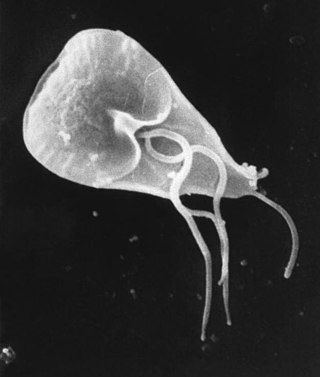
A trophozoite is the activated, feeding stage in the life cycle of certain protozoa such as malaria-causing Plasmodium falciparum and those of the Giardia group. The complementary form of the trophozoite state is the thick-walled cyst form. They are often different from the cyst stage, which is a protective, dormant form of the protozoa. Trophozoites are often found in the host's body fluids and tissues and in many cases, they are the form of the protozoan that causes disease in the host. In the protozoan, Entamoeba histolytica it invades the intestinal mucosa of its host, causing dysentery, which aid in the trophozoites traveling to the liver and leading to the production of hepatic abscesses.

Eimeria tenella is a species of Eimeria that causes hemorrhagic cecal coccidiosis in young poultry. It is found worldwide.

Eimeria is a genus of apicomplexan parasites that includes various species capable of causing the disease coccidiosis in animals such as cattle, poultry and smaller ruminants including sheep and goats. Eimeria species are considered to be monoxenous because the life cycle is completed within a single host, and stenoxenous because they tend to be host specific, although a number of exceptions have been identified. Species of this genus infect a wide variety of hosts. Thirty-one species are known to occur in bats (Chiroptera), two in turtles, and 130 named species infect fish. Two species infect seals. Five species infect llamas and alpacas: E. alpacae, E. ivitaensis, E. lamae, E. macusaniensis, and E. punonensis. A number of species infect rodents, including E. couesii, E. kinsellai, E. palustris, E. ojastii and E. oryzomysi. Others infect poultry, rabbits and cattle. For full species list, see below.
Poultry diseases occur in poultry, which are domesticated birds kept for their meat, eggs or feathers. Poultry species include the chicken, turkey, duck, goose and ostrich.
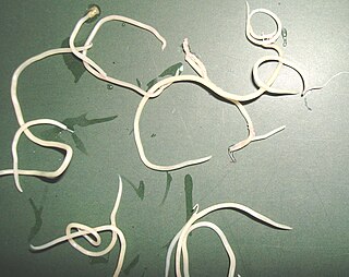
Ascaridia galli is a parasitic roundworm belonging to the phylum Nematoda. Nematodes of the genus Ascaridia are essentially intestinal parasites of birds. A. galli is the most prevalent and pathogenic species, especially in domestic fowl, Gallus domesticus. It causes ascaridiasis, a disease of poultry due to heavy worm infection, particularly in chickens and turkeys. It inhabits the small intestine, and can be occasionally seen in commercial eggs.

Balantidiasis is a protozoan infection caused by infection with Balantidium coli.

Babesia, also called Nuttallia, is an apicomplexan parasite that infects red blood cells and is transmitted by ticks. Originally discovered by Romanian bacteriologist Victor Babeș in 1888; over 100 species of Babesia have since been identified.
Chilomastix mesnili is a non-pathogenic member of primate gastrointestinal microflora, commonly associated with but not causing parasitic infections. It is found in about 3.5% of the population in the United States. In addition to humans, Chilomastix is found in chimpanzees, orangutans, monkeys, and pigs. It lives in the cecum and colon. C. mesnili has a similar life style to Giardia lamblia.
Protozoan infections are parasitic diseases caused by organisms formerly classified in the kingdom Protozoa. These organisms are now classified in the supergroups Excavata, Amoebozoa, Harosa, and Archaeplastida. They are usually contracted by either an insect vector or by contact with an infected substance or surface.

Heterakis gallinarum is a nematode parasite that lives in the cecum of some galliform birds, particularly in ground feeders such as domestic chickens and turkeys. It causes infection that is mildly pathogenic. However, it often carries a protozoan parasite Histomonas meleagridis which causes of histomoniasis. Transmission of H. meleagridis is through the H. gallinarum egg. H. gallinarum is about 1–2 cm in length with a sharply pointed tail and a preanal sucker. The parasite is a diecious species with marked sexual dimorphism. Males are smaller and shorter, measuring around 9 mm in length, with a unique bent tail. Females are stouter and longer, measuring roughly 13 mm in length, with a straight tail end.
Ascaridia is a genus of parasitic nematodes. Members of the genus are primarily intestinal parasites of birds. Three species are well known, namely, A. galli found mostly in chickens, A. dissimilis in turkeys, and A. columbae in pigeons. Lesser known species such as A. hermaphrodita, A. sergiomeirai, A. ornata, A. nicobarensis, and A. platyceri are found in parrots.

Raillietina is a genus of tapeworms that includes helminth parasites of vertebrates, mostly of birds. The genus was named in 1920 in honour of a French veterinarian and helminthologist, Louis-Joseph Alcide Railliet. Of the 37 species recorded under the genus, Raillietina demerariensis, R. asiatica, and R. formsana are the only species reported from humans, while the rest are found in birds. R. echinobothrida, R. tetragona, and R. cesticillus are the most important species in terms of prevalence and pathogenicity among wild and domestic birds.

Raillietina echinobothrida is a parasitic tapeworm belonging to the class Cestoda. It is the most prevalent and pathogenic helminth parasite in birds, particularly in domestic fowl, Gallus domesticus Linnaeus, 1758. It requires two hosts, birds and ants, for completion of its life cycle. It is a hermaphrodite worm having both the male and female reproductive organs in its body. The parasite is responsible for 'nodular tapeworm disease' in poultry.
Heterakis is a genus of parasitic nematodes. Members of the genus are minute roundworms (pinworms), hardly 1 cm long, infecting different species of gallinaceous birds, including chickens, turkeys, ducks, geese, grouse, guineafowl, partridges, pheasants, and quail. About 10 species are placed in the genus, but classification is often ambiguous due to their close resemblance, and a number of synonyms have arisen. H. gallinarumSchrank, 1788; H. isolonchevon Linstow, 1906; and H. disparSchrank, 1870 are the best understood species in terms of prevalence, pathogenicity, and biology. They inhabit the lumen of the cecum of the host.

Histomoniasis is a commercially significant disease of poultry, particularly of chickens and turkeys, due to parasitic infection of a protozoan, Histomonas meleagridis. The protozoan is transmitted to the bird by the nematode parasite Heterakis gallinarum. H. meleagridis resides within the eggs of H. gallinarum, so birds ingest the parasites along with contaminated soil or food. Earthworms can also act as a paratenic host.

Dientamoeba fragilis is a species of single-celled excavates found in the gastrointestinal tract of some humans, pigs and gorillas. It causes gastrointestinal upset in some people, but not in others. It is an important cause of traveller's diarrhoea, chronic diarrhoea, fatigue and, in children, failure to thrive. Despite this, its role as a "commensal, pathobiont, or pathogen" is still debated. D. fragilis is one of the smaller parasites that are able to live in the human intestine. Dientamoeba fragilis cells are able to survive and move in fresh feces but are sensitive to aerobic environments. They dissociate when in contact or placed in saline, tap water or distilled water.

Vasily Iakovlevich Danilewsky was a Ukrainian physician, physiologist and parasitologist. He was professor of physiology at University of Kharkiv and then at Kharkiv Medical Institute. He helped to establish the Danilevsky Institute of Endocrine Pathology Problems, which he directed until his death.

Chilomastix is a genus of pyriform excavates within the family Retortamonadidae All species within this genus are flagellated, structured with three flagella pointing anteriorly and a fourth contained within the feeding groove. Chilomastix also lacks Golgi apparatus and mitochondria but does possess a single nucleus. The genus parasitizes a wide range of vertebrate hosts, but is known to be typically non-pathogenic, and is therefore classified as harmless. The life cycle of Chilomastix lacks an intermediate host or vector. Chilomastix has a resistant cyst stage responsible for transmission and a trophozoite stage, which is recognized as the feeding stage. Chilomastix mesnili is one of the more studied species in this genus due to the fact it is a human parasite. Therefore, much of the information on this genus is based on what is known about this one species.
Eimeria falciformis is an obligate intracellular parasite that primarily infects the mouse species Mus musculus. As part of the Apicomplexa phylum, it has a complex life cycle within its host, causing tissue necrosis and inflammation, particularly in the cecum. This leads to coccidiosis, a disease characterized by symptoms like diarrhoea and weight loss. E. falciformis is one of over 1,700 species in the Eimeria genus, which infects the intestinal epithelial cells of various animals, including poultry and livestock.