
In human physiology, the lacrimal glands are paired, almond-shaped exocrine glands, one for each eye, that secrete the aqueous layer of the tear film. They are situated in the upper lateral region of each orbit, in the lacrimal fossa of the orbit formed by the frontal bone. Inflammation of the lacrimal glands is called dacryoadenitis. The lacrimal gland produces tears which then flow into canals that connect to the lacrimal sac. From that sac, the tears drain through the lacrimal duct into the nose.

The tunica media, or media for short, is the middle tunica (layer) of an artery or vein. It lies between the tunica intima on the inside and the tunica externa on the outside.
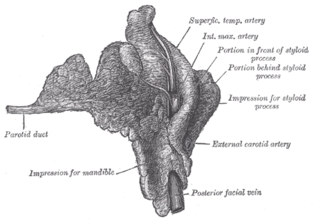
The parotid duct or Stensen duct is a duct and the route that saliva takes from the major salivary gland, the parotid gland into the mouth.
The bronchial veins are small vessels that return blood from the larger bronchi and structures at the roots of the lungs. The right side drains into the azygos vein, while the left side drains into the left superior intercostal vein or the accessory hemiazygos vein. Bronchial veins are thereby part of the bronchial circulation, carrying waste products away from the cells that constitute the lungs.

The pampiniform plexus is a network of many small veins found in the human male spermatic cord and to a lesser extent the suspensory ligament of the ovary. In the male, it is formed by the union of multiple spermatic veins from the back of the testis and tributaries from the epididymis.

The renal hilum or renal pedicle is the hilum of the kidney, that is, its recessed central fissure where its vessels, nerves and ureter pass. The medial border of the kidney is concave in the center and convex toward either extremity; it is directed forward and a little downward. Its central part presents a deep longitudinal fissure, bounded by prominent overhanging anterior and posterior lips. This fissure is a hilum that transmits the vessels, nerves, and ureter. From anterior to posterior, the renal vein exits, the renal artery enters, and the renal pelvis exits the kidney.
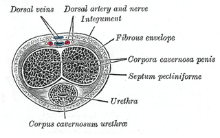
The tunica albuginea is the fibrous envelope of the corpora cavernosa penis. It consists of approximately 5% elastin, an extensible tissue that is primarily made up of the amino acids glycine, valine, alanine, and proline. The majority of the remaining tissue is collagen, which is made up of lysine, proline, glycine, alanine, and other amino acids. It is a bi-layered structure that includes an outer longitudinal layer and an inner circular layer.
The esophageal veins drain blood from the esophagus to the azygos vein, in the thorax, and to the inferior thyroid vein in the neck. It also drains, although with less significance, to the hemiazygos vein, posterior intercostal vein and bronchial veins.

Interlobular arteries are renal blood vessels given off at right angles from the side of the arcuate arteries looking toward the cortical substance. The interlobular arteries pass directly outward between the medullary rays to reach the fibrous tunic, where they end in the capillary network of this part.
The straight venules of kidney are branches from the plexuses at the apices of the medullary pyramids, formed by the terminations of the vasa recta.

The arcuate arteries of the kidney are vessels of the renal circulation. They are located at the border of the renal cortex and renal medulla.
The arcuate vein is a vessel of the renal circulation. It is located at the border of the renal cortex and renal medulla.

The trabecular veins are the largest veins inside the spleen. It drains the blood collected in the sinuses of the pulp.

A striated duct (Pflüger's ducts ) is a gland duct which connects an intercalated duct to an interlobular duct. It is characterized by the basal infoldings of its plasma membrane, characteristic of ion-pumping activity by the numerous mitochondria. Along with the intercalated ducts, they function to modify salivary fluid by secreting HCO3− and K+ and reabsorbing Na+ and Cl− using the Na-K pump and the Cl-HCO3 pump, making the saliva hypotonic.

In anatomy, a lobe is a clear anatomical division or extension of an organ that can be determined without the use of a microscope at the gross anatomy level. This is in contrast to the much smaller lobule, which is a clear division only visible under the microscope.

The central veins of liver are veins found at the center of hepatic lobules.

A hepatic lobule is a small division of the liver defined at the microscopic. The hepatic lobule is a building block of the liver matter, consisting of a portal triad, hepatocytes arranged in linear cords between a capillary network, and a central vein.

The biliary tract, refers to the liver, gall bladder and bile ducts, and how they work together to make, store and secrete bile. Bile consists of water, electrolytes, bile acids, cholesterol, phospholipids and conjugated bilirubin. Some components are synthesised by hepatocytes, the rest are extracted from the blood by the liver.
The interlobular bile ducts carry bile in the liver between the Canals of Hering and the interlobar bile ducts. They are part of the interlobular portal triad and can be easily localized by looking for the much larger portal vein. The cells of the ducts are described as cuboidal epithelium with increasing amounts of connective tissue around it.

