You can help expand this article with text translated from the corresponding article in Italian. (April 2013)Click [show] for important translation instructions.
|
| Joints in the hand | |
|---|---|
 Ligaments of wrist. Anterior view | |
 Metacarpophalangeal articulation and articulations of digit. Ulnar aspect. | |
| Details | |
| Identifiers | |
| Latin | articulationes manus |
| MeSH | D050823 |
| TA98 | A03.5.11.001 |
| TA2 | 1802 |
| Anatomical terminology | |
The joints in the hand are joints found at the distal end of the upper limb.
The joints are:
- In the wrist there is the radiocarpal joint between the radius and carpus. Between the carpal bones are the intercarpal articulations and the midcarpal joint.
- The carpometacarpal joint connects the carpal bones to the metacarpus or metacarpal bones which are joined at the intermetacarpal articulations.
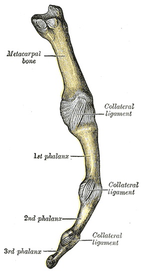
- In the fingers, finally, are the metacarpophalangeal joints (including the knuckles) between the metacarpal bones and the phalanges or finger bones which are interconnected by the interphalangeal joints.
| | This human musculoskeletal system article is a stub. You can help Wikipedia by expanding it. |