Monocentric chromosome

Last updated
Duplicated chromosome. (2) identifies the Monocentric centromere--the region that joins the two sister chromatids, or each half of the chromosome. In prophase of mitosis, specialized regions on centromeres called kinetochores attach chromosomes to spindle fibers. Chromosome.svg
Duplicated chromosome. (2) identifies the Monocentric centromere—the region that joins the two sister chromatids, or each half of the chromosome. In prophase of mitosis, specialized regions on centromeres called kinetochores attach chromosomes to spindle fibers.

The monocentric chromosome is a chromosome that has only one centromere in a chromosome and forms a narrow constriction.

Contents

Monocentric centromeres are the most common structure on highly repetitive DNA in plants and animals. [1]

Structure

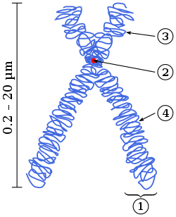
Monocentric chromosomes as compared to holocentric chromosomes where the entire length of the chromosome acts as the centromere. In monocentric chromosomes there is one primary constriction and the centromere its CenH3 loci at this location. [2]

Holocentric chromosomes are found throughout the plant and animal kingdoms such as the nematode Caenorhabditis elegans . [3] Holocentric chromosomes do have an evolutionary advantage by preventing the loss of chromosome after a DNA double-strand break. [4]

The centromere is the point of attachment for the mitotic apparatus [5]

Chromosomal aberrations

Deletions, duplications and translocations can produce a polycentric chromosome. This is troublesome for cells that divide often since at the time of anaphase the polycentric chromosome does not move to opposite poles of spindle fiber and the cell dies. [5]

See also

Related Research Articles

<span class="mw-page-title-main">Chromosome</span> DNA molecule containing genetic material of a cell

A chromosome is a long DNA molecule with part or all of the genetic material of an organism. In most chromosomes the very long thin DNA fibers are coated with packaging proteins; in eukaryotic cells the most important of these proteins are the histones. These proteins, aided by chaperone proteins, bind to and condense the DNA molecule to maintain its integrity. These chromosomes display a complex three-dimensional structure, which plays a significant role in transcriptional regulation.

<span class="mw-page-title-main">Centromere</span> Specialized DNA sequence of a chromosome that links a pair of sister chromatids

The centromere links a pair of sister chromatids together during cell division. This constricted region of chromosome connects the sister chromatids, creating a short arm (p) and a long arm (q) on the chromatids. During mitosis, spindle fibers attach to the centromere via the kinetochore.

<span class="mw-page-title-main">Meiosis</span> Cell division producing haploid gametes

Meiosis is a special type of cell division of germ cells in sexually-reproducing organisms that produces the gametes, such as sperm or egg cells. It involves two rounds of division that ultimately result in four cells with only one copy of each chromosome (haploid). Additionally, prior to the division, genetic material from the paternal and maternal copies of each chromosome is crossed over, creating new combinations of code on each chromosome. Later on, during fertilisation, the haploid cells produced by meiosis from a male and a female will fuse to create a cell with two copies of each chromosome again, the zygote.

<span class="mw-page-title-main">Mitosis</span> Process in which replicated chromosomes are separated into two new identical nuclei

In cell biology, mitosis is a part of the cell cycle in which replicated chromosomes are separated into two new nuclei. Cell division by mitosis gives rise to genetically identical cells in which the total number of chromosomes is maintained. Therefore, mitosis is also known as equational division. In general, mitosis is preceded by S phase of interphase and is often followed by telophase and cytokinesis; which divides the cytoplasm, organelles and cell membrane of one cell into two new cells containing roughly equal shares of these cellular components. The different stages of mitosis altogether define the mitotic (M) phase of a cell cycle—the division of the mother cell into two daughter cells genetically identical to each other.

<span class="mw-page-title-main">Cell division</span> Process by which living cells divide

Cell division is the process by which a parent cell divides into two daughter cells. Cell division usually occurs as part of a larger cell cycle in which the cell grows and replicates its chromosome(s) before dividing. In eukaryotes, there are two distinct types of cell division: a vegetative division (mitosis), producing daughter cells genetically identical to the parent cell, and a cell division that produces haploid gametes for sexual reproduction (meiosis), reducing the number of chromosomes from two of each type in the diploid parent cell to one of each type in the daughter cells. In cell biology, mitosis (/maɪˈtoʊsɪs/) is a part of the cell cycle, in which, replicated chromosomes are separated into two new nuclei. Cell division gives rise to genetically identical cells in which the total number of chromosomes is maintained. In general, mitosis is preceded by the S stage of interphase and is often followed by telophase and cytokinesis; which divides the cytoplasm, organelles, and cell membrane of one cell into two new cells containing roughly equal shares of these cellular components. The different stages of mitosis all together define the mitotic (M) phase of animal cell cycle—the division of the mother cell into two genetically identical daughter cells. Meiosis undergoes two divisions resulting in four haploid daughter cells. Homologous chromosomes are separated in the first division of meiosis, such that each daughter cell has one copy of each chromosome. These chromosomes have already been replicated and have two sister chromatids which are then separated during the second division of meiosis. Both of these cell division cycles are used in the process of sexual reproduction at some point in their life cycle. Both are believed to be present in the last eukaryotic common ancestor.

Non-coding DNA (ncDNA) sequences are components of an organism's DNA that do not encode protein sequences. Some non-coding DNA is transcribed into functional non-coding RNA molecules. Other functional regions of the non-coding DNA fraction include regulatory sequences that control gene expression; scaffold attachment regions; origins of DNA replication; centromeres; and telomeres. Some non-coding regions appear to be mostly nonfunctional such as introns, pseudogenes, intergenic DNA, and fragments of transposons and viruses.

Heterochromatin is a tightly packed form of DNA or condensed DNA, which comes in multiple varieties. These varieties lie on a continuum between the two extremes of constitutive heterochromatin and facultative heterochromatin. Both play a role in the expression of genes. Because it is tightly packed, it was thought to be inaccessible to polymerases and therefore not transcribed; however, according to Volpe et al. (2002), and many other papers since, much of this DNA is in fact transcribed, but it is continuously turned over via RNA-induced transcriptional silencing (RITS). Recent studies with electron microscopy and OsO4 staining reveal that the dense packing is not due to the chromatin.

<span class="mw-page-title-main">Karyotype</span> Photographic display of total chromosome complement in a cell

A karyotype is the general appearance of the complete set of chromosomes in the cells of a species or in an individual organism, mainly including their sizes, numbers, and shapes. Karyotyping is the process by which a karyotype is discerned by determining the chromosome complement of an individual, including the number of chromosomes and any abnormalities.

<span class="mw-page-title-main">Spindle apparatus</span> Feature of biological cell structure

In cell biology, the spindle apparatus is the cytoskeletal structure of eukaryotic cells that forms during cell division to separate sister chromatids between daughter cells. It is referred to as the mitotic spindle during mitosis, a process that produces genetically identical daughter cells, or the meiotic spindle during meiosis, a process that produces gametes with half the number of chromosomes of the parent cell.

<span class="mw-page-title-main">Spindle checkpoint</span> Cell cycle checkpoint

The spindle checkpoint, also known as the metaphase-to-anaphase transition, the spindle assembly checkpoint (SAC), the metaphase checkpoint, or the mitotic checkpoint, is a cell cycle checkpoint during metaphase of mitosis or meiosis that prevents the separation of the duplicated chromosomes (anaphase) until each chromosome is properly attached to the spindle. To achieve proper segregation, the two kinetochores on the sister chromatids must be attached to opposite spindle poles. Only this pattern of attachment will ensure that each daughter cell receives one copy of the chromosome. The defining biochemical feature of this checkpoint is the stimulation of the anaphase-promoting complex by M-phase cyclin-CDK complexes, which in turn causes the proteolytic destruction of cyclins and proteins that hold the sister chromatids together.

<span class="mw-page-title-main">Kinetochore</span> Protein complex that allows microtubules to attach to chromosomes during cell division

A kinetochore is a disc-shaped protein structure associated with duplicated chromatids in eukaryotic cells where the spindle fibers attach during cell division to pull sister chromatids apart. The kinetochore assembles on the centromere and links the chromosome to microtubule polymers from the mitotic spindle during mitosis and meiosis. The term kinetochore was first used in a footnote in a 1934 Cytology book by Lester W. Sharp and commonly accepted in 1936. Sharp's footnote reads: "The convenient term kinetochore has been suggested to the author by J. A. Moore", likely referring to John Alexander Moore who had joined Columbia University as a freshman in 1932.

<span class="mw-page-title-main">Isochromosome</span>

An isochromosome is an unbalanced structural abnormality in which the arms of the chromosome are mirror images of each other. The chromosome consists of two copies of either the long (q) arm or the short (p) arm because isochromosome formation is equivalent to a simultaneous duplication and deletion of genetic material. Consequently, there is partial trisomy of the genes present in the isochromosome and partial monosomy of the genes in the lost arm.

<span class="mw-page-title-main">Aurora kinase B</span> Protein

Aurora kinase B is a protein that functions in the attachment of the mitotic spindle to the centromere.

<span class="mw-page-title-main">CENPA</span> Protein-coding gene in the species Homo sapiens

Centromere protein A, also known as CENPA, is a protein which in humans is encoded by the CENPA gene. CENPA is a histone H3 variant which is the critical factor determining the kinetochore position(s) on each chromosome in most eukaryotes including humans.

<span class="mw-page-title-main">CENPC1</span> Protein-coding gene in the species Homo sapiens

Centromere protein C 1 is a protein that in humans is encoded by the CENPC1 gene.

<span class="mw-page-title-main">CENPH</span> Protein-coding gene in the species Homo sapiens

Centromere protein H is a protein that in humans is encoded by the CENPH gene. It is involved in the assembly of kinetochore proteins, mitotic progression and chromosome segregation.

In genetics, a polycentric chromosome is any chromosome featuring multiple centromeres. Polycentric chromosomes are produced by chromosomal aberrations such as deletion, duplication, or translocation. Polycentric chromosomes usually result in the death of the cell because polycentric chromosomes may fail to move to opposite poles of spindle fiber during anaphase. As a result, the chromosome is fragmented, which causes the death of the cell. In some algae, such as Spirogyra, polycentric chromosomes appear normally.

<span class="mw-page-title-main">Neocentromere</span>

Neocentromeres are new centromeres that form at a place on the chromosome that is usually not centromeric. They typically arise due to disruption of the normal centromere. These neocentromeres should not be confused with “knobs”, which were also described as “neocentromeres” in maize in the 1950s. Unlike most normal centromeres, neocentromeres do not contain satellite sequences that are highly repetitive but instead consist of unique sequences. Despite this, most neocentromeres are still able to carry out the functions of normal centromeres in regulating chromosome segregation and inheritance. This raises many questions on what is necessary versus what is sufficient for constituting a centromere.

<span class="mw-page-title-main">Kaustuv Sanyal</span>

Kaustuv Sanyal is an Indian molecular biologist, mycologist and a professor at the Molecular Biology and Genetics Unit of the Jawaharlal Nehru Centre for Advanced Scientific Research (JNCASR). He is known for his molecular and genetic studies of pathogenic yeasts such as Candida and Cryptococcus). An alumnus of Bidhan Chandra Krishi Viswavidyalaya and Madurai Kamaraj University from where he earned a BSc in agriculture and MSc in biotechnology respectively, Sanyal did his doctoral studies at Bose Institute to secure a PhD in Yeast genetics. He moved to the University of California, Santa Barbara, USA to work in the laboratory of John Carbon on the discovery of centromeres in Candida albicans. He joined JNCASR in 2005. He is a member of the Faculty of 1000 in the disciplines of Microbial Evolution and Genomics and has delivered invited speeches which include the Gordon Research Conference, EMBO conferences on comparative genomics and kinetochores. The Department of Biotechnology of the Government of India awarded him the National Bioscience Award for Career Development, one of the highest Indian science awards, for his contributions to biosciences, in 2012. He has also been awarded with the prestigious Tata Innovation Fellowship in 2017. The National Academy of Sciences, India elected him as a fellow in 2014. He is also an elected fellow of Indian Academy of Sciences (2017), and the Indian National Science Academy (2018). In 2019, he has been elected to Fellowship in the American Academy of Microbiology (AAM), the honorific leadership group within the American Society for Microbiology.

Holocentric chromosomes are chromosomes that possess multiple kinetochores along their length rather than the single centromere typical of other chromosomes. They were first described in cytogenetic experiments in 1935. Since this first observation, the term holocentric chromosome has referred to chromosomes that: i) lack the primary constriction corresponding to the centromere observed in monocentric chromosomes; and ii) possess multiple kinetochores dispersed along the entire chromosomal axis, such that microtubules bind to the chromosome along its entire length and move broadside to the pole from the metaphase plate. Holocentric chromosomes are also termed holokinetic, because, during cell division, the sister chromatids move apart in parallel and do not form the classical V-shaped figures typical of monocentric chromosomes.

References

  1. Barra, V.; Fachinetti, D. (2018). "The dark side of centromeres: Types, causes and consequences of structural abnormalities implicating centromeric DNA". Nature Communications. 9 (1): 4340. Bibcode:2018NatCo...9.4340B. doi:10.1038/s41467-018-06545-y. PMC   6194107 . PMID   30337534.
  2. Neumann, Pavel; Navrátilová, Alice; Schroeder-Reiter, Elizabeth; Koblížková, Andrea; Steinbauerová, Veronika; Chocholová, Eva; Novák, Petr; Wanner, Gerhard; Macas, Jiří (2012). "Stretching the Rules: Monocentric Chromosomes with Multiple Centromere Domains". PLOS Genetics. 8 (6): e1002777. doi: 10.1371/journal.pgen.1002777 . PMC   3380829 . PMID   22737088.
  3. Dernburg, A. F. (2001). "Here, There, and Everywhere: Kinetochore Function on Holocentric Chromosomes". The Journal of Cell Biology. 153 (6): F33–8. doi:10.1083/jcb.153.6.F33. PMC   2192025 . PMID   11402076.
  4. Friedman, Steven; Freitag, Michael (2017). Evolving Centromeres and Kinetochores. Advances in Genetics. Vol. 98. pp. 1–41. doi:10.1016/bs.adgen.2017.07.001. ISBN   9780128122808. PMID   28942791.
  5. 1 2 {{Acentric Fragment, In: Sydney Brenner and Jeffrey H. Miller, Editor(s)-in-Chief, Encyclopedia of Genetics, Academic Press, New York, 2001, Page 2, ISBN   978-0-12-227080-2, 10.1006/rwgn.2001.1750.}}