N-Formylmethionine

Last updated
N-Formylmethionine
(S)-N-Formylmethionine V.1.svg
Names
IUPAC name
N-Formylmethionine
Systematic IUPAC name
(S)-2-Formylamino-4-methylsulfanylbutanoic acid
Other names
2-Formylamino-4-methylsulfanyl-butyric acid; Formylmethionine; N-Formyl(methyl)homocysteine
Identifiers
3D model (JSmol)
AbbreviationsfMet
ChEBI
ChemSpider
EC Number
  • 224-322-8
PubChem CID
UNII
  • InChI=1S/C6H11NO3S/c1-11-3-2-5(6(9)10)7-4-8/h4-5H,2-3H2,1H3,(H,7,8)(H,9,10)
    Key: PYUSHNKNPOHWEZ-UHFFFAOYSA-N
  • CSCC[C@H](NC=O)C(O)=O
Properties
C6H11NO3S
Molar mass 177.22 g/mol
Hazards
GHS labelling: [1]
GHS-pictogram-exclam.svg
Warning
H319
P264+P265, P280, P305+P351+P338, P337+P317
Supplementary data page
N-Formylmethionine (data page)
Except where otherwise noted, data are given for materials in their standard state (at 25 °C [77 °F], 100 kPa).
Yes check.svgY  verify  (what is  Yes check.svgYX mark.svgN ?)

N-Formylmethionine (fMet, [2] HCO-Met, [3] For-Met [3] ) is a derivative of the amino acid methionine in which a formyl group has been added to the amino group. It is specifically used for initiation of protein synthesis from bacterial and organellar genes, and may be removed post-translationally.

Contents

fMet plays a crucial part in the protein synthesis of bacteria, mitochondria and chloroplasts. It is not used in cytosolic protein synthesis of eukaryotes, where eukaryotic nuclear genes are translated. It is also not used by Archaea. In the human body, fMet is recognized by the immune system as foreign material, or as an alarm signal released by damaged cells, and stimulates the body to fight against potential infection.

Function in protein synthesis

Translation

fMet is required for efficient initiaiton of protein synthesis in most groups of bacteria. The 30S ribosomemRNA complex specifically recruits tRNAs with a formylated amino acid tRNAfMet attached to fMet in the natural case. [4]

Because the fMet directs initiation, proteins in bacteria start (N-terminus) with a fMet residue instead of a methionine. Further occurrences of the "AUG" codon will result in a normal methionine, because a normal "elongating" tRNAMet is used. [4]

The addition of the formyl group to methionine is catalyzed by the enzyme methionyl-tRNA formyltransferase. This modification is done after methionine has been loaded onto tRNAfMet by aminoacyl-tRNA synthetase. Methionine itself can be loaded either onto tRNAfMet or tRNAMet. However, formyltransferase will catalyze the addition of the formyl group to methionine only if methionine has been loaded onto tRNAfMet, not onto tRNAMet. This is because the formyltransferase recognizes specific features of tRNAfMet. [4]

The mitochondria of eukaryotic cells, including those of humans, and the chloroplasts of plant cells also initiate protein synthesis with fMet. Given that mitochondria and chloroplasts have this initial protein synthesis with fMet in common with bacteria, this has been cited as evidence for the endosymbiotic theory. [5]

Unexpectedly, formyltransferase can also act upon eukaryotic initiator tRNA in living yeast cells. Even under normal conditions, the nuclear-encoded formyltransferase is not completely imported into mitochondria; even more is left in the cytosol under stress. These cytosolic formyltransferase produce fMet-tRNAi, which can be used by cytosolic ribosomes to produce proteins with a N-terminal fMet. These proteins are targeted for degredagtion by specific processes in the cell. [6]

Further processing

The N-terminal fMet is removed from majority of proteins, both host and recombinant, by a sequence of two enzymatic reactions. First, peptide deformylase (PDF) deformylates it, converting the residue back to a normal methionine. Then methionine aminopeptidase (MetAP) removes the residue from the chain. [7] MetAP only acts on proteins with second-position residues that are less bulky than valine. [8]

The N-terminal fMet, if not removed by PDF, seems to act as a degron, a signal for protein degradation. [8]

Variation

The formyl group is not strictly required for initiation. Bacteria with their formyltransferase knocked out, which prevents Met-tRNAfMet (i.e. methionine loaded onto tRNAfMet) from turning into fMet-tRNAfMet, can have varying degrees of residual ability to start protein synthesis. E. coli, S. pneumoniae and B. subtilis show almost no remaining translation ability, while P. aeruginosa, S. aureus, H. influenzae, and possibly S. faecalis still churn out plenty of protein. In P. aeruginosa, this ability is facilitated by bacterial initiation factor 2, which can carry both Met-tRNAfMet and fMet-tRNAfMet to the ribosome. [9]

Relevance to immunology

Because fMet is present in proteins made by bacteria but not in those made by eukaryotes (other than in bacterially derived organelles), the immune system might use it to help distinguish self from non-self. Polymorphonuclear cells can bind proteins starting with fMet, and use them to initiate the attraction of circulating blood leukocytes and then stimulate microbicidal activities such as phagocytosis. [10] [11] [12]

Since fMet is present in proteins made by mitochondria and chloroplasts, more recent theories do not see it as a molecule that the immune system can use to distinguish self from non-self. [13] Instead, fMet-containing oligopeptides and proteins appear to be released by the mitochondria of damaged tissues as well as by damaged bacteria, and can thus qualify as an "alarm" signal, as discussed in the Danger model of immunity. The prototypical fMet-containing oligopeptide is N-formylmethionine-leucyl-phenylalanine (FMLP) which activates leukocytes and other cell types by binding with these cells' formyl peptide receptor 1 (FPR1) and formyl peptide receptor 2 (FPR2) G protein coupled receptors (see also formyl peptide receptor 3). Acting through these receptors, the fMet-containing oligopeptides and proteins are part of the innate immune system; they function to initiate acute inflammation responses but under other conditions function to inhibit and resolve these responses. fMet-containing oligopeptides and proteins also function in other physiological and pathological responses.

See also

Related Research Articles

Protein targeting or protein sorting is the biological mechanism by which proteins are transported to their appropriate destinations within or outside the cell. Proteins can be targeted to the inner space of an organelle, different intracellular membranes, the plasma membrane, or to the exterior of the cell via secretion. Information contained in the protein itself directs this delivery process. Correct sorting is crucial for the cell; errors or dysfunction in sorting have been linked to multiple diseases.

Peptidoglycan or murein is a unique large macromolecule, a polysaccharide, consisting of sugars and amino acids that forms a mesh-like peptidoglycan layer (sacculus) that surrounds the bacterial cytoplasmic membrane. The sugar component consists of alternating residues of β-(1,4) linked N-acetylglucosamine (NAG) and N-acetylmuramic acid (NAM). Attached to the N-acetylmuramic acid is an oligopeptide chain made of three to five amino acids. The peptide chain can be cross-linked to the peptide chain of another strand forming the 3D mesh-like layer. Peptidoglycan serves a structural role in the bacterial cell wall, giving structural strength, as well as counteracting the osmotic pressure of the cytoplasm. This repetitive linking results in a dense peptidoglycan layer which is critical for maintaining cell form and withstanding high osmotic pressures, and it is regularly replaced by peptidoglycan production. Peptidoglycan hydrolysis and synthesis are two processes that must occur in order for cells to grow and multiply, a technique carried out in three stages: clipping of current material, insertion of new material, and re-crosslinking of existing material to new material.

<span class="mw-page-title-main">Ribosome</span> Intracellular organelle consisting of RNA and protein functioning to synthesize proteins

Ribosomes are macromolecular machines, found within all cells, that perform biological protein synthesis. Ribosomes link amino acids together in the order specified by the codons of messenger RNA (mRNA) molecules to form polypeptide chains. Ribosomes consist of two major components: the small and large ribosomal subunits. Each subunit consists of one or more ribosomal RNA (rRNA) molecules and many ribosomal proteins. The ribosomes and associated molecules are also known as the translational apparatus.

<span class="mw-page-title-main">Symbiogenesis</span> Evolutionary theory holding that eukaryotic organelles evolved through symbiosis with prokaryotes

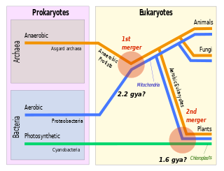
Symbiogenesis is the leading evolutionary theory of the origin of eukaryotic cells from prokaryotic organisms. The theory holds that mitochondria, plastids such as chloroplasts, and possibly other organelles of eukaryotic cells are descended from formerly free-living prokaryotes taken one inside the other in endosymbiosis. Mitochondria appear to be phylogenetically related to Rickettsiales bacteria, while chloroplasts are thought to be related to cyanobacteria.

<span class="mw-page-title-main">Translation (biology)</span> Cellular process of protein synthesis

In biology, translation is the process in living cells in which proteins are produced using RNA molecules as templates. The generated protein is a sequence of amino acids. This sequence is determined by the sequence of nucleotides in the RNA. The nucleotides are considered three at a time. Each such triple results in addition of one specific amino acid to the protein being generated. The matching from nucleotide triple to amino acid is called the genetic code. The translation is performed by a large complex of functional RNA and proteins called ribosomes. The entire process is called gene expression.

<span class="mw-page-title-main">Index of biochemistry articles</span>

Biochemistry is the study of the chemical processes in living organisms. It deals with the structure and function of cellular components such as proteins, carbohydrates, lipids, nucleic acids and other biomolecules.

The N-terminus (also known as the amino-terminus, NH2-terminus, N-terminal end or amine-terminus) is the start of a protein or polypeptide, referring to the free amine group (-NH2) located at the end of a polypeptide. Within a peptide, the amine group is bonded to the carboxylic group of another amino acid, making it a chain. That leaves a free carboxylic group at one end of the peptide, called the C-terminus, and a free amine group on the other end called the N-terminus. By convention, peptide sequences are written N-terminus to C-terminus, left to right (in LTR writing systems). This correlates the translation direction to the text direction, because when a protein is translated from messenger RNA, it is created from the N-terminus to the C-terminus, as amino acids are added to the carboxyl end of the protein.

<span class="mw-page-title-main">Start codon</span> First codon of a messenger RNA translated by a ribosome

The start codon is the first codon of a messenger RNA (mRNA) transcript translated by a ribosome. The start codon always codes for methionine in eukaryotes and archaea and a N-formylmethionine (fMet) in bacteria, mitochondria and plastids.

Bacterial translation is the process by which messenger RNA is translated into proteins in bacteria.
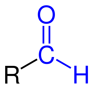
<span class="mw-page-title-main">Formylation</span>

Formylation refers to any chemical processes in which a compound is functionalized with a formyl group (-CH=O). In organic chemistry, the term is most commonly used with regards to aromatic compounds. In biochemistry the reaction is catalysed by enzymes such as formyltransferases.

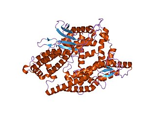
The formyl peptide receptors (FPR) belong to a class of G protein-coupled receptors involved in chemotaxis. In humans, there are three formyl peptide receptor isoforms, each encoded by a separate gene that are named FPR1, FPR2, and FPR3. These receptors were originally identified by their ability to bind N-formyl peptides such as N-formylmethionine produced by the degradation of either bacterial or host cells. Hence formyl peptide receptors are involved in mediating immune cell response to infection. These receptors may also act to suppress the immune system under certain conditions. The close phylogenetic relation of signaling in chemotaxis and olfaction was recently proved by detection formyl peptide receptor like proteins as a distinct family of vomeronasal organ chemosensors in mice.

Translocase is a general term for a protein that assists in moving another molecule, usually across a cell membrane. These enzymes catalyze the movement of ions or molecules across membranes or their separation within membranes. The reaction is designated as a transfer from “side 1” to “side 2” because the designations “in” and “out”, which had previously been used, can be ambiguous. Translocases are the most common secretion system in Gram positive bacteria.

<span class="mw-page-title-main">Formyl peptide receptor 2</span> Protein-coding gene in the species Homo sapiens

N-formyl peptide receptor 2 (FPR2) is a G-protein coupled receptor (GPCR) located on the surface of many cell types of various animal species. The human receptor protein is encoded by the FPR2 gene and is activated to regulate cell function by binding any one of a wide variety of ligands including not only certain N-Formylmethionine-containing oligopeptides such as N-Formylmethionine-leucyl-phenylalanine (FMLP) but also the polyunsaturated fatty acid metabolite of arachidonic acid, lipoxin A4 (LXA4). Because of its interaction with lipoxin A4, FPR2 is also commonly named the ALX/FPR2 or just ALX receptor.

<i>N</i>-Formylmethionine-leucyl-phenylalanine Chemical compound

N-Formylmethionyl-leucyl-phenylalanine is an N-formylated tripeptide and sometimes simply referred to as chemotactic peptide is a potent polymorphonuclear leukocyte (PMN) chemotactic factor and is also a macrophage activator.

<span class="mw-page-title-main">Formyl peptide receptor 3</span> Protein-coding gene in the species Homo sapiens

N-formyl peptide receptor 3 (FPR3) is a receptor protein that in humans is encoded by the FPR3 gene.
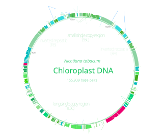
Translational regulation refers to the control of the levels of protein synthesized from its mRNA. This regulation is vastly important to the cellular response to stressors, growth cues, and differentiation. In comparison to transcriptional regulation, it results in much more immediate cellular adjustment through direct regulation of protein concentration. The corresponding mechanisms are primarily targeted on the control of ribosome recruitment on the initiation codon, but can also involve modulation of peptide elongation, termination of protein synthesis, or ribosome biogenesis. While these general concepts are widely conserved, some of the finer details in this sort of regulation have been proven to differ between prokaryotic and eukaryotic organisms.

The N-end rule is a rule that governs the rate of protein degradation through recognition of the N-terminal residue of proteins. The rule states that the N-terminal amino acid of a protein determines its half-life. The rule applies to both eukaryotic and prokaryotic organisms, but with different strength, rules, and outcome. In eukaryotic cells, these N-terminal residues are recognized and targeted by ubiquitin ligases, mediating ubiquitination thereby marking the protein for degradation. The rule was initially discovered by Alexander Varshavsky and co-workers in 1986. However, only rough estimations of protein half-life can be deduced from this 'rule', as N-terminal amino acid modification can lead to variability and anomalies, whilst amino acid impact can also change from organism to organism. Other degradation signals, known as degrons, can also be found in sequence.

A target peptide is a short peptide chain that directs the transport of a protein to a specific region in the cell, including the nucleus, mitochondria, endoplasmic reticulum (ER), chloroplast, apoplast, peroxisome and plasma membrane. Some target peptides are cleaved from the protein by signal peptidases after the proteins are transported.

<span class="mw-page-title-main">Formyl peptide receptor 1</span> Protein-coding gene in the species Homo sapiens

Formyl peptide receptor 1 is a cell surface receptor protein that in humans is encoded by the formyl peptide receptor 1 (FPR1) gene. This gene encodes a G protein-coupled receptor cell surface protein that binds and is activated by N-Formylmethionine-containing oligopeptides, particularly N-Formylmethionine-leucyl-phenylalanine (FMLP). FPR1 is prominently expressed by mammalian phagocytic and blood leukocyte cells where it functions to mediate these cells' responses to the N-formylmethionine-containing oligopeptides which are released by invading microorganisms and injured tissues. FPR1 directs these cells to sites of invading pathogens or disrupted tissues and then stimulates these cells to kill the pathogens or to remove tissue debris; as such, it is an important component of the innate immune system that operates in host defense and damage control.

<span class="mw-page-title-main">Thimet oligopeptidase</span>

Thimet oligopeptidases, also known as TOPs, are a type of M3 metallopeptidases. These enzymes can be found in animals and plants, showing distinctive functions. In animals and humans, they are involved in the degradation of peptides, such as bradykinin, neurotensin, angiotensin I, and Aβ peptide, helping to regulate physiological processes. In plants, their role is related to the degradation of targeting peptides and the immune response to pathogens through Salicylic Acid (SA)-dependent stress signaling. In Arabidopsis thaliana—recognized as a model plant for scientific studies—two thimet oligopeptidases, known as TOP1 and TOP2, have been identified as targets for salicylic acid binding in the plant. These TOP enzymes are key components to understand the SA-mediated signaling where interactions exist with different components and most of the pathways are unknown.

References

  1. "N-Formyl-DL-methionine". pubchem.ncbi.nlm.nih.gov.
  2. PubChem. "N-Formyl-DL-methionine". pubchem.ncbi.nlm.nih.gov. Retrieved 2020-10-24.
  3. 1 2 Nomenclature and Symbolism for Amino Acids and Peptides, 3AA-18 and 3AA-19
  4. 1 2 3 Shetty, S; Shah, RA; Chembazhi, UV; Sah, S; Varshney, U (28 February 2017). "Two highly conserved features of bacterial initiator tRNAs license them to pass through distinct checkpoints in translation initiation". Nucleic Acids Research. 45 (4): 2040–2050. doi:10.1093/nar/gkw854. PMC   5389676 . PMID   28204695.
  5. Alberts, Bruce (18 November 2014). Molecular biology of the cell (Sixth ed.). New York, NY. p. 800. ISBN   978-0-8153-4432-2. OCLC   887605755.{{cite book}}: CS1 maint: location missing publisher (link)
  6. Varshavsky, Alexander (8 January 2019). "N-degron and C-degron pathways of protein degradation". Proceedings of the National Academy of Sciences. 116 (2): 358–366. doi:10.1073/pnas.1816596116. PMC   6329975 . PMID   30622213.
  7. Sherman F, Stewart JW, Tsunasawa S (July 1985). "Methionine or not methionine at the beginning of a protein". BioEssays . 3 (1): 27–31. doi:10.1002/bies.950030108. PMID   3024631. S2CID   33735710.
  8. 1 2 Piatkov, KI; Vu, TT; Hwang, CS; Varshavsky, A (2015). "Formyl-methionine as a degradation signal at the N-termini of bacterial proteins". Microbial cell (Graz, Austria). 2 (10): 376–393. doi:10.15698/mic2015.10.231. PMC   4745127 . PMID   26866044.
  9. Piatkov, KI; Vu, TT; Hwang, CS; Varshavsky, A (2015). "Formyl-methionine as a degradation signal at the N-termini of bacterial proteins". Microbial cell (Graz, Austria). 2 (10): 376–393. doi: 10.15698/mic2015.10.231 . PMID   26866044.
  10. Immunology at MCG 1/phagstep
  11. "The Innate Immune System: Pattern-Recognition Receptors, Antigen-Nonspecific Antimicrobial Body Molecules, and Cytokines". Archived from the original on 2010-07-27.
  12. Detmers PA, Wright SD, Olsen E, Kimball B, Cohn ZA (September 1987). "Aggregation of complement receptors on human neutrophils in the absence of ligand". The Journal of Cell Biology . 105 (3): 1137–45. doi:10.1083/jcb.105.3.1137. PMC   2114803 . PMID   2958480.
  13. Zhang Q, Raoof M, Chen Y, Sumi Y, Sursal T, Junger W, Brohi K, Itagaki K, Hauser CJ (Mar 4, 2010). "Circulating mitochondrial DAMPs cause inflammatory responses to injury". Nature. 464 (7285): 104–107. Bibcode:2010Natur.464..104Z. doi:10.1038/nature08780. PMC   2843437 . PMID   20203610.