Paired receptors

Last updated
The siglec-like immunoglobulin domains of two paired receptors, PILRA (red) and PILRB (yellow), illustrating the similarities in structure between two ligand-binding domains of receptors with opposing signaling function. From PDB: 4NFC and PDB: 4NFB . 4nfb red PILRA 4nfc yellow PILRB.png
The siglec-like immunoglobulin domains of two paired receptors, PILRA (red) and PILRB (yellow), illustrating the similarities in structure between two ligand-binding domains of receptors with opposing signaling function. From PDB: 4NFC and PDB: 4NFB .

Paired receptors are pairs or clusters of receptor proteins that bind to extracellular ligands but have opposing activating and inhibitory signaling effects. [2] [3] [4] Traditionally, paired receptors are defined as homologous pairs with similar extracellular domains and different cytoplasmic regions, whose genes are located together in the genome as part of the same gene cluster and which evolved through gene duplication. [3] [5] Homologous paired receptors often, but not always, have a shared ligand in common. [5] [6] More broadly, pairs of receptors have been identified that exhibit paired functional behavior - responding to a shared ligand with opposing intracellular signals - but are not closely homologous or co-located in the genome. [4] Paired receptors are highly expressed in the cells of the immune system, especially natural killer (NK) and myeloid cells, and are involved in immune regulation. [5] [7]

Contents

Structure

The C-type lectin domain of an NKG2 receptor. From PDB: 3CDG . 3cdg chainF.png
The C-type lectin domain of an NKG2 receptor. From PDB: 3CDG .

Paired receptors are membrane proteins with extracellular domains that interact with extracellular ligands. The extracellular region may contain multiple repeating protein domains and may be members of either the immunoglobulin or C-type lectin families. [5] The extracellular domains of homologous paired receptors are typically very similar in sequence but have different binding affinity for their shared ligands, with the inhibitory member of the pair binding more tightly. [4]

Homologous paired receptors have characteristic differences in their transmembrane and cytoplasmic regions that distinguish the activating and inhibiting members of the pair. Inhibitory receptors have a cytoplasmic sequence typically containing at least one immunoreceptor tyrosine-based inhibitory motif (ITIM). Activating receptors have a truncated cytoplasmic sequence compared to their corresponding inhibitory receptor and feature a positively charged amino acid residue in their transmembrane domain, enabling protein-protein interaction with an adaptor protein that possesses a immunoreceptor tyrosine-based activation motif (ITAM). [3]

Genetics and evolution

Homologous paired receptors are located in the same gene cluster and are thought to have evolved through gene duplication. [3] [5] Sequence features such as the presence of an ITIM-like sequence in the 3' untranslated region of some activating receptors imply that the activating members of the pair likely evolved from the inhibitory members. [4] [9] A number of pathogens interact with the inhibitory member of a pair as a means of immune evasion or viral entry, suggesting that activating members with similar binding competencies may be an evolutionary response to this mechanism. [4] [10] This hypothesis is known as the "counterbalance theory" [11] and these evolutionary dynamics represent an evolutionary arms race between pathogens and the host immune system. [12] The evolutionary pressures on some paired-receptor families have been described as examples of the "Red Queen" effect. [5]

Including non-paired examples, over 300 potential immune inhibitory receptors have been identified in the human genome. [6] There are strong indications that paired receptors are rapidly and recently evolving. These genetic regions have high levels of gene polymorphism, and the gene repertoires found in the genomes of closely related lineages vary significantly. [5] The selective pressure experienced by the host from pathogens is thought to underlie this rapid evolution. [4] [5]

Although paired receptors are best characterized as part of the human and mouse immune systems, [4] they have also been studied in other organisms. The chicken (Gallus gallus domesticus) genome contains a number of examples including a very large family, the chicken Ig-like receptors (CHIR) with over 100 members. [13] Paired receptor evolution has also been studied in Xenopus (clawed frog) species. [14] [15] The adaptive immune system is unique to jawed vertebrates, but an example of a paired receptor family has been identified in a jawless vertebrate, termed agnathan paired receptors resembling Ag receptors (APAR) in the hagfish. [16]

Expression

Expression of paired receptors is common in many types of leukocytes, especially myeloid cells and natural killer (NK) cells. [4] [5] [7] Activation of NK cells is a complex regulatory process modulated by a number of different paired receptor families coexpressed in this cell type. [7] In some cases, only one member of the pair is expressed in a cell type. Expression of the paired members in a single cell type may vary with time, or the proteins may differ in subcellular localization, resulting in variations in signaling. [4] Expression in NK cells can be stochastic, resulting in unique variations in receptor repertoire. [4] [12]

Some paired receptors are expressed outside the immune system, for example in neurons, [3] [12] endothelium, and epithelium [5] but in many examples, wide tissue distribution can be observed. [4]

Function

The immunoglobulin domains of the inhibitory receptor KIR2DL1 (blue) interacting with the MHC class I ligand HLA-Cw4 (red), an HLA-Cw4-specific peptide (yellow), and beta-2 microglobulin (green). From PDB: 1IM4 . 1im9 chainABCD.png
The immunoglobulin domains of the inhibitory receptor KIR2DL1 (blue) interacting with the MHC class I ligand HLA-Cw4 (red), an HLA-Cw4-specific peptide (yellow), and beta-2 microglobulin (green). From PDB: 1IM4 .

Paired receptors transduce extracellular signals through opposing intracellular signaling pathways. Canonically, inhibitory receptors recruit phosphatases through their ITIM motifs, inhibiting the function of cells in which they are expressed. By contrast, activating receptors interact with adaptor proteins such as DAP-12 bearing an ITAM motif, which in turn recruit kinases such as Syk and ZAP70. [5]

Ligands for paired receptors can be very diverse. They are often proteins; the best-characterized are the MHC class I molecules, but a number of other endogenous molecules have been described as ligands for at least one family of paired receptors, and in a few cases in the LILR family, even intact bacteria or viruses can serve as ligands. [18] Lipids such as phosphatidylethanolamine and phosphatidylserine, sugars and sialylated glycans, and nucleic acids can all serve as ligands for some paired receptors. [4]

The binding affinity of paired receptors' extracellular domains for their ligands is generally fairly weak, with dissociation constants (Kd) in the micromolar (μM) range. However, the inhibitory member of a pair usually binds with higher affinity than the activating member. [3] [4] This can produce a competitive inhibition effect, in which the inhibitory member of the pair out-competes its activating counterpart for ligand binding; other mechanisms of interference with activation, such as disrupting dimerization, have also been described. [4] Thus the net baseline signal from the pair is usually inhibitory, but may be modulated through differences in expression, surface density, subcellular localization, or other factors. [4]

In NK cells, ligands for inhibitory receptors are often MHC class I (MHC-I) molecules, while those for activating receptors may include signals of abnormality or infection such as proteins from pathogens or tumors, or molecules associated with cell stress. [5] Endogenous ligands for inhibitory receptors are better characterized than those for activating receptors. [3] Paired receptor signaling may represent maintenance of homeostasis such that immune responses to normal host cells are inhibited, while responses to abnormal or pathogenic molecules in the environment are activating. NK activation in the absence of inhibitory receptor signals from endogenous ligands is a molecular mechanism for the missing-self hypothesis of NK activation. [3] [5] [12]

Interaction with pathogens

A number of examples of molecular mimicry by pathogens, emulating natural endogenous ligands of paired receptors for immune evasion, have been described in the literature. Such interactions are particularly common with the inhibitory members of receptor pairs, bolstering the hypothesis that activating partners are a later evolutionary response to this immune escape strategy. [4] [10]

The first described interaction between a paired receptor and a viral protein identified ILT-2 and ILR-4 (LILRB1 and LILRB2) as targets for herpes simplex virus UL18 protein, which resembles an MHC-I molecule. [5] Variations in susceptibility to mouse cytomegalovirus infection due to differences in Ly49-family paired receptors among mouse strains are well-characterized, and are attributed to the structural resemblance between the viral protein m157 and MHC-I molecules. [5] The pathogenic bacterium Escherichia coli K1 exposes surface polysialic acid molecules that serve as a molecular mimic for the native ligand of the inhibitory receptor Siglec-11, but induces an opposing response through interactions with the paired activating receptor Siglec-16, exemplifying the benefit of activating receptors as defense mechanisms against molecular mimicry by pathogens. [10]

Paired receptors are also used as viral entry receptors by a number of viruses and occasionally as entry mechanisms for other pathogens. [4] Sialylation is common among mammalian cell-surface proteins and a number of pathogens use sialic acid - either self-synthesized or obtained from the host cell - to evade host immunity, including by interacting with inhibitory siglec receptors. [5]

Families

There are two main groups of paired receptors, distinguished by extracellular regions containing immunoglobulin or C-type lectin domains. Nomenclature within these families is complex and has changed over time as new members were identified. [19] In general, the example of the LILR family applies; genes designated A represent the inhibitory receptor and genes designated B represent the activating receptor. [18]

Immunoglobulin-like receptors

Immunoglobulin-like receptors are members of the immunoglobulin superfamily and have one or more 70-110 residue immunoglobulin domains (Ig) in their extracellular region, typically multiple such domains in tandem. Many of the genes encoding these proteins occur in the leukocyte receptor complex (LRC), a large gene cluster on human chromosome 19. [3] Members of this group found in the human genome include:

A protein complex consisting of NKG2 (blue), CD94 (orange), their ligand HLA-E (red), and beta-2 microglobulin (green). From PDB: 3CDG . 3cdg chainCDEF.png
A protein complex consisting of NKG2 (blue), CD94 (orange), their ligand HLA-E (red), and beta-2 microglobulin (green). From PDB: 3CDG .

C-type lectin-like receptors

C-type lectin-like receptors (CLRs) contain one or more C-type lectin (Ca2+ dependent carbohydrate-binding lectin) domains. Example pairs include:

Related Research Articles

<span class="mw-page-title-main">Fc receptor</span> Surface protein important to the immune system

In immunology, an Fc receptor is a protein found on the surface of certain cells – including, among others, B lymphocytes, follicular dendritic cells, natural killer cells, macrophages, neutrophils, eosinophils, basophils, human platelets, and mast cells – that contribute to the protective functions of the immune system. Its name is derived from its binding specificity for a part of an antibody known as the Fc region. Fc receptors bind to antibodies that are attached to infected cells or invading pathogens. Their activity stimulates phagocytic or cytotoxic cells to destroy microbes, or infected cells by antibody-mediated phagocytosis or antibody-dependent cell-mediated cytotoxicity. Some viruses such as flaviviruses use Fc receptors to help them infect cells, by a mechanism known as antibody-dependent enhancement of infection.

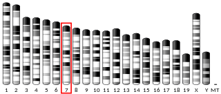
Killer-cell immunoglobulin-like receptors (KIRs), are a family of type I transmembrane glycoproteins expressed on the plasma membrane of natural killer (NK) cells and a minority of T cells. At least 15 genes and 2 pseudogenes encoding KIR map in a 150-kb region of the leukocyte receptor complex (LRC) on human chromosome 19q13.4.

Siglecs(Sialic acid-binding immunoglobulin-type lectins) are cell surface proteins that bind sialic acid. They are found primarily on the surface of immune cells and are a subset of the I-type lectins. There are 14 different mammalian Siglecs, providing an array of different functions based on cell surface receptor-ligand interactions.

<span class="mw-page-title-main">CD22</span> Lectin molecule

CD22, or cluster of differentiation-22, is a molecule belonging to the SIGLEC family of lectins. It is found on the surface of mature B cells and to a lesser extent on some immature B cells. Generally speaking, CD22 is a regulatory molecule that prevents the overactivation of the immune system and the development of autoimmune diseases.

An immunoreceptor tyrosine-based inhibitory motif (ITIM), is a conserved sequence of amino acids that is found intracellularly in the cytoplasmic domains of many inhibitory receptors of the non-catalytic tyrosine-phosphorylated receptor family found on immune cells. These immune cells include T cells, B cells, NK cells, dendritic cells, macrophages and mast cells. ITIMs have similar structures of S/I/V/LxYxxI/V/L, where x is any amino acid, Y is a tyrosine residue that can be phosphorylated, S is the amino acid serine, I is the amino acid isoleucine, and V is the amino acid valine. ITIMs recruit SH2 domain-containing phosphatases, which inhibit cellular activation. ITIM-containing receptors often serve to target immunoreceptor tyrosine-based activation motif (ITAM)-containing receptors, resulting in an innate inhibition mechanism within cells. ITIM bearing receptors have important role in regulation of immune system allowing negative regulation at different levels of the immune response.

Ly49 is a family of membrane C-type lectin-like receptors expressed mainly on NK cells but also on other immune cells. Their primary role is to bind MHC-I molecules to distinguish between self healthy cells and infected or altered cells. Ly49 family is coded by Klra gene cluster and include genes for both inhibitory and activating paired receptors, but most of them are inhibitory. Inhibitory Ly49 receptors play a role in the recognition of self cells and thus maintain self-tolerance and prevent autoimmunity by suppressing NK cell activation. On the other hand, activating receptors recognise ligands from cancer or viral infected cells and are used when cells lack or have abnormal expression of MHC-I molecules, which activate cytokine production and cytotoxic activity of NK and immune cells.

<span class="mw-page-title-main">LILRB1</span> Protein-coding gene in the species Homo sapiens

Leukocyte immunoglobulin-like receptor subfamily B member 1 is a protein that in humans is encoded by the LILRB1 gene.

<span class="mw-page-title-main">CD244</span> Protein found in humans

CD244 also known as 2B4 or SLAMF4 is a protein that in humans is encoded by the CD244 gene.

<span class="mw-page-title-main">KIR3DL1</span> Protein-coding gene in the species Homo sapiens

Killer cell immunoglobulin-like receptor 3DL1 is a protein that in humans is encoded by the KIR3DL1 gene.

<span class="mw-page-title-main">KIR2DL4</span> Protein-coding gene in the species Homo sapiens

Killer cell immunoglobulin-like receptor 2DL4 is a protein that in humans is encoded by the KIR2DL4 gene.

<span class="mw-page-title-main">CLEC7A</span> Protein-coding gene in humans

C-type lectin domain family 7 member A or Dectin-1 is a protein that in humans is encoded by the CLEC7A gene. CLEC7A is a member of the C-type lectin/C-type lectin-like domain (CTL/CTLD) superfamily. The encoded glycoprotein is a small type II membrane receptor with an extracellular C-type lectin-like domain fold and a cytoplasmic domain with a partial immunoreceptor tyrosine-based activation motif. It functions as a pattern-recognition receptor for a variety of β-1,3-linked and β-1,6-linked glucans from fungi and plants, and in this way plays a role in innate immune response. Expression is found on myeloid dendritic cells, monocytes, macrophages and B cells. Alternate transcriptional splice variants, encoding different isoforms, have been characterized. This gene is closely linked to other CTL/CTLD superfamily members on chromosome 12p13 in the natural killer gene complex region.

<span class="mw-page-title-main">LILRB2</span> Protein-coding gene in the species Homo sapiens

Leukocyte immunoglobulin-like receptor subfamily B member 2 is a protein that in humans is encoded by the LILRB2 gene.

<span class="mw-page-title-main">LILRB4</span> Protein-coding gene in the species Homo sapiens

Leukocyte immunoglobulin-like receptor subfamily B member 4 is a protein that in humans is encoded by the LILRB4 gene.

<span class="mw-page-title-main">KLRB1</span> Protein-coding gene in humans

Killer cell lectin-like receptor subfamily B, member 1, also known as KLRB1, NKR-P1A or CD161, is a human gene.

<span class="mw-page-title-main">KIR2DS4</span> Protein-coding gene in the species Homo sapiens

Killer cell immunoglobulin-like receptor 2DS4 is a protein that in humans is encoded by the KIR2DS4 gene.

<span class="mw-page-title-main">FCGR2B</span>

Fc fragment of IgG receptor IIb is a low affinity inhibitory receptor for the Fc region of immunoglobulin gamma (IgG). FCGR2B participates in the phagocytosis of immune complexes and in the regulation of antibody production by B lymphocytes.

The following outline is provided as an overview of and topical guide to immunology:

<span class="mw-page-title-main">Killer activation receptor</span> Class of protein

Killer Activation Receptors (KARs) are receptors expressed on the plasmatic membrane of Natural Killer cells. KARs work together with inhibitory receptors, which inactivate them in order to regulate the NK cells functions on hosted or transformed cells. These two kinds of specific receptors have some morphological features in common, such as being transmembrane proteins. The similarities are specially found in the extracellular domains and, the differences tend to be in the intracellular domains. KARs and KIRs can have tyrosine containing activatory or inhibitory motifs in the intracellular part of the receptor molecule.

<span class="mw-page-title-main">Killer cell immunoglobulin-like receptor 2DL3</span>

KIR2DL3, Killer cell immunoglobulin-like receptor 2DL3 is a transmembrane glycoprotein expressed by the natural killer cells and the subsets of the T cells. The KIR genes are polymorphic, which means that they have many different alleles. The KIR genes are also extremely homologous, which means that they are similar in position, structure and evolutionary origin, but not necessarily in function.

<span class="mw-page-title-main">SIGLEC6</span> Protein-coding gene in the species Homo sapiens

Sialic acid-binding Ig-like lectin 6 is a protein that in humans is encoded by the SIGLEC6 gene. The gene was originally named CD33L (CD33-like) due to similarities between these genes but later became known as OB-BP1 due to its ability to bind to this factor and, finally, SIGLEC6 as the sixth member of the SIGLEC family of receptors to be identified. The protein has also been given the CD designation CD327.

References

  1. 1 2 Lu, Q.; Lu, G.; Qi, J.; Wang, H.; Xuan, Y.; Wang, Q.; Li, Y.; Zhang, Y.; Zheng, C.; Fan, Z.; Yan, J.; Gao, G. F. (2014-06-03). "PILR and PILR have a siglec fold and provide the basis of binding to sialic acid". Proceedings of the National Academy of Sciences. 111 (22): 8221–8226. doi: 10.1073/pnas.1320716111 . PMC   4050567 . PMID   24843130.
  2. Lanier, Lewis L (June 2001). "Face off — the interplay between activating and inhibitory immune receptors". Current Opinion in Immunology. 13 (3): 326–331. doi:10.1016/S0952-7915(00)00222-3. PMID   11406364.
  3. 1 2 3 4 5 6 7 8 9 10 11 12 13 14 Kuroki, Kimiko; Furukawa, Atsushi; Maenaka, Katsumi (2012). "Molecular Recognition of Paired Receptors in the Immune System". Frontiers in Microbiology. 3: 429. doi: 10.3389/fmicb.2012.00429 . PMC   3533184 . PMID   23293633.
  4. 1 2 3 4 5 6 7 8 9 10 11 12 13 14 15 16 17 18 Levi-Schaffer, Francesca; Mandelboim, Ofer (February 2018). "Inhibitory and Coactivating Receptors Recognising the Same Ligand: Immune Homeostasis Exploited by Pathogens and Tumours". Trends in Immunology. 39 (2): 112–122. doi:10.1016/j.it.2017.10.001. PMC   7106362 . PMID   29066058.
  5. 1 2 3 4 5 6 7 8 9 10 11 12 13 14 15 16 17 18 19 Akkaya, Munir; Barclay, A. Neil (February 2013). "How do pathogens drive the evolution of paired receptors?: HIGHLIGHTS". European Journal of Immunology. 43 (2): 303–313. doi: 10.1002/eji.201242896 . PMID   23280392. S2CID   677081.
  6. 1 2 Rumpret, Matevž; Drylewicz, Julia; Ackermans, Laura J. E.; Borghans, José A. M.; Medzhitov, Ruslan; Meyaard, Linde (December 2020). "Functional categories of immune inhibitory receptors". Nature Reviews Immunology. 20 (12): 771–780. doi:10.1038/s41577-020-0352-z. PMID   32612208. S2CID   220293404.
  7. 1 2 3 4 Martinet, Ludovic; Smyth, Mark J. (April 2015). "Balancing natural killer cell activation through paired receptors". Nature Reviews Immunology. 15 (4): 243–254. doi:10.1038/nri3799. PMID   25743219. S2CID   20825600.
  8. 1 2 Petrie, Emma J.; Clements, Craig S.; Lin, Jie; Sullivan, Lucy C.; Johnson, Darryl; Huyton, Trevor; Heroux, Annie; Hoare, Hilary L.; Beddoe, Travis; Reid, Hugh H.; Wilce, Matthew C.J.; Brooks, Andrew G.; Rossjohn, Jamie (2008-03-17). "CD94-NKG2A recognition of human leukocyte antigen (HLA)-E bound to an HLA class I leader sequence". Journal of Experimental Medicine. 205 (3): 725–735. doi:10.1084/jem.20072525. PMC   2275392 . PMID   18332182.
  9. Arase, Hisashi; Lanier, Lewis L. (March 2004). "Specific recognition of virus-infected cells by paired NK receptors: Paired NK receptors". Reviews in Medical Virology. 14 (2): 83–93. doi: 10.1002/rmv.422 . PMID   15027001. S2CID   22034323.
  10. 1 2 3 Schwarz, Flavio; Landig, Corinna S; Siddiqui, Shoib; Secundino, Ismael; Olson, Joshua; Varki, Nissi; Nizet, Victor; Varki, Ajit (2017-03-15). "Paired Siglec receptors generate opposite inflammatory responses to a human‐specific pathogen". The EMBO Journal. 36 (6): 751–760. doi:10.15252/embj.201695581. PMC   5350563 . PMID   28100677.
  11. Barclay, A. Neil; Hatherley, Deborah (November 2008). "The Counterbalance Theory for Evolution and Function of Paired Receptors". Immunity. 29 (5): 675–678. doi:10.1016/j.immuni.2008.10.004. PMC   3128988 . PMID   19006692.
  12. 1 2 3 4 Miletić, Antonija; Krmpotić, Astrid; Jonjić, Stipan (April 2013). "The evolutionary arms race between NK cells and viruses: Who gets the short end of the stick?: HIGHLIGHTS". European Journal of Immunology. 43 (4): 867–877. doi: 10.1002/eji.201243101 .
  13. Viertlboeck, Birgit C.; Hanczaruk, Matthias A.; Amann, Barbara; Bader, Sophie R.; Schmitt, Ramona; Sperling, Beatrice; Schwarz, Susanne C.N.; Schmahl, Wolfgang; Deeg, Cornelia A.; Göbel, Thomas W. (November 2013). "Chicken immunoregulatory Ig-like receptor families: An overview and expression details on ggTREM-A1". Developmental & Comparative Immunology. 41 (3): 403–412. doi:10.1016/j.dci.2013.04.017. PMID   23648646.
  14. Guselnikov, Sergey V; Ramanayake, Thaminda; Erilova, Aleksandra Y; Mechetina, Ludmila V; Najakshin, Alexander M; Robert, Jacques; Taranin, Alexander V (2008). "The Xenopus FcR family demonstrates continually high diversification of paired receptors in vertebrate evolution". BMC Evolutionary Biology. 8 (1): 148. doi: 10.1186/1471-2148-8-148 . PMC   2413239 . PMID   18485190.
  15. Zimmermann, Wolfgang; Kammerer, Robert (December 2016). "Coevolution of paired receptors in Xenopus carcinoembryonic antigen-related cell adhesion molecule families suggests appropriation as pathogen receptors". BMC Genomics. 17 (1): 928. doi: 10.1186/s12864-016-3279-9 . PMC   5112662 . PMID   27852220.
  16. Suzuki, Takashi; Shin-I, Tadasu; Fujiyama, Asao; Kohara, Yuji; Kasahara, Masanori (2005-03-01). "Hagfish Leukocytes Express a Paired Receptor Family with a Variable Domain Resembling Those of Antigen Receptors". The Journal of Immunology. 174 (5): 2885–2891. doi: 10.4049/jimmunol.174.5.2885 . PMID   15728499.
  17. Fan, Qing R.; Long, Eric O.; Wiley, Don C. (May 2001). "Crystal structure of the human natural killer cell inhibitory receptor KIR2DL1–HLA-Cw4 complex". Nature Immunology. 2 (5): 452–460. doi:10.1038/87766. PMID   11323700. S2CID   24707532.
  18. 1 2 3 Burshtyn, Deborah N.; Morcos, Chris (2016-02-01). "The Expanding Spectrum of Ligands for Leukocyte Ig-like Receptors". The Journal of Immunology. 196 (3): 947–955. doi: 10.4049/jimmunol.1501937 . PMID   26802060.
  19. Yamada, Eriko; McVicar, Daniel W. (May 2008). "Paired Receptor Systems of the Innate Immune System". Current Protocols in Immunology. 81 (1): Appendix-1X. doi:10.1002/0471142735.ima01xs81. PMC   2771913 . PMID   18491293.
  20. Bruijnesteijn, Jesse; van der Wiel, Marit K. H.; de Groot, Nanine; Otting, Nel; de Vos-Rouweler, Annemiek J. M.; Lardy, Neubury M.; de Groot, Natasja G.; Bontrop, Ronald E. (2018-12-04). "Extensive Alternative Splicing of KIR Transcripts". Frontiers in Immunology. 9: 2846. doi: 10.3389/fimmu.2018.02846 . PMC   6288254 . PMID   30564240.
  21. Bruijnesteijn, Jesse; de Groot, Natasja G.; Bontrop, Ronald E. (2020-09-11). "The Genetic Mechanisms Driving Diversification of the KIR Gene Cluster in Primates". Frontiers in Immunology. 11: 582804. doi: 10.3389/fimmu.2020.582804 . PMC   7516082 . PMID   33013938.
  22. Lewis Marffy, Alexander L.; McCarthy, Alex J. (2020-05-13). "Leukocyte Immunoglobulin-Like Receptors (LILRs) on Human Neutrophils: Modulators of Infection and Immunity". Frontiers in Immunology. 11: 857. doi: 10.3389/fimmu.2020.00857 . PMC   7237751 . PMID   32477348.
  23. Takahashi, Shinichiro (2018-05-23). "Molecular functions of SIRPα and its role in cancer (Review)". Biomedical Reports. 9 (1): 3–7. doi:10.3892/br.2018.1102. PMC   6006759 . PMID   29930800.
  24. 1 2 Barclay, A. Neil; van den Berg, Timo K. (2014-03-21). "The Interaction Between Signal Regulatory Protein Alpha (SIRP α ) and CD47: Structure, Function, and Therapeutic Target". Annual Review of Immunology. 32 (1): 25–50. doi:10.1146/annurev-immunol-032713-120142. PMID   24215318.
  25. 1 2 3 Han, Zi-Wen; Lyv, Zhi-Wu; Cui, Bin; Wang, Ying-Ying; Cheng, Jun-Ting; Zhang, Ying; Cai, Wen-Qi; Zhou, Yang; Ma, Zhao-Wu; Wang, Xian-Wang; Peng, Xiao-Chun; Cui, Shu-Zhong; Xiang, Ying; Yang, Mo; Xin, Hong-Wu (December 2020). "The old CEACAMs find their new role in tumor immunotherapy". Investigational New Drugs. 38 (6): 1888–1898. doi:10.1007/s10637-020-00955-w. PMID   32488569. S2CID   219176219.
  26. Kuespert, Katharina; Pils, Stefan; Hauck, Christof R (October 2006). "CEACAMs: their role in physiology and pathophysiology". Current Opinion in Cell Biology. 18 (5): 565–571. doi:10.1016/j.ceb.2006.08.008. PMC   7127089 . PMID   16919437.
  27. Lenza, María Pia; Atxabal, Unai; Oyenarte, Iker; Jiménez-Barbero, Jesús; Ereño-Orbea, June (2020-12-15). "Current Status on Therapeutic Molecules Targeting Siglec Receptors". Cells. 9 (12): 2691. doi: 10.3390/cells9122691 . PMC   7765293 . PMID   33333862.
  28. Toyonaga, Kenji; Yamasaki, Sho (2020). "Recognition of Mycobacteria by Dendritic Cell Immunoactivating Receptor". C-Type Lectins in Immune Homeostasis. Current Topics in Microbiology and Immunology. 429: 103–115. doi:10.1007/82_2020_203. ISBN   978-3-030-62236-7. PMID   32300915. S2CID   215804099.