
Trematoda is a class of flatworms known as flukes or trematodes. They are obligate internal parasites with a complex life cycle requiring at least two hosts. The intermediate host, in which asexual reproduction occurs, is usually a snail. The definitive host, where the flukes sexually reproduce, is a vertebrate. Infection by trematodes can cause disease in all five traditional vertebrate classes: mammals, birds, amphibians, reptiles, and fish.

Clonorchis sinensis, the Chinese liver fluke, is a liver fluke belonging to the class Trematoda, phylum Platyhelminthes. It infects fish-eating mammals, including humans. In humans, it infects the common bile duct and gall bladder, feeding on bile. It was discovered by British physician James McConnell at the Medical College Hospital in Calcutta (Kolkata) in 1874. The first description was given by Thomas Spencer Cobbold, who named it Distoma sinense. The fluke passes its lifecycle in three different hosts, namely freshwater snail as first intermediate hosts, freshwater fish as second intermediate host, and mammals as definitive hosts.

Digenea is a class of trematodes in the Platyhelminthes phylum, consisting of parasitic flatworms with a syncytial tegument and, usually, two suckers, one ventral and one oral. Adults commonly live within the digestive tract, but occur throughout the organ systems of all classes of vertebrates. Once thought to be related to the Monogenea, it is now recognised that they are closest to the Aspidogastrea and that the Monogenea are more closely allied with the Cestoda. Around 6,000 species have been described to date.
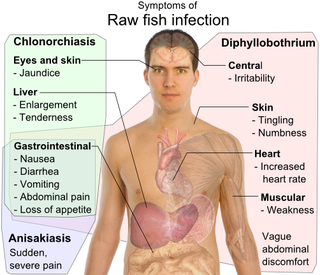
Clonorchiasis is an infectious disease caused by the Chinese liver fluke and two related species. Clonorchiasis is a known risk factor for the development of cholangiocarcinoma, a neoplasm of the biliary system.

Fasciola hepatica, also known as the common liver fluke or sheep liver fluke, is a parasitic trematode of the class Trematoda, phylum Platyhelminthes. It infects the livers of various mammals, including humans, and is transmitted by sheep and cattle to humans all over the world. The disease caused by the fluke is called fasciolosis or fascioliasis, which is a type of helminthiasis and has been classified as a neglected tropical disease. Fasciolosis is currently classified as a plant/food-borne trematode infection, often acquired through eating the parasite's metacercariae encysted on plants. F. hepatica, which is distributed worldwide, has been known as an important parasite of sheep and cattle for decades and causes significant economic losses in these livestock species, up to £23 million in the UK alone. Because of its relatively large size and economic importance, it has been the subject of many scientific investigations and may be the best-known of any trematode species. The closest relative of Fasciola hepatica is F. gigantica. These two flukes are sister species; they share many morphological features and can mate with each other.

Trematodes are parasitic flatworms of the class Trematoda, specifically parasitic flukes with two suckers: one ventral and the other oral. Trematodes are covered by a tegument, that protects the organism from the environment by providing secretory and absorptive functions.

Paragonimus westermani is the most common species of lung fluke that infects humans, causing paragonimiasis. Human infections are most common in eastern Asia and in South America. Paragonimiasis may present as a sub-acute to chronic inflammatory disease of the lung. It was discovered by Dutch zoologist Coenraad Kerbert in 1878.

Echinostoma is a genus of trematodes (flukes), which can infect both humans and other animals. These intestinal flukes have a three-host life cycle with snails or other aquatic organisms as intermediate hosts, and a variety of animals, including humans, as their definitive hosts.

Paragonimiasis is a food-borne parasitic disease caused by several species of lung flukes belonging to genus Paragonimus. Infection is acquired by eating crustaceans such as crabs and crayfishes which host the infective forms called metacercariae, or by eating raw or undercooked meat of mammals harboring the metacercariae from crustaceans.

Dicrocoelium dendriticum, the lancet liver fluke, is a parasite fluke that tends to live in cattle or other grazing mammals.

Fasciolopsis is a genus of trematodes. They are also known as giant intestinal flukes.

Heterophyes heterophyes, or the intestinal fish fluke, was discovered by Theodor Maximaillian Bilharz in 1851. This parasite was found during an autopsy of an Egyptian mummy. H. heterophyes is found in the Middle East, West Europe and Africa. They use different species to complete their complex lifestyle. Humans and other mammals are the definitive host, first intermediate host are snails, and second intermediate are fish. Mammals that come in contact with the parasite are dogs, humans, and cats. Snails that are affected by this parasite are the Cerithideopsilla conica. Fish that come in contact with this parasite are Mugil cephalus, Tilapia milotica, Aphanius fasciatus, and Acanthgobius sp. Humans and mammals will come in contact with this parasite by the consumption of contaminated or raw fish. This parasite is one of the smallest endoparasite to infect humans. It can cause intestinal infection called heterophyiasis.

Clinostomum marginatum is a species of parasitic fluke. It is commonly called the "Yellow grub". It is found in many freshwater fish in North America, and no fish so far is immune to this parasite. It is also found in frogs. Clinostomum marginatum can also be found in the mouth of aquatic birds such as herons and egrets. They are commonly present in the esophagus of fish-eating birds and reptiles. Eggs of these trematodes are shed in the feces of aquatic birds and released into water. Aquatic birds become hosts of this parasite by ingesting infected freshwater fish. The metacercariae are found right beneath the skin or in the muscles of host fish.
Megalodiscus temperatus is a Digenean in the phylum Platyhelminthes. This parasite belongs to the Cladorchiidae family and is a common parasite located in the urinary bladder and rectum of frogs. The primary host is frogs and the intermediate hosts of Megalodiscus temeperatus are freshwater snails in the genus Helisoma.
Philophthalmus gralli, commonly known as the Oriental avian eye fluke, parasitises the conjunctival sac of the eyes of many species of birds, including birds of the orders Galliformes and Anseriformes. In Brazil this parasite was reported in native Anseriformes species. It was first discovered by Mathis and Leger in 1910 in domestic chickens from Hanoi, Vietnam. Birds are definitive hosts and freshwater snail species are intermediate hosts. Human cases of philophthalmosis are rare, but have been previously reported in Europe, Asia, and America.

Alaria is a genus of flatworms, or trematodes, in the family Diplostomidae.
Paragonimus kellicotti, the North American lung fluke, is a species of parasitic trematode in the genus Paragonimus. This species of Paragonimus has an intricate lifecycle, and although its name may suggest that it is only a health concern in North America, it is also prominent in Southeast Asia and China.
Paragonimus skrjabini is classified as a species in the genus Paragonimus, which consists of many species of lung flukes that result in the food-borne parasitic disease paragonimiasis.

Trematodiasis is a group of parasitic infections caused by different species of flukes, in humans mainly by digenean trematodes. Symptoms can range from mild to severe depending on the species, number and location of trematodes in the infected organism. Symptoms depend on the type of trematode present, and include chest and abdominal pain, high temperature, digestion issues, cough and shortness of breath, diarrhoea and change in appetite.

Gastropod-borne parasitic diseases (GPDs) are a group of infectious diseases that require a gastropod species to serve as an intermediate host for a parasitic organism that can infect humans upon ingesting the parasite or coming into contact with contaminated water sources. These diseases can cause a range of symptoms, from mild discomfort to severe, life-threatening conditions, with them being prevalent in many parts of the world, particularly in developing regions. Preventive measures such as proper sanitation and hygiene practices, avoiding contact with infected gastropods and cooking or boiling food properly can help to reduce the risk of these diseases.

