Wright's stain

Last updated
Wright's stain, with red blood cells taking up eosin Y, azure B giving nuclei a purple color, and methylene blue coloring the cytoplasm of this plasmablast. Wright's staining of multiple myeloma, plasmablastic type.png
Wright's stain, with red blood cells taking up eosin Y, azure B giving nuclei a purple color, and methylene blue coloring the cytoplasm of this plasmablast.

Wright's stain is a hematologic stain that facilitates the differentiation of blood cell types. It is classically a mixture of eosin (red) and methylene blue dyes. It is used primarily to stain peripheral blood smears, urine samples, and bone marrow aspirates, which are examined under a light microscope. In cytogenetics, it is used to stain chromosomes to facilitate diagnosis of syndromes and diseases.

It is named for James Homer Wright, who devised the stain, a modification of the Romanowsky stain, in 1902. Because it distinguishes easily between blood cells, it became widely used for performing differential white blood cell counts, which are routinely ordered when conditions such as infection or leukemia are suspected.

The related stains are known as the buffered Wright stain, the Wright-Giemsa stain (a combination of Wright and Giemsa stains), and the buffered Wright-Giemsa stain, and specific instructions depend on the solutions being used, which may include eosin Y, azure B, and methylene blue (some commercial preparations combine solutions to simplify staining). [1] The May–Grünwald stain, which produces a more intense coloration, also takes a longer time to perform.

Urine samples stained with Wright's stain will identify eosinophils, which can indicate interstitial nephritis or urinary tract infection. [2]

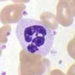
White blood cells stained with Wright's stain:

See also

Related Research Articles

Cystoscopy Medical procedure; endoscopy of the urinary bladder via the urethra

Cystoscopy is endoscopy of the urinary bladder via the urethra. It is carried out with a cystoscope.

Romanowsky stain Family of related stains for examination of blood including the detection of parasites

Romanowsky staining, also known as Romanowsky–Giemsa staining, is a prototypical staining technique that was the forerunner of several distinct but similar stains widely used in hematology and cytopathology. Romanowsky-type stains are used to differentiate cells for microscopic examination in pathological specimens, especially blood and bone marrow films, and to detect parasites such as malaria within the blood. Stains that are related to or derived from the Romanowsky-type stains include Giemsa, Jenner, Wright, Field, May–Grünwald and Leishman stains. The staining technique is named after the Russian physician Dmitri Leonidovich Romanowsky (1861–1921), who was one of the first to recognize its potential for use as a blood stain.

Methylthioninium chloride, commonly called methylene blue, is a salt used as a dye and as a medication. Methylene blue is a thiazine dye. As a medication, it is mainly used to treat methemoglobinemia by converting the ferric iron in hemoglobin to ferrous iron. Specifically, it is used to treat methemoglobin levels that are greater than 30% or in which there are symptoms despite oxygen therapy. It has previously been used for cyanide poisoning and urinary tract infections, but this use is no longer recommended.

Staining Technique used to enhance visual contrast of specimens observed under a microscope

Staining is a technique used to enhance contrast in samples, generally at the microscopic level. Stains and dyes are frequently used in histology and in the medical fields of histopathology, hematology, and cytopathology that focus on the study and diagnoses of disease at a microscopic level. Stains may be used to define biological tissues, cell populations, or organelles within individual cells.

Blood smear Stained blood on microscope slide

A blood smear, peripheral blood smear or blood film is a thin layer of blood smeared on a glass microscope slide and then stained in such a way as to allow the various blood cells to be examined microscopically. Blood smears are examined in the investigation of hematological (blood) disorders and are routinely employed to look for blood parasites, such as those of malaria and filariasis.

Urinalysis Array of tests performed on urine

Urinalysis, a portmanteau of the words urine and analysis, is a panel of medical tests that includes physical (macroscopic) examination of the urine, chemical evaluation using urine test strips, and microscopic examination. Macroscopic examination targets parameters such as color, clarity, odor, and specific gravity; urine test strips measure chemical properties such as pH, glucose concentration, and protein levels; and microscopy is performed to identify elements such as cells, urinary casts, crystals, and organisms.

Giemsa stain

Giemsa stain, named after German chemist and bacteriologist Gustav Giemsa, is a nucleic acid stain used in cytogenetics and for the histopathological diagnosis of malaria and other parasites.

Periodic acid–Schiff stain Histological staining method

Periodic acid–Schiff (PAS) is a staining method used to detect polysaccharides such as glycogen, and mucosubstances such as glycoproteins, glycolipids and mucins in tissues. The reaction of periodic acid oxidizes the vicinal diols in these sugars, usually breaking up the bond between two adjacent carbons not involved in the glycosidic linkage or ring closure in the ring of the monosaccharide units that are parts of the long polysaccharides, and creating a pair of aldehydes at the two free tips of each broken monosaccharide ring. The oxidation condition has to be sufficiently regulated so as to not oxidize the aldehydes further. These aldehydes then react with the Schiff reagent to give a purple-magenta color. A suitable basic stain is often used as a counterstain.

Papanicolaou stain Histological staining method

Papanicolaou stain is a multichromatic (multicolored) cytological staining technique developed by George Papanicolaou in 1942. The Papanicolaou stain is one of the most widely used stains in cytology, where it is used to aid pathologists in making a diagnosis. Although most notable for its use in the detection of cervical cancer in the Pap test or Pap smear, it is also used to stain non-gynecological specimen preparations from a variety of bodily secretions and from small needle biopsies of organs and tissues. Papanicolaou published three formulations of this stain in 1942, 1954, and 1960.

Urinary cast Cylindrical protein structure in urine in certain disease states

Urinary casts are microscopic cylindrical structures produced by the kidney and present in the urine in certain disease states. They form in the distal convoluted tubule and collecting ducts of nephrons, then dislodge and pass into the urine, where they can be detected by microscopy.

Leishman stain Diagnostic technique

Leishman stain, also known as Leishman's stain, is used in microscopy for staining blood smears. It is generally used to differentiate between and identify white blood cells, malaria parasites, and trypanosomas. It is based on a methanolic mixture of "polychromed" methylene blue and eosin. The methanolic stock solution is stable and also serves the purpose of directly fixing the smear eliminating a prefixing step. If a working solution is made by dilution with an aqueous buffer, the resulting mixture is very unstable and cannot be used for long. Leishman stain is named after its inventor, the Scottish pathologist William Boog Leishman. It is a version of the Romanowsky stain, and is thus similar to and partially replaceable by Giemsa stain, Jenner's stain, and Wright's stain.

Field stain

Field stain is a histological method for staining of blood smears. It is used for staining thick blood films in order to discover malarial parasites. Field's stain is a version of a Romanowsky stain, used for rapid processing of the specimens.

Thionine Chemical compound and histologic stain C12H10N3S

Thionine, also known as Lauth's violet, is the salt of a heterocyclic compound. It was firstly synthesised by Charles Lauth. A variety of salts are known including the chloride and acetate, called respectively thionine chloride and thionine acetate. The dye is structurally related to methylene blue, which also features a phenothiazine core. The dye's name is frequently misspelled with omission of the e, and is not to be confused with the plant protein thionin. The -ine ending indicates that the compound is an amine.

Gustav Giemsa German scientist

Gustav Giemsa was a German chemist and bacteriologist who was a native of Medar-Blechhammer. He is remembered for creating a dye solution commonly known as "Giemsa stain". This dye is used for the histopathological diagnosis of malaria and parasites such as Plasmodium, Trypanosoma, and Chlamydia.

Diff-Quik

Diff-Quik is a commercial Romanowsky stain variant used to rapidly stain and differentiate a variety of pathology specimens. It is most frequently used for blood films and cytopathological smears, including fine needle aspirates. The Diff-Quik procedure is based on a modification of the Wright-Giemsa stain pioneered by Harleco in the 1970s, and has advantages over the routine Wright-Giemsa staining technique in that it reduces the 4-minute process into a much shorter operation and allows for selective increased eosinophilic or basophilic staining depending upon the time the smear is left in the staining solutions.

Tzanck test

In dermatopathology, the Tzanck test, also Tzanck smear, is scraping of an ulcer base to look for Tzanck cells. It is sometimes also called the chickenpox skin test and the herpes skin test. It is a simple, low-cost, and rapid office based test.

Urine test strip

A urine test strip or dipstick is a basic diagnostic tool used to determine pathological changes in a patient's urine in standard urinalysis.

White blood cell Type of cells of the immunological system

White blood cells, also called leukocytes or leucocytes, are the cells of the immune system that are involved in protecting the body against both infectious disease and foreign invaders. All white blood cells are produced and derived from multipotent cells in the bone marrow known as hematopoietic stem cells. Leukocytes are found throughout the body, including the blood and lymphatic system.

Jaswant Singh–Bhattacharji stain, commonly referred to as JSB stain, is a rapid staining method for detection of malaria. It is useful for the diagnosis of malaria in thick smear samples of blood. The JSB stain is commonly used throughout India, but rarely used in other countries.

White blood cell differential Blood test

A white blood cell differential is a medical laboratory test that provides information about the types and amounts of white blood cells in a person's blood. The test, which is usually ordered as part of a complete blood count (CBC), measures the amounts of the five normal white blood cell types – neutrophils, lymphocytes, monocytes, eosinophils and basophils – as well as abnormal cell types if they are present. These results are reported as percentages and absolute values, and compared against reference ranges to determine whether the values are normal, low, or high. Changes in the amounts of white blood cells can aid in the diagnosis of many health conditions, including viral, bacterial, and parasitic infections and blood disorders such as leukemia.

References

  1. "Wright Giemsa Stains". Sigma-Aldrich. Retrieved 2016-10-28.
  2. Corwin, H. L.; Bray, R. A.; Haber, M. H. (1989-11-01). "The detection and interpretation of urinary eosinophils". Archives of Pathology & Laboratory Medicine. 113 (11): 1256–1258. ISSN   0003-9985. PMID   2479358.